. 2022 Dec 8;66(6):781-795.
doi: 10.1042/EBC20220042.

The premetazoan ancestry of the synaptic toolkit and appearance of first neurons

Affiliations

The premetazoan ancestry of the synaptic toolkit and appearance of first neurons

Jeffrey Colgren et al. Essays Biochem. .

Abstract

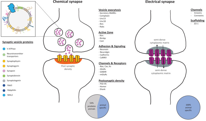
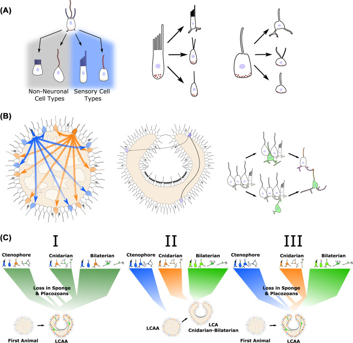
Neurons, especially when coupled with muscles, allow animals to interact with and navigate through their environment in ways unique to life on earth. Found in all major animal lineages except sponges and placozoans, nervous systems range widely in organization and complexity, with neurons possibly representing the most diverse cell-type. This diversity has led to much debate over the evolutionary origin of neurons as well as synapses, which allow for the directed transmission of information. The broad phylogenetic distribution of neurons and presence of many of the defining components outside of animals suggests an early origin of this cell type, potentially in the time between the first animal and the last common ancestor of extant animals. Here, we highlight the occurrence and function of key aspects of neurons outside of animals as well as recent findings from non-bilaterian animals in order to make predictions about when and how the first neuron(s) arose during animal evolution and their relationship to those found in extant lineages. With advancing technologies in single cell transcriptomics and proteomics as well as expanding functional techniques in non-bilaterian animals and the close relatives of animals, it is an exciting time to begin unraveling the complex evolutionary history of this fascinating animal cell type.

Keywords: choanoflagellates; cnidarians; ctenophores; placozoans; sponges; synapses.


Conflict of interest statement

The authors declare that there are no competing interests associated with the manuscript.

Figures

Figure 1
Figure 1. Major components of chemical and electrical synapses
Chemical synapses are composed of a rich repertoire of conserved proteins involved in bringing the pre- and post-synapse into close proximity, tightly regulating vesicle exocytosis, and clustering of receptors on the post-synapse. Many of these proteins originated before animal multicellularity [33–3533–35]. Electrical synapses are primarily established through the interaction of the innexins or connexins, neither of which have been identified outside of animals.
Figure 2
Figure 2. Origin of neurons and neuronal circuits
(A) Simplified diagram for division of labor of apical structures of a choanoflagellate-like ancestral cell type into villi- (blue) and cilia-based (red) structures on non-neuronal cells and sensory cells. Following the initial division, further specialization can occur under different pressures. (B) Establishment of early neuronal circuits based on diffusion based signaling from peptidergic sensory cells. Distinct sensory cells (blue and orange) release different peptides, which act on cells expressing specific receptors, based on [167]. As bodyplans increase in size and complexity, volumetric signaling is inefficient for full integration and rapid response. Right panel shows hypothetical transitions to directed signaling through interneurons. Synapses are indicated in red. (C) Hypothetical scenarios giving rise to the diversity of neurons and nervous systems seen in extant lineages. First showing a single origin, which diversified in each lineage and was lost in placozoans and possibly sponges. Next, independent origins in major lineages with neurons from a LCAA that did not have true neurons. Third, independent specialization of systems in extant lineages from different aspects of a LCAA with neurons. Better resolving the relationships between the cell types present in extant lineages will help understand which of these occurred.

References

    1. Martinez-Arca S., Coco S., Mainguy G., Schenk U., Alberts P., Bouillé P.et al. (2001) A common exocytotic mechanism mediates axonal and dendritic outgrowth. J. Neurosci. 21, 3830–3838 10.1523/JNEUROSCI.21-11-03830.2001 - DOI - PMC - PubMed
    1. Zhou Q., Xiao J. and Liu Y. (2000) Participation of syntaxin 1A in membrane trafficking involving neurite elongation and membrane expansion. J. Neurosci. Res. 61, 321–328 10.1002/1097-4547(20000801)61:3<321::AID-JNR10>3.0.CO;2-L - DOI - PubMed
    1. Urbina F.L., Gomez S.M. and Gupton S.L. (2018) Spatiotemporal organization of exocytosis emerges during neuronal shape change. J. Cell Biol. 217, 1113–1128 10.1083/jcb.201709064 - DOI - PMC - PubMed
    1. Vega I.E. and Hsu S.C. (2001) The exocyst complex associates with microtubules to mediate vesicle targeting and neurite outgrowth. J. Neurosci. 21, 3839–3848 10.1523/JNEUROSCI.21-11-03839.2001 - DOI - PMC - PubMed
    1. Wu B. and Guo W. (2015) The exocyst at a glance. J. Cell Sci. 128, 2957–2964 10.1242/jcs.156398 - DOI - PMC - PubMed

Publication types