. 2022 Oct 21;14(40):15141-15155.
doi: 10.1039/d2nr03792k.

Substrate stiffness reduces particle uptake by epithelial cells and macrophages in a size-dependent manner through mechanoregulation

Affiliations

Substrate stiffness reduces particle uptake by epithelial cells and macrophages in a size-dependent manner through mechanoregulation

Aaron Lee et al. Nanoscale. .

Abstract

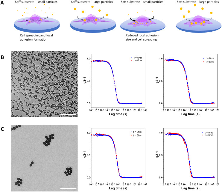
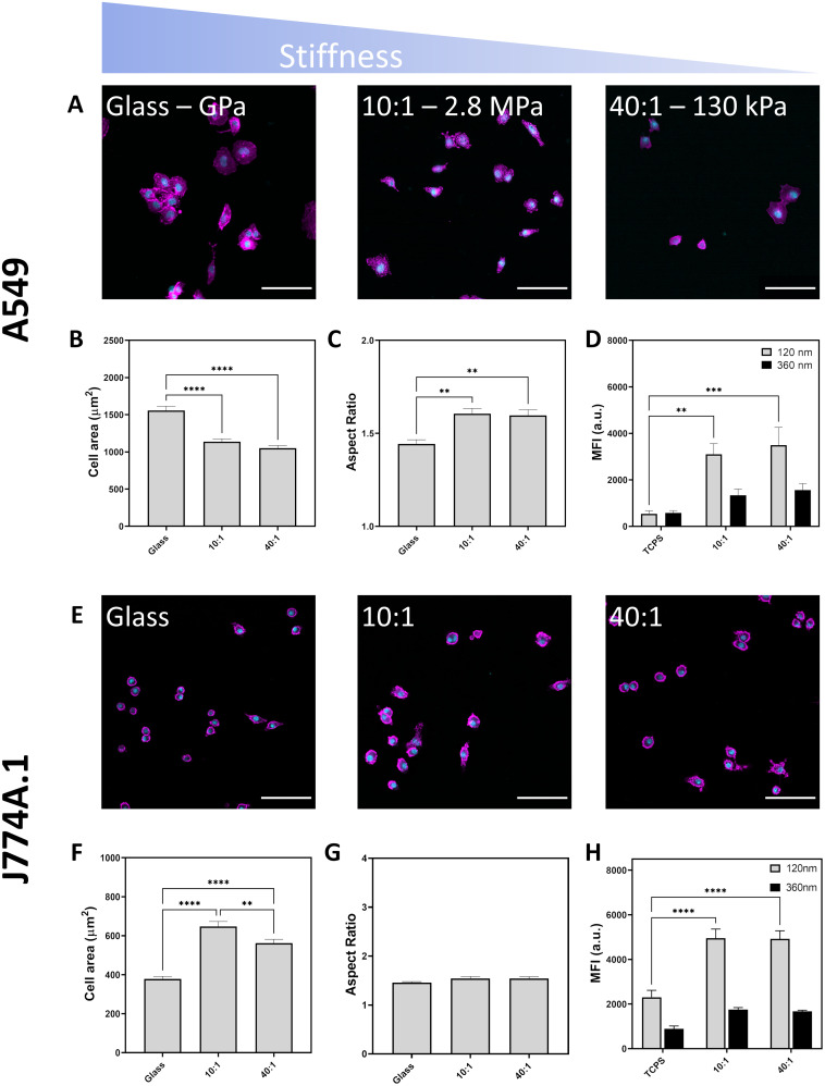
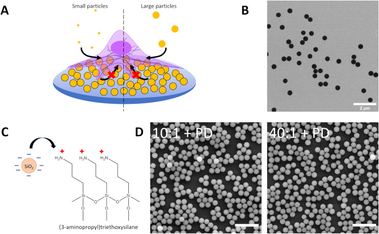
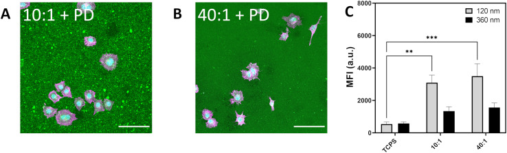
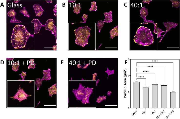
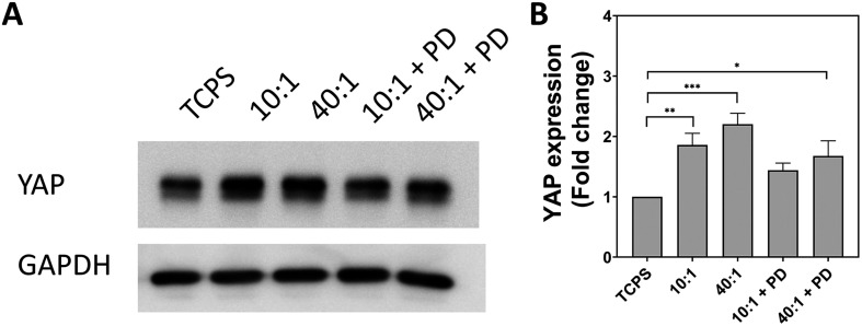
Cells continuously exert forces on their environment and respond to changes in mechanical forces by altering their behaviour. Many pathologies such as cancer and fibrosis are hallmarked by dysregulation in the extracellular matrix, driving aberrant behaviour through mechanotransduction pathways. We demonstrate that substrate stiffness can be used to regulate cellular endocytosis of particles in a size-dependent fashion. Culture of A549 epithelial cells and J774A.1 macrophages on polystyrene/glass (stiff) and polydimethylsiloxane (soft) substrates indicated that particle uptake is increased up to six times for A549 and two times for macrophages when cells are grown in softer environments. Furthermore, we altered surface characteristics through the attachment of submicron-sized particles as a method to locally engineer substrate stiffness and topography to investigate the biomechanical changes which occurred within adherent epithelial cells, i.e. characterization of A549 cell spreading and focal adhesion maturation. Consequently, decreasing substrate rigidity and particle-based topography led to a reduction of focal adhesion size. Moreover, expression levels of Yes-associated protein were found to correlate with the degree of particle endocytosis. A thorough appreciation of the mechanical cues may lead to improved solutions to optimize nanomedicine approaches for treatment of cancer and other diseases with abnormal mechanosignalling.


Conflict of interest statement

The authors have no conflicts of interest to declare.

Figures

Fig. 1
Fig. 1. (A) Schematic representation of the influence of substrate stiffness on cell endocytosis of 120 nm and 360 nm Cy5-labelled silica particles. Cell spreading and focal adhesion maturation on tissue culture polystyrene is abrogated when cultures are performed on softer fibronectin-coated PDMS 10 : 1 and fibronectin-coated PDMS 40 : 1. Increased internalization of particles was observed for cells on more mechanically compliant substrates. (B) Transmission electron micrograph of silica nanoparticles (dTEM = 120 nm). Corrected time course dynamic light scattering measurements performed in water and supplemented RPMI at 0 h and 6 h indicate particle stability. (C) Transmission electron micrograph of silica particles (dTEM = 360 nm). Corrected time course dynamic light scattering measurements performed in water and supplemented RPMI at 0 h and 6 h indicate no particle aggregation. Scale bars represent 2 μm.
Fig. 2
Fig. 2. Effect of substrate stiffness on cell morphology imaged by confocal laser scanning microscopy. Nuclei labelled in cyan and F-actin labelled in magenta. Scale bars represent 100 μm. (A) A549 attachment to glass, PDMS 10 : 1 and PDMS 40 : 1 following 24 hours of culture. (B) Cell area of A549 cultured on different substrates. (C) Aspect ratio of A549 cultured on mechanically distinct substrates. (D) Median fluorescence intensity of A549 cells upon exposure to fluorescent silica particles (120 nm and 360 nm) at a concentration of 20 μg mL−1 for six hours (E) light microscope images of J774A.1 cells showing their attachment and spreading on various surfaces. (F) Quantification of J774A.1 cell area on different substrates, (G) quantification of J774A.1 aspect ratio on different substrates. (H) Median fluorescent intensity of J774A.1 reflecting particle uptake via flow cytometry. All experiments performed with three biological replicates. Statistical significance determined by one-way ANOVA with Tukey's test, *p < 0.05, **p < 0.01, ***p < 0.001, ****p < 0.0001.
Fig. 3
Fig. 3. (A) Schematic depicting uptake of small (120 nm) and large (360 nm) Cy5-labelled silica particles by A549 on particle-decorated substrates to assess the influence of particle-generated topography on particle internalisation in a competition co-exposure system. Particle-decorated substrates consist of 420 nm BDP-labelled silica electrostatically adsorbed to APTES-modified PDMS 10 : 1 (10 : 1 + PD) and APTES-modified PDMS 40 : 1 (40 : 1 + PD). (B) Transmission electron microscope image of silica particles (dTEM = 420 nm) used to form the particle-decorated (PD). (C) Schematic of PDMS surface functionalization with 3-aminopropyltriethyoxysilane (APTES) to facilitate adsorption of negatively charged silica particles. (D) Scanning electron microscope image of PDMS 10 : 1 and PDMS 40 : 1 treated with APTES and decorated with silica particles (dTEM = 420 nm) to form a particle-decorated surface. Scale bar represents 2 μm.
Fig. 4
Fig. 4. (A) Light microscope image depicting A549 spreading on PDMS 10 : 1 decorated with silica particles. (B) A549 spreading on silica decorated PDMS 40 : 1. F-actin labelled in magenta, nuclei labelled in cyan and silica particle-decorated PDMS (BDP, 420 nm) in green. Scale bar represents 100 μm. (C) Median fluorescence intensity of A549 cultured on silica decorated PDMS showing uptake of Cy5-labelled silica 120 nm and 360 nm exposed for 6 h at 20 μg mL−1. Data expressed as mean + standard error of the mean. Statistical significance determined by one-way ANOVA with Tukey post hoc test for multiple comparisons. *p < 0.05.
Fig. 5
Fig. 5. Effect of substrate stiffness and topography on formation of focal adhesion plaques as determined by paxillin labelling and visualization by confocal laser scanning microscopy. A549 on (A) glass, (B) PDMS 10 : 1, (C) PDMS 40 : 1, (D) PDMS 10 : 1 + PD, (E) PDMS 40 : 1 + PD. F-actin labelled in magenta while paxillin is labelled with rabbit anti-paxillin and goat anti-rabbit IgG conjugated with AlexaFluor 555 in yellow. Scale bar represents 50 μm. (F) Paxillin area assessed over n > 100 cells over three biological replicates. Data expressed as mean + sem. Statistical analysis performed using a one-way ANOVA with Tukey's post hoc test, *****p < 0.0001. PD – particle-decorated surface.
Fig. 6
Fig. 6. Evaluation of YAP expression in A549 cells cultured for 24 hours on substrates with different stiffness and topography. (A) Bands obtained from western blot for YAP and GAPDH. GAPDH used as a loading control. (B) Fold change in YAP expression expressed as mean + standard error of the mean from three biologically independent replicates. Statistical significance determined by one-way ANOVA with Tukey's post hoc test. *p < 0.05, **p < 0.01, ***p < 0.001. TCPS – tissue cell culture plastic. PD – particle-decorated.

References

    1. Puech P. H. Bongrand P. Mechanotransduction as a major driver of cell behaviour: Mechanisms, and relevance to cell organization and future research. Open Biol. 2021;11:210256. doi: 10.1098/rsob.210256. - DOI - PMC - PubMed
    1. Selman M. Pardo A. Fibroageing: An ageing pathological feature driven by dysregulated extracellular matrix-cell mechanobiology. Ageing Res. Rev. 2021;70:101393. doi: 10.1016/j.arr.2021.101393. - DOI - PubMed
    1. Zhou Y. W. Wu Y. Substrate Viscoelasticity Amplifies Distinctions between Transient and Persistent LPS-Induced Signals. Adv. Healthcare Mater. 2021;11:2102271. doi: 10.1002/adhm.202102271. - DOI - PubMed
    1. Amer M. Shi L. Wolfenson H. The ‘yin and yang’ of cancer cell growth and mechanosensing. Cancers. 2021;13:4754. doi: 10.3390/cancers13194754. - DOI - PMC - PubMed
    1. Yang C. et al., Spatially patterned matrix elasticity directs stem cell fate. Proc. Natl. Acad. Sci. U. S. A. 2016;113:E4439–E4445. - PMC - PubMed