. 2022 Nov:85:104296.
doi: 10.1016/j.ebiom.2022.104296. Epub 2022 Oct 4.

The fatal trajectory of pulmonary COVID-19 is driven by lobular ischemia and fibrotic remodelling

Affiliations

The fatal trajectory of pulmonary COVID-19 is driven by lobular ischemia and fibrotic remodelling

Maximilian Ackermann et al. EBioMedicine. 2022 Nov.

Abstract

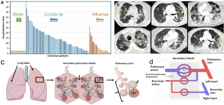
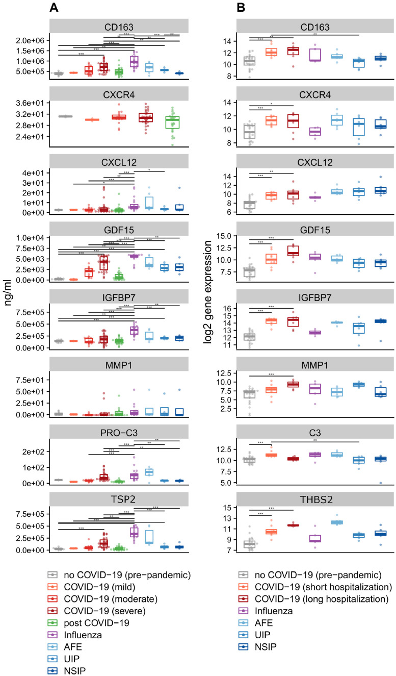
Background: COVID-19 is characterized by a heterogeneous clinical presentation, ranging from mild symptoms to severe courses of disease. 9-20% of hospitalized patients with severe lung disease die from COVID-19 and a substantial number of survivors develop long-COVID. Our objective was to provide comprehensive insights into the pathophysiology of severe COVID-19 and to identify liquid biomarkers for disease severity and therapy response.

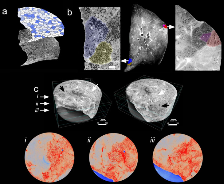
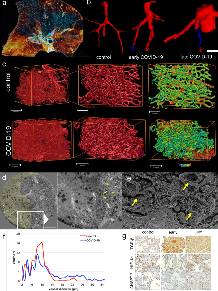
Methods: We studied a total of 85 lungs (n = 31 COVID autopsy samples; n = 7 influenza A autopsy samples; n = 18 interstitial lung disease explants; n = 24 healthy controls) using the highest resolution Synchrotron radiation-based hierarchical phase-contrast tomography, scanning electron microscopy of microvascular corrosion casts, immunohistochemistry, matrix-assisted laser desorption ionization mass spectrometry imaging, and analysis of mRNA expression and biological pathways. Plasma samples from all disease groups were used for liquid biomarker determination using ELISA. The anatomic/molecular data were analyzed as a function of patients' hospitalization time.

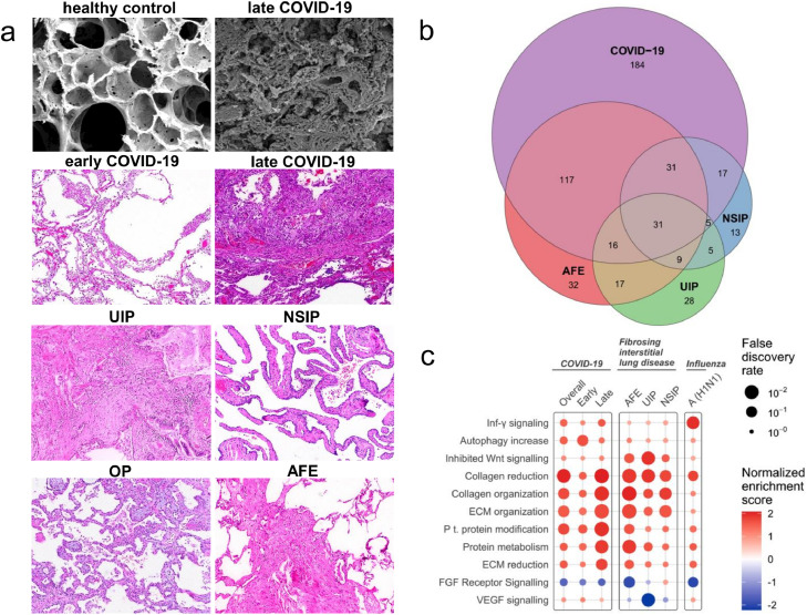
Findings: The observed patchy/mosaic appearance of COVID-19 in conventional lung imaging resulted from microvascular occlusion and secondary lobular ischemia. The length of hospitalization was associated with increased intussusceptive angiogenesis. This was associated with enhanced angiogenic, and fibrotic gene expression demonstrated by molecular profiling and metabolomic analysis. Increased plasma fibrosis markers correlated with their pulmonary tissue transcript levels and predicted disease severity. Plasma analysis confirmed distinct fibrosis biomarkers (TSP2, GDF15, IGFBP7, Pro-C3) that predicted the fatal trajectory in COVID-19.

Interpretation: Pulmonary severe COVID-19 is a consequence of secondary lobular microischemia and fibrotic remodelling, resulting in a distinctive form of fibrotic interstitial lung disease that contributes to long-COVID.

Funding: This project was made possible by a number of funders. The full list can be found within the Declaration of interests / Acknowledgements section at the end of the manuscript.

Keywords: Biomarkers; COVID-19; Fibrogenesis; Intussusceptive angiogenesis.


Conflict of interest statement

Declaration of interests HHK received fees for lectures and consultations from Roche Pharma AG, Novartis, AstraZeneca, Genomic Health, Pfizer, and Amgen, all outside the present study. MMH received fees for lectures and consultations from Acceleron, Actelion, Bayer, GSK, Janssen, MSD, and Pfizer, all outside the present study. TW declares funding by the German Ministry of Research and Education. MAK and DJL declare the possession of “Nordic Bioscience” stock options. BS received fees for lectures from Boehringer Ingelheim. The other authors have no potential conflicts of interest to report.

Figures

Figure 1
Figure 1
(a) Patient population with fatal COVID-19. Lung tissue and/or plasma samples were collected from 75 hospitalized COVID-19 patients, 17 hospitalized influenza A patients, and 19 healthy lung transplant donors. In most patients, serum and imaging studies were performed; a subset of patients was also analyzed via histopathology and gene expression analyses (dark blue and dark orange columns). (b) Dual energy computed tomography (CT) showed the characteristic features of severe COVID-19 pneumonia as bilateral ground-glass opacities, peribronchial consolidations, and a diffuse crazy-paving pattern characteristic of COVID-19 (yellow ellipses). (c) Illustration of anatomy and morphology of secondary lobules as an anatomical and functional subunit. (d) Schematic of pulmonary vascular supply consisting of pulmonary and bronchial circulation. The bronchial circulation primarily arises directly from the thoracic aorta and the intercostal arteries and constitutes less than 2% of the left cardiac output volume.
Figure 2
Figure 2
Synchrotron imaging of COVID-19 autopsy lungs. (a) Hierarchical phase-contrast tomography (HiP-CT) reconstructions segmented for secondary pulmonary lobules involved with microischemia. (b) Segmentation of individual secondary pulmonary lobules via HiP-CT depicts the presence of severely abnormal-appearing secondary lobules immediately juxtaposed to nearly normal-appearing lobules. (c) HiP-CT demonstrates the dramatic paucity of blood flow in diseased secondary lobules in the vicinity of fibrotic remodelled areas.
Figure 3
Figure 3
Microvascular alterations in COVID-19 lungs. (a) Volume rendering of a representative hierarchical phase contrast tomography (HiP-CT) slice demonstrates the spatial involvement of microischemia and fibrotic remodelling of airways. Blue color shows regular-shaped airways, whereas yellow depicts the subpleural hot spots of condensation and fibrotic remodelling. (b) Micro-CT-based 3D reconstruction of subsegmental pulmonary arteries (red) and airways (blue) demonstrated (sub-)total occlusion of the arteries in COVID-19-lungs of early and late hospitalized patients as compared to uninfected controls (scale bar equals 200 µm). (c) Three-dimensional evaluation of microvascular corrosion casts by Synchrotron radiation tomographic microscopy illustrating the altered and increased alveolar vascularity in COVID-19 lungs. (d) Representative slices of high-resolution scans from a HiP-CT image of a lung lobe affected by COVID-19 reveals the margin of well-demarcated secondary pulmonary lobules; left with signs of early fibrotic remodelling and microischemia and right with mostly preserved lung parenchyma and increased vascularity (arrows) at the vicinity of the lobular septum. (e) Scanning electron micrograph depicts the dilated blood vessels (arrows) and fibrotic strands along this lobular septum. (f) Volumetric assessment of vessel calibers based on Synchrotron radiation tomographic microscopy of microvascular corrosion casts underlines the occurrence of increased vessel diameters in COVID-19 lungs. (g) Immunohistochemical detection of transforming growth factor beta 1 (TGF-β1), hypoxia inducible factor 1 alpha (HIF1-α), and angiopoietin 2 (ANGPT-2) expression in COVID-19 lungs in the phase of early and late hospitalization compared to controls. Staining results demonstrate the spatial heterogeneity of markers of microischemia. Scale bars equal 600 µm.
Figure 4
Figure 4
Comparison of COVID-19 with common forms of fibrosing interstitial lung disease (ILD). (a) Different morphological patterns in lungs of early and late hospitalized COVID-19 patients compared to lungs of patients with ILD [usual interstitial pneumonia (UIP), non-specific interstitial pneumonia (NSIP), organizing pneumonia (OP), and alveolar fibroelastosis (AFE) lungs]. Scanning electron microscopy was performed using a Philips XL30 microscope (Philips, Eindhoven, the Netherlands) at 15 keV and 21 μA. Histopathological analysis was performed after hematoxylin and eosin staining. (b) Venn diagram of differentially expressed genes of COVID-19 and influenza lung samples compared with expression in controls (COVID-19, n = 15; influenza A lung samples, n = 7; ILD lung samples, n = 18 (UIP, n = 6; NSIP, n = 6; AFE, n = 6); healthy control lung samples, n = 25). (c) Functional pathway analysis highlights the differential functional gene expression in lung tissue of patients with COVID-19, ILD, and influenza A. Color indicates up- (red) and down (blue)-regulation; Circle size depicts the false discovery rate (FDR). Only significantly up- or down-regulated pathways are shown.
Figure 5
Figure 5
Biomarkers of early fibrotic remodelling, collagen synthesis, and inflammation. (a) Plasma levels of CD163 and the matricellular markers GDF15, MMP1, TSP2, and IGFBP7 were measured in patients with COVID-19, influenza A, and with interstitial lung diseases (UIP, NSIP, and AFE). Mean levels ± standard errors for all time points. Asterisks indicate statistical significance as indicated by bars (*p < 0·05, **p < 0·01, ***p < 0·001). (b) Differential regulation of mRNA expression in lung autopsy tissue of COVID-19 (n = 15) and influenza A (n = 7), and in lung explants from patients with interstitial lung diseases (UIP, n = 6; NSIP, n = 6; AFE, n = 6). Boxplots showing normalized log2 counts of mRNA expression ± standard errors of the mean. Asterisks indicate statistical significance as indicated by bars (*p < 0·05, **p < 0·01, ***p < 0·001).

Comment in

References

    1. Wölfel R, Corman VM, Guggemos W, et al. Virological assessment of hospitalized patients with COVID-2019. Nature. 2020;581(7809):465–469. doi: 10.1038/s41586-020-2196-x. Epub 2020 Apr 1. Erratum in: Nature. 2020 Dec;588(7839):E35. - DOI - PubMed
    1. Wu F, Zhao S, Yu B, et al. A new coronavirus associated with human respiratory disease in China. Nature. 2020;579(7798):265–269. doi: 10.1038/s41586-020-2008-3. Epub 2020 Feb 3. Erratum in: Nature. 2020 Apr;580(7803):E7. - DOI - PMC - PubMed
    1. Lim ZJ, Subramaniam A, Ponnapa Reddy M, et al. Case fatality rates for patients with COVID-19 requiring invasive mechanical ventilation. A meta-analysis. Am J Respir Crit Care Med. 2021;203(1):54–66. doi: 10.1164/rccm.202006-2405OC. - DOI - PMC - PubMed
    1. Sanyaolu A, Okorie C, Marinkovic A, et al. Comorbidity and its impact on patients with COVID-19. SN Compr Clin Med. 2020;2(8):1069–1076. doi: 10.1007/s42399-020-00363-4. Epub 2020 Jun 25. - DOI - PMC - PubMed
    1. Sattar N, Valabhji J. Obesity as a risk factor for severe COVID-19: summary of the best evidence and implications for health care. Curr Obes Rep. 2021;10(3):282–289. doi: 10.1007/s13679-021-00448-8. Epub 2021 Aug 10. - DOI - PMC - PubMed