. 2022 Oct 7;13(1):5909.
doi: 10.1038/s41467-022-33669-z.

Marker-free co-selection for successive rounds of prime editing in human cells

Affiliations

Marker-free co-selection for successive rounds of prime editing in human cells

Sébastien Levesque et al. Nat Commun. .

Abstract

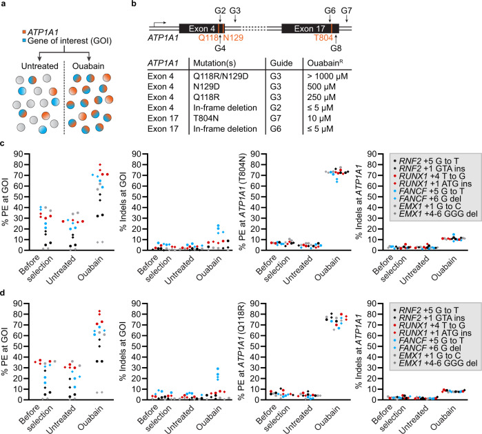
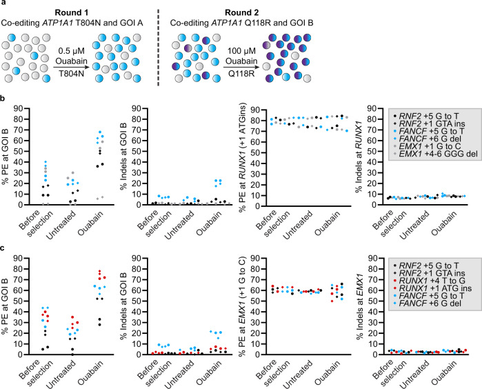
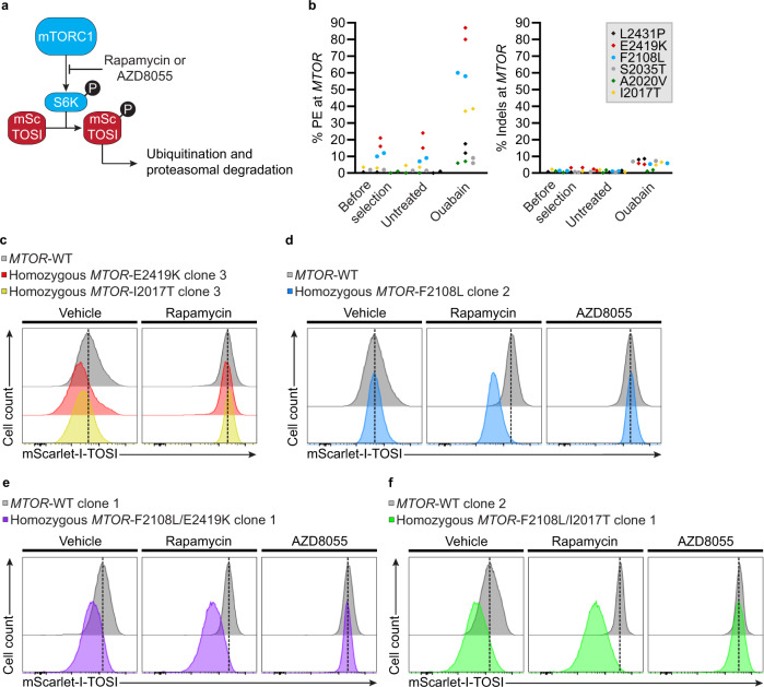
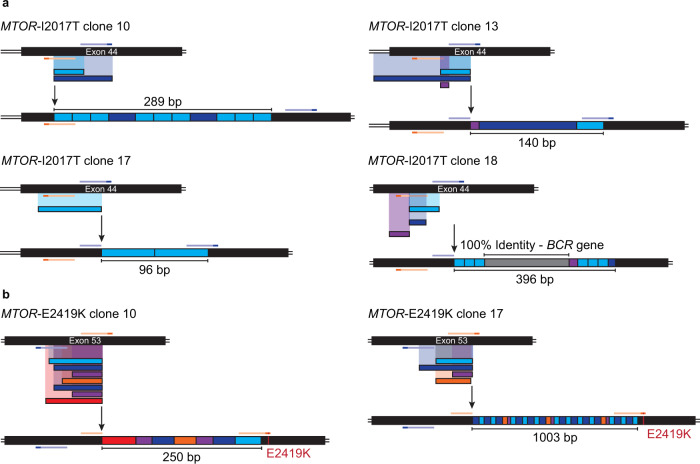
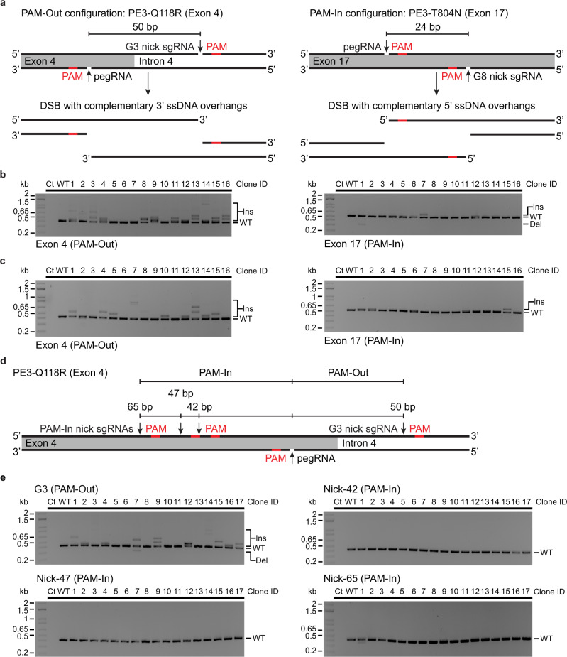
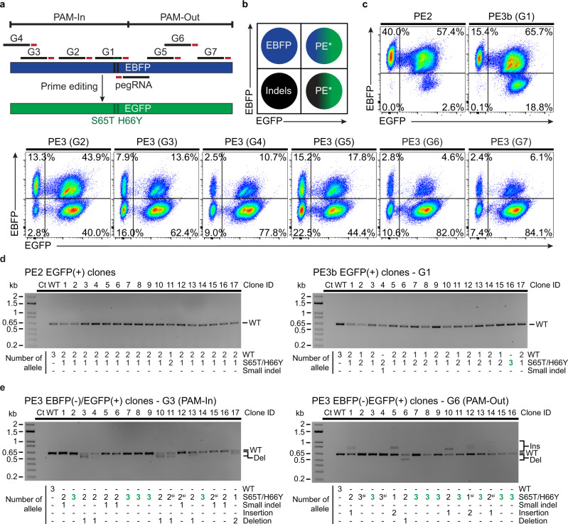
Prime editing enables the introduction of precise point mutations, small insertions, or short deletions without requiring donor DNA templates. However, efficiency remains a key challenge in a broad range of human cell types. In this work, we design a robust co-selection strategy through coediting of the ubiquitous and essential sodium/potassium pump (Na+/K+ ATPase). We readily engineer highly modified pools of cells and clones with homozygous modifications for functional studies with minimal pegRNA optimization. This process reveals that nicking the non-edited strand stimulates multiallelic editing but often generates tandem duplications and large deletions at the target site, an outcome dictated by the relative orientation of the protospacer adjacent motifs. Our approach streamlines the production of cell lines with multiple genetic modifications to create cellular models for biological research and lays the foundation for the development of cell-type specific co-selection strategies.


Conflict of interest statement

The authors declare no competing interests.

Figures

Fig. 1
Fig. 1. Robust co-selection for prime editing.
a Schematic for the co-enrichment of CRISPR-driven editing events at a GOI. b Schematic representation of the ATP1A1 locus regions targeted by SpCas9. The first and third extracellular loops of the Na+/K+ ATPase are encoded by ATP1A1 exon 4 and 17, respectively. The relative levels of resistance to ouabain conferred by different ATP1A1 mutations in K562 cells are shown. c PE and small indels quantification as determined by BEAT and TIDE analysis from Sanger sequencing. K562 cells were transfected with PE3 vectors targeting ATP1A1 exon 17 (T804N) and the indicated GOI. Genomic DNA was harvested 3 days post-transfection (before selection) and cells were treated (ouabain) or not (untreated) with 0.5 µM ouabain until all non-resistant cells were eliminated. d Same as in c but co-targeting was performed via ATP1A1 exon 4 (Q118R). n = 2 independent biological replicates performed at different times. Source data are provided in the Source Data file.
Fig. 2
Fig. 2. Robust co-selection for successive rounds of prime editing.
a Schematic of the strategy for performing successive rounds of co-selection. Cells harboring modifications at ATP1A1 (T804N) and GOI A are first co-selected with 0.5 µM ouabain. Following the first round, a subsequent round of co-selection occurs at GOI B via modification at ATP1A1 (Q118R) with 100 µM ouabain. b PE and small indels quantification as determined by BEAT and TIDE analysis from Sanger sequencing. K562 cells harboring the ATP1A1-T804N and RUNX1 + 1 ATG insertion (GOI A) modifications (Fig. 1c) were transfected with PE3 vectors targeting ATP1A1 exon 4 (Q118R) and the indicated GOI B. Genomic DNA was harvested 3 days post-transfection (before selection) and cells were treated (ouabain) or not (untreated) with 100 µM ouabain until all non-resistant cells were eliminated. c Same as in b with K562 cells harboring the ATP1A1-T804N and EMX1 + 1 G to C (GOI A) modifications (See Fig. 1c). n = 2 independent biological replicates performed at different times. Source data are provided in the Source Data file.
Fig. 3
Fig. 3. Co-selection for the installation of clinically relevant mutations at the MTOR locus.
a Schematic of mScarlet-I mTOR Signaling Indicator (mSc-TOSI) reporter degradation under mTORC1 signaling. b PE and small indels quantification as determined by BEAT and TIDE analysis from Sanger sequencing. K562 cells stably expressing the mSc-TOSI reporter were transfected with PE3 vectors targeting ATP1A1 exon 4 (Q118R) and MTOR. Genomic DNA was harvested 3 days post-transfection (before selection) and cells were treated (ouabain) or not (untreated) with 100 µM ouabain until all non-resistant cells were eliminated. n = 2 independent biological replicates performed at different times. c Histogram plot of mSc-TOSI intensity in homozygous single cell-derived K562 clones harboring MTOR hyperactivating mutations. d Same as in c with a homozygous clone harboring the MTOR-F2108L rapamycin resistance mutation. e Same as in c with a homozygous clone harboring the MTOR-F2108L rapamycin resistance mutation and the MTOR-E2419K hyperactivating mutation. f Same as in c with a homozygous clone harboring the MTOR-F2108L rapamycin resistance mutation and the MTOR-I2017T hyperactivating mutation. Where indicated, cells were treated for 24 h with 50 nM rapamycin or 50 nM AZD8055 before FACS analysis. Representative images are from one of two independent biological replicates performed at different times with equivalent results (see Supplementary Figs. 15 and 16). Source data are provided in the Source Data file.
Fig. 4
Fig. 4. Identification and characterization of large PE3-mediated insertions in single cell-derived clones.
a Schematic representation of the large insertions observed at MTOR exon 44 from single cell-derived K562 clones harboring the MTOR-I2017T hyperactivating mutation. K562 cells stably expressing the mSc-TOSI reporter were transfected with PE3 vectors targeting ATP1A1 exon 4 (Q118R) and MTOR. Single cell-derived clones were isolated in methylcellulose-based semi-solid RPMI media supplemented with 100 µM ouabain and genomic DNA was harvested after co-selection. TOPO cloning and Sanger sequencing were performed to characterize the large insertions. The pegRNA and its PAM sequence are represented in orange and dark orange, respectively. The nick sgRNA and its PAM sequence are represented in blue and dark blue, respectively. b Same as in a for large insertions at MTOR exon 53 from single cell-derived K562 clones harboring the MTOR-E2419K hyperactivating mutation.
Fig. 5
Fig. 5. Nick location dictates the type of PE3 editing byproducts at ATP1A1.
a Schematic representation of the complementary 5′ and 3′ single-stranded DNA overhangs generated with the PE3-T804N strategy at ATP1A1 exon 17 (PAM-In configuration) and the PE3-Q118R strategy at ATP1A1 exon 4 (PAM-Out configuration), respectively. b PCR-based genotyping of ATP1A1 exon 17 and 4 from single cell-derived MTOR-F2108L/I2017T K562 clones. Single cell-derived clones were isolated in methylcellulose-based semi-solid RPMI media supplemented with 100 µM ouabain and genomic DNA was harvested after co-selection. n = 16 single cell-derived clones from one experiment. c Same as in b with single cell-derived MTOR-F2108L/E2419K K562 clones. n = 16 single cell-derived clones from one experiment. d Schematic representation of pegRNA and nick sgRNA target sites with PAMs facing inwards (PAM-In configuration) and outwards (PAM-Out configuration) at ATP1A1 exon 4. e PCR-based genotyping of ATP1A1 exon 4 from single cell-derived K562 clones targeted with PAM-out and PAM-in configurations using different nick sgRNAs. Single cell-derived clones were isolated in methylcellulose-based semi-solid RPMI media supplemented with 0.5 µM ouabain and genomic DNA was harvested after co-selection. n = 17 single cell-derived clones for each condition from one experiment. Ins, insertion. Del, deletion.
Fig. 6
Fig. 6. Assessing the impact of complementary-strand nick locations on prime editing outcomes.
a Schematic representation of the EBFP to EGFP reporter system using different nick sgRNAs with PAMs facing outwards (PAM-Out) or inwards (PAM-In) towards the pegRNA. b Schematic of the FACS-based quantification of EBFP to EGFP conversion via PE. In K562 cells homozygous for EBFP integration at ATP1A1 (triallelic), FACS-based analysis reports PE outcomes indirectly; (i) EBFP(+)/EGFP(+) cells result from monoallelic or biallelic PE, (ii) EBFP(−)/EGFP(+) cells are generated by either triallelic PE or combinations of mono- and biallelic PE along with indels*, (iii) EBFP(−)/EGFP(−) occur from indels on the three copies of the reporter. Indels are defined broadly in this context as any edits that inactivates the reporter. c FACS-based quantification of EBFP to EGFP conversion via PE after co-selection. K562 cells stably expressing the EBFP reporter from the ATP1A1 locus were transfected with PE3max vectors targeting ATP1A1 exon 4 (pegRNA-Q118R_v1) and EBFP (epegRNA). Cells were treated with 100 µM ouabain starting 3 days post-transfection until all non-resistant cells were eliminated. n = 3 independent biological replicates performed at different times with equivalent results (see Supplementary Figs. 22,23). d PCR-based genotyping of EGFP(+) single cell-derived K562 clones targeted with the PE2 or PE3b strategy. Single cell-derived clones were isolated in methylcellulose-based semi-solid RPMI media supplemented with 100 µM ouabain and genomic DNA from EGFP(+) clones was harvested after co-selection. The number of precise prime edited alleles was determined from BEAT Sanger sequencing trace analysis and small indels were analysed with DECODR. n = 17 single cell-derived clones for each condition from one experiment. e PCR-based genotyping of EBFP from single K562 cell-derived clones after sorting for EBFP(−)/EGFP(+) cells. The number of precise prime edited alleles was determined from BEAT Sanger sequencing trace analysis and small indels were analysed with DECODR. Larger insertions and deletions are indicated, and homozygous single cell-derived clones are highlighted in bold and green. PE alleles harboring pegRNA scaffold incorporation are indicated (si). n = 17 and 16 single cell-derived clones from one experiment for the PE3-G3 and PE3-G6 conditions, respectively. Ins, insertion. Del, deletion.

References

    1. Anzalone AV, et al. Search-and-replace genome editing without double-strand breaks or donor DNA. Nature. 2019;576:149–157. doi: 10.1038/s41586-019-1711-4. - DOI - PMC - PubMed
    1. Yan J, Cirincione A, Adamson B. Prime editing: precision genome editing by reverse transcription. Mol. Cell. 2020;77:210–212. doi: 10.1016/j.molcel.2019.12.016. - DOI - PubMed
    1. Anzalone AV, Koblan LW, Liu DR. Genome editing with CRISPR–Cas nucleases, base editors, transposases and prime editors. Nat. Biotechnol. 2020;38:824–844. doi: 10.1038/s41587-020-0561-9. - DOI - PubMed
    1. Jinek M, et al. A programmable dual-RNA-guided DNA endonuclease in adaptive bacterial immunity. Science. 2012;337:816–822. doi: 10.1126/science.1225829. - DOI - PMC - PubMed
    1. Kim HK, et al. Predicting the efficiency of prime editing guide RNAs in human cells. Nat. Biotechnol. 2021;39:198–206. doi: 10.1038/s41587-020-0677-y. - DOI - PubMed

Publication types

Grants and funding