Review
. 2022 Dec;52(13):2452-2466.
doi: 10.1007/s00247-022-05408-5. Epub 2022 Oct 8.

Neuroimaging features of genetic syndromes associated with CNS overgrowth

Affiliations
Review

Neuroimaging features of genetic syndromes associated with CNS overgrowth

Anthony R Zamary et al. Pediatr Radiol. 2022 Dec.

Abstract

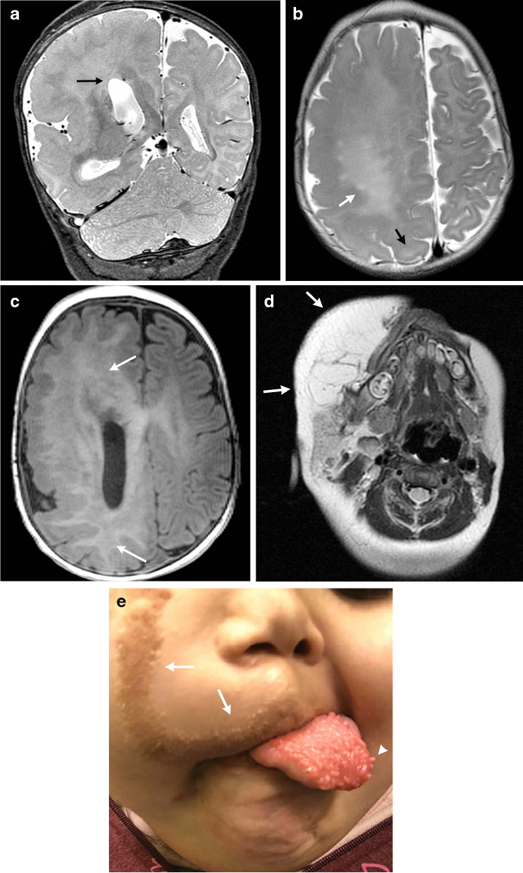
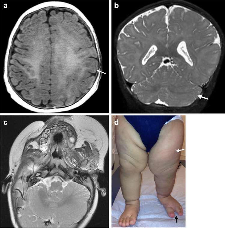
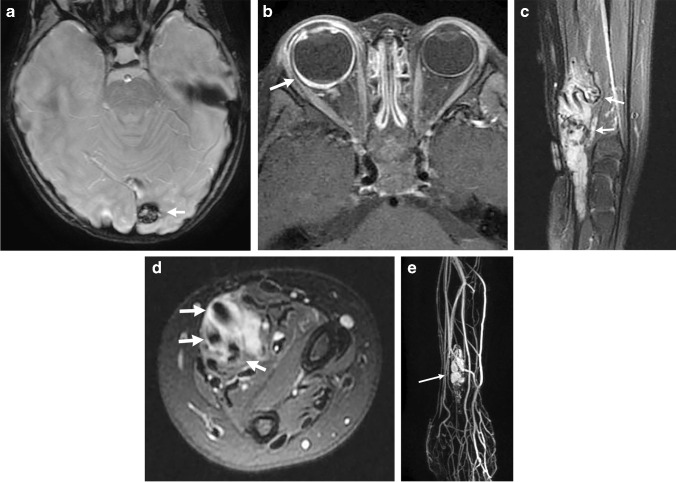
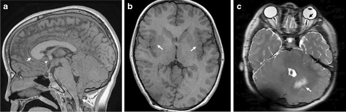
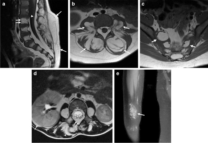
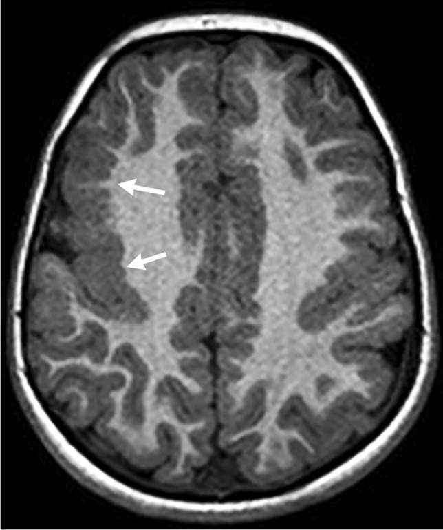
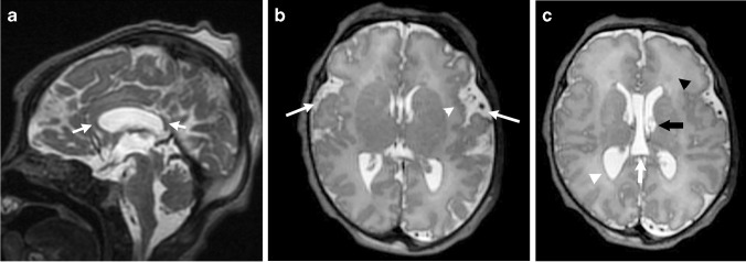
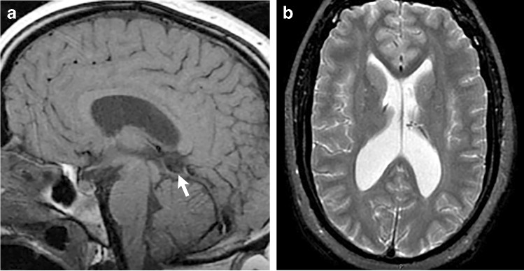
Overgrowth syndromes can manifest with enlargement of the brain and other body parts and are associated with malignancy. Much of the current literature focuses on the imaging findings of the somatic overgrowth, while there is relatively little describing the overgrowth of the central nervous system. In this pictorial essay, we discuss common syndromes with central nervous system overgrowth, highlight key imaging features, and review the underlying genetics, including the PI3K-AKT-mTOR pathway as well as other syndromes from various genes.

Keywords: Central nervous system; Children; Hemimegalencephaly; Magnetic resonance imaging; Overgrowth syndromes; PIK3CA; Vascular malformations.


Conflict of interest statement

None

Figures

Fig. 1
Fig. 1
CLOVES (congenital lipomatous overgrowth, vascular anomalies, epidermal nevi, and scoliosis/spinal deformities) syndrome in a 4-month-old girl with a genetically confirmed PIK3CA mutation with hemimegalencephaly and facial overgrowth. a A coronal T2-weighted image shows right hemimegalencephaly in the cerebrum and cerebellum. The right lateral ventricular body is large and superiorly directed (arrow). b An axial T2-weighted image shows increased signal within the white matter (white arrow). Pachygyria is noted (black arrow). c An axial spoiled gradient recalled echo image shows increased signal throughout the white matter (arrows). d An axial T2-weighted image shows substantial lipomatous overgrowth in the right face (arrows). e A clinical photograph at 5 years of age shows the corresponding right facial overgrowth. Linear epidermal nevi are seen (arrows). The tongue is also enlarged with verrucous papules (arrowhead). Consent for the photograph was obtained from the patient’s legal guardian
Fig. 2
Fig. 2
CLOVES syndrome in a 5-month-old boy with genetically confirmed PIK3CA mutation with mild hemimegalencephaly and a normal head circumference. a An axial spoiled gradient recalled echo image shows mild hemimegalencephaly and subtle pachygyria in the left perirolandic region (arrow). b A coronal T2-weighted image shows associated enlargement of the left cerebellar hemisphere (arrow). c An axial T2-weighted image shows lipomatous overgrowth of the left facial soft tissues. d A clinical photograph shows subtle overgrowth of the left lower extremity with associated capillary malformations (white arrow) and a sandal gap deformity (black arrow)
Fig. 3
Fig. 3
CLOVES syndrome in a 16-year-old girl with a genetically confirmed PIK3CA mutation with thoracic lipomatous overgrowth. a A clinical photograph shows left upper back overgrowth (arrows) that is typical of the CLOVES phenotype. Skin-colored papules (arrowhead) were also present that were consistent with collagenomas on biopsy. b An axial contrast-enhanced chest computed tomography (CT) shows enlargement of the left anterior and posterior chest wall muscles (white arrows) and subcutaneous fat (white arrowhead). Venous malformations are present in the left mediastinum (black arrowhead) as well as the left back with an associated phlebolith (wide white arrow). c A posteroanterior radiograph of the hand shows deformity diagnostic of arthrogryposis. d A coronal contrast-enhanced CT abdomen shows multiple splenic lesions (arrows) that also represent vascular malformations
Fig. 4
Fig. 4
Megalencephaly-Capillary Malformation (MCAP) syndrome in a 19-month-old-boy with a genetically confirmed PIK3CA mutation presenting with macrocephaly, facial dysmorphism, somatic overgrowth, capillary malformations, generalized hypotonia and developmental delay. a-f Axial and coronal T2-weighted images show an enlarged right cerebral hemisphere compared to the left with associated ventricular enlargement and hyperintense signal within the periventricular white matter (a, white arrow; d, black arrow). There is enlargement of the right superior cerebellar vermis (b, arrow), right olfactory bulb (c, arrow), and right cerebral cortical veins (d, white arrows) compared to the normal left cortical veins (d, arrowhead). Subependymal heterotopia in the right temporal and occipital lobes is present (e, arrows). Hypertelorism (f). g A clinical photo shows enlargement of the right upper and lower extremities (arrows) with associated deformities and bilateral club feet. h A clinical photo shows capillary malformation in a cutis marmorata-like appearance
Fig. 5
Fig. 5
Cowden syndrome in a 14-year-old girl with a genetically confirmed PTEN gene mutation with multiple systemic anomalies. a, b Sagittal and axial spoiled gradient recalled echo images show a mildly enlarged corpus callosum and excess white matter. c, d Axial T2-weighted and coronal contrast-enhanced T1-weighted images show a striated T2 hyperintense nonenhancing lesion in the left cerebellar hemisphere, which was consistent with a dysplastic gangliocytoma—Lhermitte-Duclos disease—after resection. e An axial contrast-enhanced chest computed tomography scan shows a large left chest wall lipoma (arrow). f A longitudinal ultrasound shows a left thyroid nodule (arrow) with irregular borders and punctate echogenic foci that was compatible with a papillary thyroid cancer after surgical resection. g A conventional spinal angiogram in the anteroposterior projection with the patient supine with injection from the right L1 lumbar artery shows a spinal arteriovenous malformation (arrow)
Fig. 6
Fig. 6
Cowden syndrome in a 3-year-old girl with a genetically confirmed PTEN mutation who presented with autism, macrocephaly and a vascular malformation. a An axial susceptibility weighted image shows a left occipital cavernous malformation (arrow). b An axial contrast-enhanced fat-suppressed T1-weighted image of the orbits shows an enlarged right-side globe with abnormal choroidal enhancement (arrow) consistent with uveal ganglioneuroma after biopsy and surgical removal. c, d Sagittal and axial fat-suppressed T2-weighted images show a predominantly hyperintense intramuscular lesion in the distal forearm and wrist with multiple flow voids (arrows). e A coronal contrast-enhanced time-resolved magnetic resonance angiogram/magnetic resonance venogram shows arterial enhancement within the lesion (arrow), but there are no large draining veins to suggest an arteriovenous malformation. This mass represents a PTEN hamartoma of soft-tissue (PHOST) lesion
Fig. 7
Fig. 7
Cowden syndrome in a 10-year-old boy with genetically confirmed PTEN mutation. a A sagittal spoiled gradient recalled echo (SPGR) image shows an enlarged corpus callosum that is greatest at the anterior aspect (short arrow), along with an enlarged anterior commissure (arrowhead). The cerebellum is enlarged, and there is tonsillar ectopia (long arrow). b An axial SPGR image shows bilateral perisylvian polymicrogyria (arrows). Note the increased volume of the white matter. c An axial T2-weighted image shows marked enlargement of the cerebellar hemispheres with a dysplastic appearance and hyperintense signal on the left (white arrow), which likely represents Lhermitte-Duclos disease although it is not classic in appearance. The orbital globes show hypertelorism and papilledema (black arrow) from increased intracranial pressure. Susceptibility artifact is noted in the right posterior fossa from a ventriculoperitoneal shunt catheter
Fig. 8
Fig. 8
Bannayan-Riley-Ruvalcaba syndrome in a 3-year-old boy with a genetically confirmed PTEN mutation with multiple anomalies. a-c Sagittal and axial T1-weighted images show excessive fat throughout the lumbosacral spine. In (a), there is cutaneous and subcutaneous dorsal lipomatous overgrowth (arrows), extensive dorsal and ventral epidural fat (arrowhead) and abnormal fat within the vertebral body bone marrow (double arrows). In (b, c), extensive fatty replacement is observed within the psoas and paraspinal muscles (arrows in b) and along the sacral neural foramina (arrows in c). d An axial T2-weighted image shows right-side nephromegaly (arrow) as well as the excess fat in the paraspinal tissues (arrowhead) and epidural fat (double arrows). e A coronal contrast-enhanced T1-weighted image shows a solid enhancing mass in the ventral forearm consistent with a venous malformation
Fig. 9
Fig. 9
AKT3 genetically confirmed mutation. An axial spoiled gradient recalled echo image shows extensive polymicrogyria within the right frontal lobe (arrows). Image courtesy of A.J. Barkovich, MD
Fig. 10
Fig. 10
Sotos syndrome in a 4-day-old boy with genetically confirmed NSD1 gene mutation. a A sagittal T2-weighted image shows a small corpus callosum (arrows). Scalp swelling is noted from recent delivery. b An axial T2-weighted image shows widened frontal opercula (arrows) and left perisylvian polymicrogyria (arrowhead). c An axial T2-weighted image shows ventriculomegaly with large trigones (white arrowhead), cavum septum pellucidum and vergae (white arrow), and diffusely increased T2 signal within the white matter (black arrowhead). Periventricular cysts are also noted (black arrow) that could be related to the syndrome or be sequala from previous germinal matrix hemorrhages
Fig. 11
Fig. 11
Sotos syndrome in a 17-year-old-girl with developmental delay. a A sagittal T1-weighted image shows a midline cyst (arrow) superior to the cerebellar vermis. b An axial T2-weighted image shows ventriculomegaly with large trigones

References

    1. Brioude F, Toutain A, Giabicani E, et al. Overgrowth syndromes - clinical and molecular aspects and tumour risk. Nat Rev Endocrinol. 2019;15:299–311. doi: 10.1038/s41574-019-0180-z. - DOI - PubMed
    1. Kamien B, Ronan A, Poke G, et al. A clinical review of generalized overgrowth syndromes in the era of massively parallel sequencing. Mol Syndromol. 2018;9:70–82. doi: 10.1159/000484532. - DOI - PMC - PubMed
    1. Severino M, Geraldo AF, Utz N, et al. Definitions and classification of malformations of cortical development: practical guidelines. Brain. 2020;143:2874–2894. doi: 10.1093/brain/awaa174. - DOI - PMC - PubMed
    1. Bertino F, Braithwaite KA, Hawkins CM, et al. Congenital limb overgrowth syndromes associated with vascular anomalies. Radiographics. 2019;39:491–515. doi: 10.1148/rg.2019180136. - DOI - PubMed
    1. Mirzaa G, Graham JM, Jr, Keppler-Noreuil K, et al. PIK3CA-related overgrowth spectrum. In: Adam MP, Ardinger HH, Pagon RA, et al., editors. GeneReviews. Seattle: University of Washington; 2013.

Substances

LinkOut - more resources