

doi: 10.1007/s12350-022-03114-1.
Epub 2022 Oct 7.
Cases from a busy nuclear cardiology laboratory
Affiliations
- PMID: 36207574
- PMCID: PMC9542470
- DOI: 10.1007/s12350-022-03114-1
Item in Clipboard
Cases from a busy nuclear cardiology laboratory
J Nucl Cardiol.
2023 Jun.
No abstract available
Conflict of interest statement
All authors have no disclosures in relation to this manuscript.
Figures
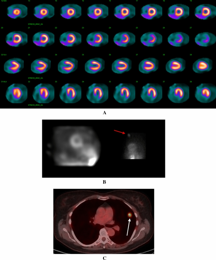
A Regadenoson SPECT MPI study demonstrating homogenous uptake of radiotracer throughout the left ventricular myocardium. B Right image: One of the 19 raw projection images revealed significant focal radiotracer uptake in the superior left thorax (red arrow). Left image: The simulated rotating planar projection image did not reproduce this finding. C FDG-PET scan revealed a hypermetabolic nodule (white arrow) in the superior left upper lobe of the lung which was determined to be poorly differentiated adenocarcinoma on biopsy

A Left panel: The simulated rotating projection image did not demonstrate an IECF. Right panel: A single raw projection planar image revealed focal radiotracer uptake (white arrow) in superior right thorax. B Cine rotating planar projection image on an Anger camera obtained 3 years prior did not show radiotracer uptake in that region. C FDG-PET scan showed a hypermetabolic nodule (SUV = 4.2) in the right lung apex without evidence of nodal metastasis
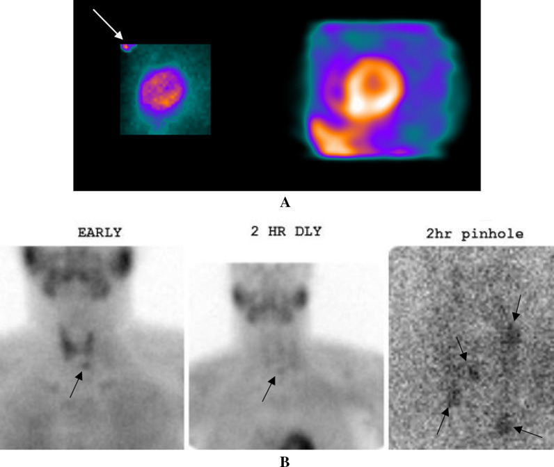
A Right panel: The simulated rotating planar projection images showed no IECF. Left panel: A raw projection planar image revealed significant radiotracer uptake in the regions of the thyroid and parathyroid glands (white arrow). The exact location of radiotracer uptake is difficult to discern, necessitating biochemical evaluation of thyroid and parathyroid function. B. Early, 2 h delayed, and 2 h delayed pinhole magnified Sestamibi parathyroid images show multigland parathyroid hyperplasia (black arrows)

A CT scan of the chest with contrast, at the level of the neck. Imaging demonstrates the presence of a 6.6 × 5.3 cm mass (red arrow) originating from the left lobe of the thyroid gland. There is extension of the mass into the superior mediastinum. B. FDG-PET scan with a four-chamber cardiac view. Notable cardiac findings seen in this view included a hypermetabolic pericardiophrenic lymph node (blue arrow) and a right atrial metastatic lesion (white arrow)
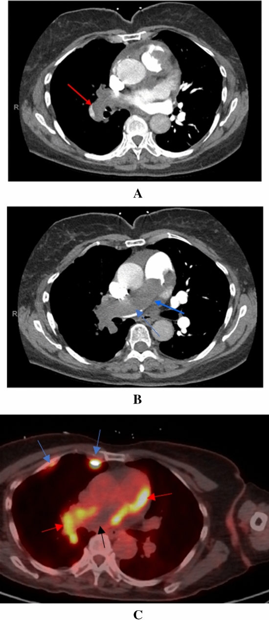
A CT pulmonary angiogram at the level of the right main pulmonary artery. There is extension of the mass into the right segmental pulmonary artery (red arrow). B CT pulmonary angiogram at the level of the main pulmonary artery. The mass extends completely through the main pulmonary artery (blue arrow). C FDG-PET scan revealing the presence of a hypermetabolic tumor thrombus (red arrows) and hypometabolic bland thrombus (black arrow) in the main and right pulmonary arteries. Post right-sided pleurodesis inflammatory plaques are incidentally seen (blue arrows)
References
-
- Dorbala S, Ananthasubramaniam K, Armstrong IS, Chareonthaitawee P, DePuey EG, Einstein AJ, et al. Single photon emission computed tomography (SPECT) myocardial perfusion imaging guidelines: Instrumentation, acquisition, processing, and interpretation. J Nucl Cardiol. 2018;25(5):1784–1846. doi: 10.1007/s12350-018-1283-y. - DOI - PubMed
-
- Saab R, Farag A, White S, Hage FG, Bhambhvani P. Artifacts and Incidental Findings on Myocardial Perfusion Imaging. In: Hage FG (ed) Myocardial Perfusion Imaging (MPI): Performance, Potential Risks and Outcomes. Nova Science Publishers. 2018. ISBN: 978-1-53613-476-6.
MeSH terms
LinkOut - more resources
Full Text Sources
