. 2022 Dec 16;3(4):101678.
doi: 10.1016/j.xpro.2022.101678. Epub 2022 Oct 7.

Protocol for renal ischemia-reperfusion injury by flank incisions in mice

Affiliations

Protocol for renal ischemia-reperfusion injury by flank incisions in mice

Yung-Ting Cheng et al. STAR Protoc. .

Abstract

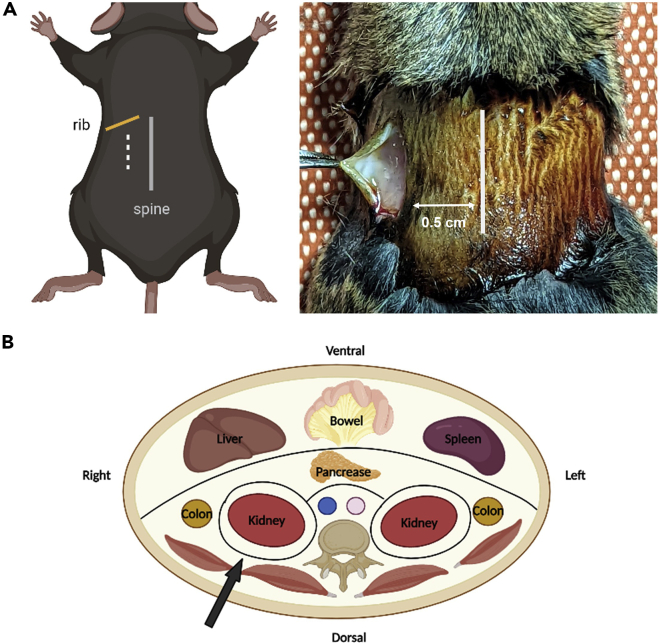
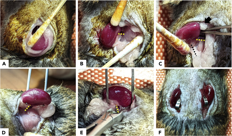
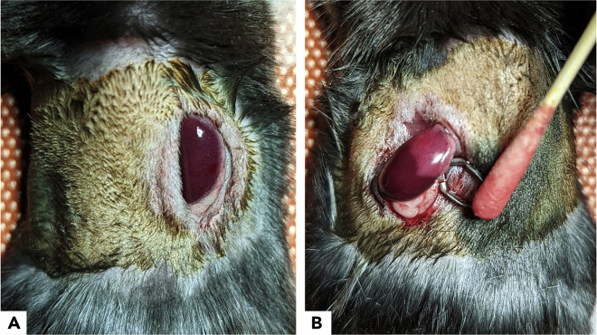
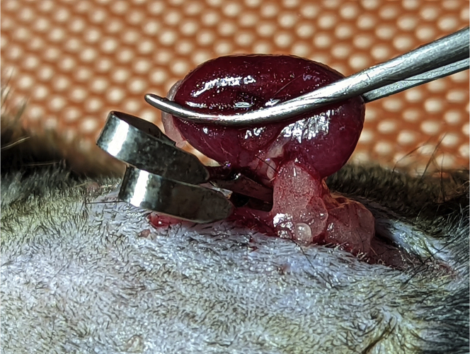
Ischemia-reperfusion injury (IRI) contributes to acute kidney injury (AKI) and development of chronic kidney disease. We describe an IRI protocol for mice via flank incisions approach, using a pedicle clamp to cause ischemic injury. Compared with trans-abdominal approach, it is technically easier with lesser fluid loss and organ injury. Technical challenges during the dissection of renal pedicles are highlighted. For complete details on the execution of this protocol, please refer to Lai et al. (2014).

Keywords: Health Sciences; Metabolism; Model Organisms; Molecular Biology.


Conflict of interest statement

Declaration of interests The authors have disclosed that they do not have any potential conflicts of interest.

Figures

None
Graphical abstract
Figure 1
Figure 1
Anatomical landmarks for flank approach to the mouse kidney (A) Landmark of flank incision. (B) Anatomy of mouse kidney. Black arrow indicates the route of dissection towards the kidney.
Figure 2
Figure 2
Main steps of the exposure of mouse renal hilum (A) Expose the kidney from the retroperitoneal space by cotton swab in an outward direction. (B–D) Liberate the renal pedicle (yellow dotted line) from the renal sinus fat with prevention of ureteral (black dotted line) and adrenal gland (black arrow) injury. (E) Placement of a pedicle clamp over the renal pedicle. (F) Repeat at the contralateral side as an optional procedure depending on the study design.
Figure 3
Figure 3
Expected changes of the kidney after ischemia-reperfusion injury (A) Appearance of the kidney after successful clamping. (B) Ineffective clamp leads to incomplete renal ischemia.
Figure 4
Figure 4
Stable body temperature intraoperatively with temperature controller
Figure 5
Figure 5
Expected changes of the kidney after 30-min unilateral ischemia-reperfusion injury (A) Histopathological changes of kidney by Hematoxylin and Eosin (H&E) stain. Significant sloughing epithelial cells (yellow arrow), tubular necrosis and cast formation of the post-IRI kidney with H&E stain on day 1. Tubular injury score grade 2. Scale bar: 50 μm, magnification: 40×. (B) Reduced mass of the post-IRI kidney compared with contralateral kidney (CLK) on day 14. (C and D) Histopathological changes of the CLK (C) and post-IRI kidney (D) on day 14. Injured and atrophic renal tubules (black arrow) with interstitial inflammatory cells infiltration (yellow arrow) are evident in post-IRI kidneys. Tubular injury score grade 4. Scale bar: 50 μm, magnification: 40×.
Figure 6
Figure 6
Immunofluorescence and picrosirius red staining of the kidney at 7 days after 30-min unilateral ischemia-reperfusion injury (A) Platelet-derived growth factor receptor-β (PDGFR-β) were upregulated in renal mesenchymal cells during renal fibrosis. (B) α-smooth muscle actin (α-SMA) as a marker of myofibroblasts expressed on fibrosis tissue. (C) F4/80 antibody for macrophage staining. (D) Picrosirius red staining could visualize collagen I and III fibers in renal fibrosis. Scale bar: 25 μm, magnification: 40×. CLK: Contralateral kidney.
Figure 7
Figure 7
Two pedicle clamps to confirm complete occlusion of the renal pedicle

References

    1. Beker B.M., Corleto M.G., Fieiras C., Musso C.G. Novel acute kidney injury biomarkers: their characteristics, utility and concerns. Int. Urol. Nephrol. 2018;50:705–713. - PubMed
    1. Burne M.J., Haq M., Matsuse H., Mohapatra S., Rabb H. Genetic susceptibility to renal ischemia reperfusion injury revealed in a murine model. Transplantation. 2000;69:1023–1025. - PubMed
    1. Chou Y.H., Pan S.Y., Shao Y.H., Shih H.M., Wei S.Y., Lai C.F., Chiang W.C., Schrimpf C., Yang K.C., Lai L.C., et al. Methylation in pericytes after acute injury promotes chronic kidney disease. J. Clin. Invest. 2020;130:4845–4857. - PMC - PubMed
    1. Dong Y., Zhang Q., Wen J., Chen T., He L., Wang Y., Yin J., Wu R., Xue R., Li S., et al. Ischemic duration and frequency determines AKI-to-CKD progression monitored by dynamic changes of tubular biomarkers in IRI mice. Front. Physiol. 2019;10:153. - PMC - PubMed
    1. Goujon J.M., Hauet T., Menet E., Levillain P., Babin P., Carretier M. Histological evaluation of proximal tubule cell injury in isolated perfused pig kidneys exposed to cold ischemia. J. Surg. Res. 1999;82:228–233. - PubMed

Publication types

LinkOut - more resources