Review
. 2022 Oct 9;29(1):79.
doi: 10.1186/s12929-022-00865-4.

Delivering genes with human immunodeficiency virus-derived vehicles: still state-of-the-art after 25 years

Affiliations
Review

Delivering genes with human immunodeficiency virus-derived vehicles: still state-of-the-art after 25 years

Jonas Holst Wolff et al. J Biomed Sci. .

Abstract

Viruses are naturally endowed with the capacity to transfer genetic material between cells. Following early skepticism, engineered viruses have been used to transfer genetic information into thousands of patients, and genetic therapies are currently attracting large investments. Despite challenges and severe adverse effects along the way, optimized technologies and improved manufacturing processes are driving gene therapy toward clinical translation. Fueled by the outbreak of AIDS in the 1980s and the accompanying focus on human immunodeficiency virus (HIV), lentiviral vectors derived from HIV have grown to become one of the most successful and widely used vector technologies. In 2022, this vector technology has been around for more than 25 years. Here, we celebrate the anniversary by portraying the vector system and its intriguing properties. We dive into the technology itself and recapitulate the use of lentiviral vectors for ex vivo gene transfer to hematopoietic stem cells and for production of CAR T-cells. Furthermore, we describe the adaptation of lentiviral vectors for in vivo gene delivery and cover the important contribution of lentiviral vectors to basic molecular research including their role as carriers of CRISPR genome editing technologies. Last, we dwell on the emerging capacity of lentiviral particles to package and transfer foreign proteins.

Keywords: CRISPR; Gene editing; Gene therapy; Genome engineering; HIV; IDLV; Integrase-defective lentiviral vectors; Lentiviral vectors; Lentivirus.


Conflict of interest statement

No competing financial interests exist.

Figures
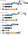
Fig. 1
Fig. 1
Schematic representation of the three generations of lentiviral vectors. A First generation vector systems include all HIV-1 genes, except env, in a single packaging plasmid. The env gene is replaced with VSV-G and provided in a separate plasmid. The vector plasmid contains an internal promotor-driven transgene cassette flanked by the HIV-1 LTRs. B In second-generation lentiviral vector systems, genes encoding accessory proteins Vif, Vpr, Vpu, and Nef are removed from the packaging plasmid. C In third-generation lentiviral vector system, the rev gene is placed on a separate plasmid, giving rise to a total of four separate plasmids required for production. Replacement of the U3 with a heterologous promoter (usually CMV or RSV) in the 5′ LTR allows the tat gene to be removed from the packaging plasmid, while a partial deletion of the U3 region from the 3′ LTR results in so-called ‘self-inactivating’ (SIN) lentiviral vectors. State-of-the art third-generation lentiviral transfer vectors usually also contains additional cis-acting elements such as cPPT/FLAP and WPRE for increased transduction efficiency and transgene expression, respectively. All vectors are shown schematically, and elements such as genes and promoters are not shown to scale
Fig. 2
Fig. 2
Derivation of current third-generation lentiviral vectors. The HIV-1 genome encodes three structural genes (gag, pol and env) as well as regulatory (rev and tat) and accessory (vif, vpr, vpu and nef) genes. The Gag precursor contains the viral core proteins, which are the matrix (MA), capsid (CA), nucleocapsid (NC) and p6 proteins, whereas the GagPol precursor also contains the protease (PR), reverse transcriptase (RT) and the integrase (IN) proteins. The entire HIV-1 genome is flanked by long terminal repeats (LTRs), responsible for viral transcription, reverse transcription and integration. For the production of current third-generation lentiviral vectors, the essential parts of the HIV-1 genome have been split into four separate plasmids; (i) the packaging plasmid encoding the GagPol polyprotein, (ii) the envelope plasmid encoding the viral glycoprotein (here VSV-G), (iii) the rev plasmid encoding Rev, and (iv) the transfer vector, carrying the transgene flanked by LTRs
Fig. 3
Fig. 3
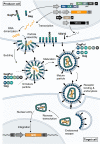
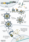
Gene transfer using lentiviral vectors. Third-generation lentiviral vectors are produced by transfecting producer cells with the four packaging plasmids, which will initiate transcription of Gag and GagPol polyprotein precursors, the envelope glycoprotein (e.g. VSV-G), Rev and the transfer vector carrying the (trans)gene of interest (GOI) to be inserted into the target cells. Nascent lentiviral particles are packaged together with an RNA dimer encoding the transgene flanked by viral cis-elements required for RNA packaging and reverse transcription. Budding of lentiviral particles results in immature particles, which are then matured in a process involving cleavage of the Gag and GagPol polyproteins as well as formation of the viral core. Uptake into target cells is achieved through receptor-mediated endocytosis, following which the viral core is released into the cytoplasm. Reverse transcription of the transfer vector single-stranded RNA then occurs, resulting in double-stranded DNA, which is then transported into the nucleus and integrated into the genome of the target cell
Fig. 4
Fig. 4
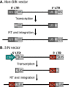
Self-inactivating (SIN) lentiviral vectors. A Non-SIN lentiviral vectors contain two wild-type HIV-1 LTRs, each comprised of a U3, R and U5 region. The U3 region contains promoter/enhancer regions from which transcription initiates at the junction between U3 and R in the 5′ LTR and terminates at the junction between R and U5 in the 3′ LTR. Viral transcripts thus lack the 5′ U3 and the 3′ U5, which are regained by duplication from each end upon reverse transcription. B In SIN lentiviral vectors, the 5′ U3 region is replaced by a heterologous promotor, such as CMV, whereas part of the 3′ U3 region has been deleted. When the 3′ U3 region of SIN vectors are duplicated, the deletion in the 3′ U3 (ΔU3) is transferred to the 5′ LTR, rendering the integrated lentiviral vector remote of viral promoter/enhancer regions
Fig. 5
Fig. 5
Schematic representation of MLV and HIV-1 integration profiles. The integration profile of retroviral vectors derived from murine leukemia virus (MLV) exhibits a strong bias for regions flanking transcription start sites (TSS), whereas lentiviral vectors derived from HIV-1 does not exhibit the same preference for integration near transcription start sites, but instead have a preference towards actively transcribed regions. Schematic representation based on data presented by Cattoglio et al. [74]
Fig. 6
Fig. 6
Schematic overview of lentiviral gene therapies used for the treatment of SCID-X1. The strategy employed to treat X-linked severe combined immunodeficiency (SCID-X1) is based on the delivery of a normal copy of the IL2γc gene into the genome of a patient’s own hematopoietic stem cells (HSCs). HSCs are isolated from the patient and expanded prior to transduction using a lentiviral vector carrying a normal cDNA copy of the IL2γc gene expressed from an EF1α promoter. In addition to using the SIN-configuration, the lentiviral vector contains a 400-bp chicken β-globin insulator element, which aids in the safety of the vector by contributing enhancer-blocking activity. Integration of the ‘healthy’ IL2γc gene into the patient's HSCs restores IL2γc expression in HSCs, which upon autologous transplantation are able to reconstitute functional immunity
Fig. 7
Fig. 7
Examples of lentiviral vectors used in clinical studies. A The CG1711 vector used by Cartier et al. for the treatment of ALD. B The HPV569 and BB305 LentiGlobin vectors used for the treatment of β-thallassemia. C The LV-w1.6WASp used to treat WAS. D The LV-EFS-ADA vector used for the treatment of ADA-SCID. E The G1XCDG vector used for the treatment of X-CGD. See text for details. All vectors are shown schematically, and elements such as genes and promoters are not shown to scale
Fig. 8
Fig. 8
Schematic overview of CRISPR/Cas9 delivery using lentiviral vectors. A Cas9 and sgRNA are delivered within a single lentiviral transfer vector (lentiCRISPR-v2). Integration of the lentiCRISPR-v2 vector in the genome of target cells results in persisting expression of both Cas9 and sgRNA, which can then act on the genome of target cells. B Using integrase-defective lentiviral vectors (IDLVs), the lentiCRISPR-v2 vector remains episomal upon transduction, which results in only transient expression of both Cas9 and sgRNA, thus minimizing potential unwanted off-target effects. C In addition to delivering Cas9 and sgRNA, IDLVs can also be utilized to deliver a repair template (donor) for homology-directed repair (HDR)
Fig. 9
Fig. 9
CRISPR-based lentiviral knockout (KO) libraries. A library of pooled sgRNAs are cloned into a suitable lentiviral vector (here exemplified by a lentiCRISPR-v2 vector), resulting in a plasmid library, which is then used for production of a pooled lentiviral sgRNA library. Transduction of target cells is made at a low MOI ensuring that each cell receives a single unique sgRNA. Transcription of sgRNA and Cas9 results in KO of the target gene in each transduced cell, which results in a large population of cells collectively carrying knockout mutations in all library-targeted genes. Selection (e.g. a chemotherapy drug) is then applied to the pool of cells, which makes it possible to detect genes that affect the drug response. Genes involved in the given drug response is identified by next-generation sequencing (NGS) of sgRNA-containing cassettes and downstream bioinformatic analysis
Fig. 10
Fig. 10
Schematic representation of lentiviral protein transduction. Lentiviral vectors can be manipulated to package a protein of interest (POI). This can be done either by fusing the POI to the C-terminal integrase (IN) protein of the GagPol polypeptide, or as shown in this illustration, by fusing it to the N-terminal matrix (MA) protein separated by a Pleckstrin homology domain (PH) to aid the anchorage to the plasma membrane. The integrase harbors the D64V mutation rendering the viral particles integrase-defective. By including a protease recognition site between the POI and MA protein, the POI is released in mature viral particles and delivered to the nucleus of the target cell by mechanisms currently unknown. Two possible mechanisms have been suggested: Transport of the POI within the viral core or diffusion through the cytoplasm. An optional vector RNA genome can be included in the viral particles, which in interest of CRISPR-Cas9 genome editing, could be a donor template for HDR. In this case, the lentiviral particles would carry all necessary components for CRISPR-based HDR within a single lentiviral particle

Similar articles

Cited by

References

    1. Centers for Disease C. Pneumocystis pneumonia--Los Angeles. MMWR Morb Mortal Wkly Rep. 1981;30(21):250–2. - PubMed
    1. Hymes KB, Cheung T, Greene JB, Prose NS, Marcus A, Ballard H, et al. Kaposi's sarcoma in homosexual men—a report of eight cases. Lancet. 1981;2(8247):598–600. doi: 10.1016/S0140-6736(81)92740-9. - DOI - PubMed
    1. Klatzmann D, Barre-Sinoussi F, Nugeyre MT, Danquet C, Vilmer E, Griscelli C, et al. Selective tropism of lymphadenopathy associated virus (LAV) for helper-inducer T lymphocytes. Science. 1984;225(4657):59–63. doi: 10.1126/science.6328660. - DOI - PubMed
    1. Broder S, Gallo RC. A pathogenic retrovirus (HTLV-III) linked to AIDS. N Engl J Med. 1984;311(20):1292–1297. doi: 10.1056/NEJM198411153112006. - DOI - PubMed
    1. Coffin J, Haase A, Levy JA, Montagnier L, Oroszlan S, Teich N, et al. What to call the AIDS virus? Nature. 1986;321(6065):10. doi: 10.1038/321010a0. - DOI - PubMed