Review
. 2022 Oct 8;15(1):140.
doi: 10.1186/s13045-022-01365-6.

The emerging role of pyroptosis in pediatric cancers: from mechanism to therapy

Affiliations
Review

The emerging role of pyroptosis in pediatric cancers: from mechanism to therapy

Hua Wang et al. J Hematol Oncol. .

Abstract

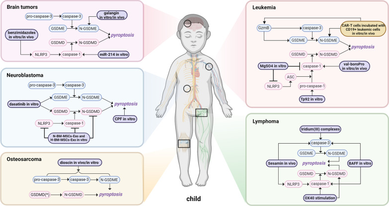
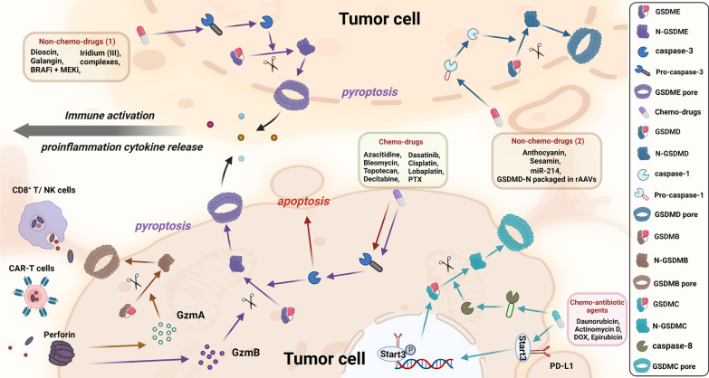
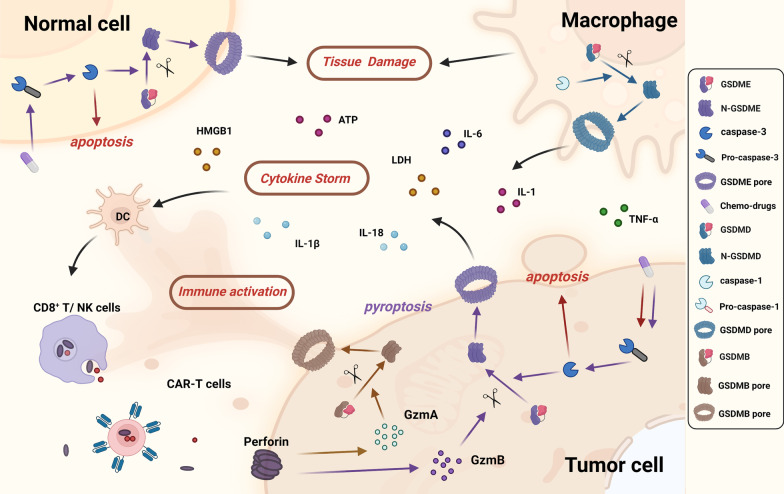
Pediatric cancers are the driving cause of death for children and adolescents. Due to safety requirements and considerations, treatment strategies and drugs for pediatric cancers have been so far scarcely studied. It is well known that tumor cells tend to progressively evade cell death pathways, which is known as apoptosis resistance, one of the hallmarks of cancer, dominating tumor drug resistance. Recently, treatments targeting nonapoptotic cell death have drawn great attention. Pyroptosis, a newly specialized form of cell death, acts as a critical physiological regulator in inflammatory reaction, cell development, tissue homeostasis and stress response. The action in different forms of pyroptosis is of great significance in the therapy of pediatric cancers. Pyroptosis could be induced and consequently modulate tumorigenesis, progression, and metastasis if treated with local or systemic therapies. However, excessive or uncontrolled cell death might lead to tissue damage, acute inflammation, or even cytokine release syndrome, which facilitates tumor progression or recurrence. Herein, we aimed to describe the molecular mechanisms of pyroptosis, to highlight and discuss the challenges and opportunities for activating pyroptosis pathways through various oncologic therapies in multiple pediatric neoplasms, including osteosarcoma, neuroblastoma, leukemia, lymphoma, and brain tumors.

Keywords: Cytokine release syndrome; Osteosarcoma; Pediatric cancer; Programmed cell death; Pyroptosis.


Conflict of interest statement

The authors declare no conflict of interest.

Figures

Fig. 1
Fig. 1
The timeline of various cell deaths
Fig. 2
Fig. 2
The cross talk between pyroptosis and apoptosis or necroptosis. a–b Interplay between necroptosis and pyroptosis. MLKL is the terminal executioner of necroptosis, which is also a key intermedium between necroptosis and pyroptosis. RIPK1-RIPK3 association or cytosolic ZBP1 activation results in phosphorylation of MLKL, thus forming pores on the membrane and engaging necroptosis. Plasma and K+ efflux mediated by MLKL can ultimately lead to cellular stress, triggering NLRP3 activation, inflammasome assembly, and caspase-1 cleavage, which is the canonical pyroptosis pathway. ZBP1 can directly activate NLRP3 inflammasome in response to virus infection. Additionally, when TNF binding to TNFR on the cell membrane, complex I is assembled and activated, further forming ripoptosome complex. Caspase-8 from ripoptosome complex can in turn promote the initiation of caspase-3 and -7 to execute GSDME-mediated pyroptosis. bc Interplay between pyroptosis and apoptosis. Caspase family of proteases and its targeting downstream molecules connect apoptosis with pyroptosis. In extrinsic apoptosis, recruitment of FADD and caspase-8 promotes the initiation of the death-inducing signaling complex (DISC) when death receptor is activated. Then, activated caspase-8 from DISC can promote the initiation of caspase-3 and -7 and execute GSDME-mediated pyroptosis. In intrinsic apoptosis, Bcl-2 family member Bid can be cleaved by caspase-8 and pyroptosis-inducing caspase-1 into proapoptotic tBID, together with intracellular stress, mitochondrial outer membrane permeabilization (MOMP) is induced, subsequently triggering cytochrome c release, apoptosome formation and caspase-9 activation, which in turn promotes activation of caspase-3 and -7
Fig. 3
Fig. 3
The canonical and noncanonical pathway of pyroptosis. In the canonical pathway, PRRs like TLRs and NLRs recognize intracellular and extracellular signals such as DAMPs and PAMPs; then, they assemble with pro-caspase-1 and ASC to form inflammasomes and active caspase-1. Afterward, GSDMD and pro-IL-1β/18 are cleaved into N-GSDMD and IL-1β/18. N-GSDMD perforates the cell membrane by forming nonselective pores, and IL-1β and IL-18 are secreted from the pores, eventually resulting in cell swelling and lysis. In the noncanonical pathway, cytosolic LPS activates caspase-4/5 in human and caspase-11 in mice, respectively. Then, with a process of GSDMD cleavage, cell membrane pores formation, and osmotic cell lysis, pyroptosis is forming. Additionally, the activated caspase-11 can cleave pannexin-1, resulting in the release of ATP and P2X7-mediated pyroptotic cell death
Fig. 4
Fig. 4
Other pathways of pyroptosis. In the caspase-3/8-mediated pathway, the inhibition of TAK1 activates caspase-8, resulting in GSDMD cleavage and pyroptosis. With the help of p-Stat3, PD-L1 is transferred to the nucleus and upregulates the transcription of GSDMC under hypoxia conditions. Activated by TNF-α, caspase-8 specifically cleaves GSDMC into GSDMC-N and eventually forms pores on the cell membrane, causing cell swelling, lysis and death. Chemotherapeutic drugs could induce caspase-3-mediated GSDME cleavage with high GSDME expression and form N-GSDME termini, which caused pyroptosis of tumor cells. In the granzyme-mediated pathway, CAR-T cells activate caspase-3 in target cells and release GzmB, causing GSDME-mediated pyroptosis, while GzmA secreted from CD8+ T cells and NK cells induces GSDMB-mediated pyroptosis
Fig. 5
Fig. 5
Mechanisms of pyroptosis across several common pediatric cancers, including osteosarcoma, neuroblastoma, leukemia, lymphoma, and brain tumors
Fig. 6
Fig. 6
Mechanism of chemotherapy drugs and nonchemotherapy drugs in pyroptosis pathway in tumor cells. a Chemotherapy drugs mainly induce GSDME-mediated pyroptosis via activation of pro-caspase-3, while caspase-3 can further promote apoptosis. Formation of GSDME pore leads to cytolysis, cytokine release, and activation of immune cells like dendritic cells, CD8+ T cells, and NK cells. GzmA secreted from CD8+ T cells and NK cells induce GSDMB-mediated pyroptosis while CAR-T cells can activate caspase-3 in target cells and release GzmB, promoting GSDME-mediated pyroptosis. Chemo-antibiotic drugs help increase the expression of GSDMC and nuclear PD-L1, and with the help of p-Stat3, they together upregulate the expression of GSDMC. Later, caspase-8 specifically cleaves GSDMC and eventually induces pyroptosis. b Mechanisms of nonchemotherapy drugs are more complex. Drugs like dioscin, galangin, BRAFi/MEKi, etc. induce GSDME-mediated pyroptosis and release proinflammatory cytokines from pyroptotic or apoptotic pores, which could subsequently initiate the activation of immune systems. Meanwhile, other reagents like anthocyanin and sesamin mainly exert their role in pyroptosis via GSDMD pathway in tumor cells
Fig. 7
Fig. 7
Adverse effects of chemotherapy drugs and nonchemotherapy drugs in pyroptosis pathway. a Tissue damage: pyroptosis triggered by chemotherapy drugs in normal cells with high GSDME-expression contributes to their cytotoxicity mainly via GSDME-mediated pyroptosis. b Cytokine storm: immune cells like CD8 + T cells and NK cells release a large amount of perforin, GzmA and GzmB, thus promoting pyroptosis and pore-forming process. Cytokines like IL-1β, IL-18, ATP, LDH, and HMGB1 are released into the intercellular substance which subsequently activate immune cells, resulting in severe positive feedback regulatory between immune response and pyroptosis. Activation of pyroptosis in macrophages can release IL-1, IL-6, and TNF-α, thus further exacerbating cytokine storm

Similar articles

Cited by

References

    1. Galluzzi L, Vitale I, Aaronson SA, Abrams JM, Adam D, Agostinis P, et al. Molecular mechanisms of cell death: recommendations of the Nomenclature Committee on Cell Death 2018. Cell Death Differ. 2018;25(3):486–541. doi: 10.1038/s41418-017-0012-4. - DOI - PMC - PubMed
    1. Zebell SG, Dong X. Cell-cycle regulators and cell death in immunity. Cell Host Microbe. 2015;18(4):402–407. doi: 10.1016/j.chom.2015.10.001. - DOI - PMC - PubMed
    1. Deter RL, Baudhuin P, De Duve C. Participation of lysosomes in cellular autophagy induced in rat liver by glucagon. J Cell Biol. 1967;35(2):C11–C16. doi: 10.1083/jcb.35.2.C11. - DOI - PMC - PubMed
    1. Deter RL, De Duve C. Influence of glucagon, an inducer of cellular autophagy, on some physical properties of rat liver lysosomes. J Cell Biol. 1967;33(2):437–449. doi: 10.1083/jcb.33.2.437. - DOI - PMC - PubMed
    1. Liu X, Xia S, Zhang Z, Wu H, Lieberman J. Channelling inflammation: gasdermins in physiology and disease. Nat Rev Drug Discov. 2021;20(5):384–405. doi: 10.1038/s41573-021-00154-z. - DOI - PMC - PubMed

Publication types