. 2022 Oct 8;13(10):860.
doi: 10.1038/s41419-022-05287-6.

Inhibition of p38 MAPK or immunoproteasome overcomes resistance of chronic lymphocytic leukemia cells to Bcl-2 antagonist venetoclax

Affiliations

Inhibition of p38 MAPK or immunoproteasome overcomes resistance of chronic lymphocytic leukemia cells to Bcl-2 antagonist venetoclax

Damjan Avsec et al. Cell Death Dis. .

Abstract

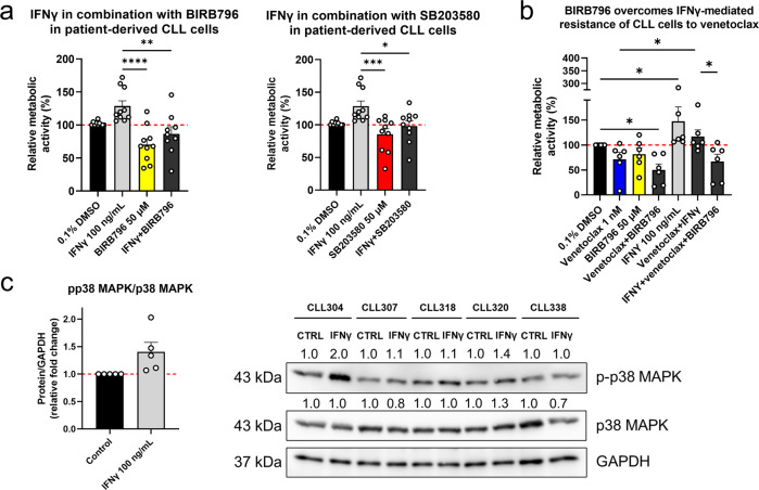
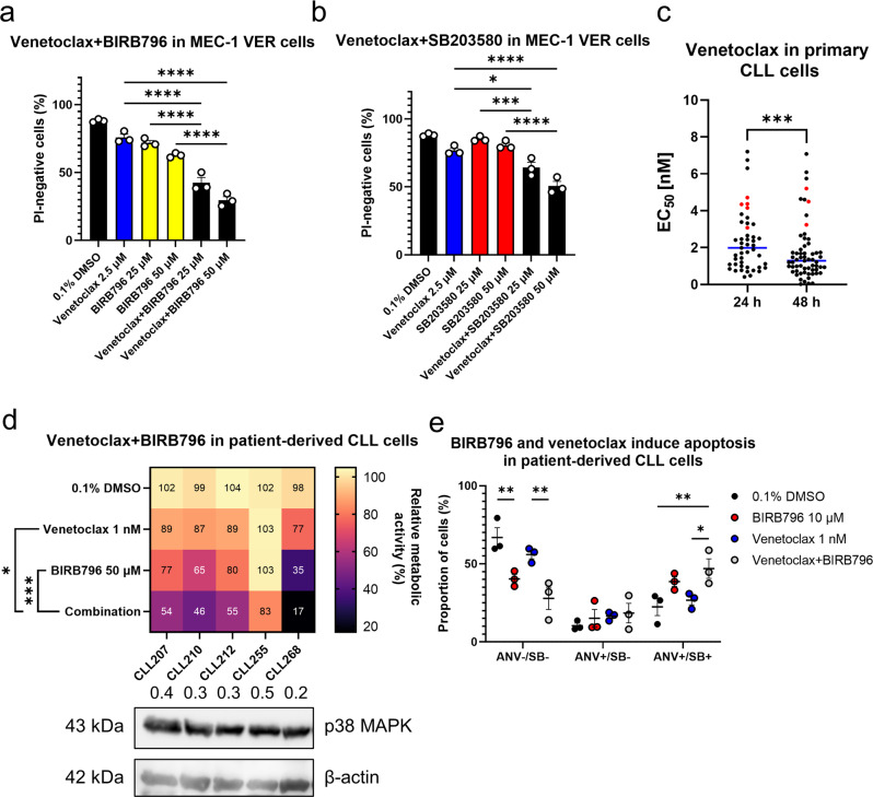
Chronic lymphocytic leukemia (CLL) is a hematological neoplasm of CD19-positive mature-appearing B lymphocytes. Despite the clinical success of targeted therapies in CLL, the development of resistance diminishes their therapeutic activity. This is also true for the Bcl-2 antagonist venetoclax. We investigated the molecular mechanisms that drive venetoclax resistance in CLL, with a clear focus to provide new strategies to successfully combat it. Activation of CLL cells with IFNγ, PMA/ionomycin, and sCD40L diminished the cytotoxicity of venetoclax. We demonstrated that the metabolic activity of cells treated with 1 nM venetoclax alone was 48% of untreated cells, and was higher for cells co-treated with IFNγ (110%), PMA/ionomycin (78%), and sCD40L (62%). As of molecular mechanism, we showed that PMA/ionomycin and sCD40L triggered translocation of NFκB in primary CLL cells, while IFNγ activated p38 MAPK, suppressed spontaneous and venetoclax-induced apoptosis and induced formation of the immunoproteasome. Inhibition of immunoproteasome with ONX-0914 suppressed activity of immunoproteasome and synergized with venetoclax against primary CLL cells. On the other hand, inhibition of p38 MAPK abolished cytoprotective effects of IFNγ. We demonstrated that venetoclax-resistant (MEC-1 VER) cells overexpressed p38 MAPK and p-Bcl-2 (Ser70), and underexpressed Mcl-1, Bax, and Bak. Inhibition of p38 MAPK or immunoproteasome triggered apoptosis in CLL cells and overcame the resistance to venetoclax of MEC-1 VER cells and venetoclax-insensitive primary CLL cells. In conclusion, the p38 MAPK pathway and immunoproteasome represent novel targets to combat venetoclax resistance in CLL.


Conflict of interest statement

Author H.P. has been involved as consultant for Abbvie. The other authors declare no conflict of interest. The funders had no role in the design of the study; in the collection, analyses, or interpretation of data; in the writing of the manuscript; or in the decision to publish the results.

Figures

Fig. 1
Fig. 1. IFNγ rescues CLL cells from spontaneous and venetoclax-induced cell death.
a Diverse stimulants abrogate venetoclax cytotoxicity against patient-derived CLL cells. Primary cells (1 × 10⁶ cells/mL) obtained from nine patients with CLL were treated with 0.1% DMSO (vehicle control), and 1 nM venetoclax alone and in combination with 100 ng/mL IFNγ, 10 nM PMA/1 µM ionomycin, and 100 ng/mL sCD40L for 24 h. The metabolic activities of the cells were determined using the PrestoBlue assay. Data are means ± SEM of ≥ three independent experiments, each carried out in duplicate. Two-way ANOVA (post hoc Tukey); *, **, and **** denote p < 0.05, p < 0.01, and p < 0.0001, respectively; b Cytoprotective effects of IFNγ in the patient-derived CLL cells undergoing spontaneous and venetoclax-induced apoptosis. CLL cells (1 × 10⁶ cells/mL) were treated with 0.1% DMSO (vehicle control) and 1 nM venetoclax, 100 ng/mL IFNγ, and their combination for 24 h. Apoptosis was assessed using the SYTOX Blue/annexin V assay. Representative dot plots (left) and means ± SEM of three independent experiments (right) are shown. Two-way ANOVA (post hoc Tukey); *, **, ***, and **** denote p < 0.05, p < 0.01, p < 0.001, and p < 0.0001, respectively; c Nuclear translocation of NFκB in the primary CLL cells. CLL cells (1 × 10⁶ cells/mL) from three patients with CLL were treated with 0.1% DMSO (vehicle control), 100 ng/mL IFNγ, 10 nM PMA/1 µM ionomycin, and 100 ng/mL sCD40L for 1 h. Quantification (left) and representative images (right) are shown for the translocation of NFκB to the nucleus, using imaging flow cytometry. One-way ANOVA (post hoc Dunnett); **** denotes p < 0.0001.
Fig. 2
Fig. 2. IFNγ activates the immunoproteasome in CLL cells.
a Schematic depiction of immunoproteasome formation in the presence of IFNγ; b MEC-1 cells (1 × 10⁶ cells/mL) were treated with 0.1% DMSO (vehicle control) or 100 ng/mL IFNγ for 8 h, 16 h, and 24 h. The cells (4 × 10⁶) were harvested and lysed, and the activities of LMP2, MECL1, and LMP7 were determined in whole-cell lysates using subunit-specific substrates. Fluorescence of the cleaved substrates at 20 min was normalized to the total protein concentration. Data are means ± SEM of ≥ three independent experiments, each carried out in duplicate. One-way ANOVA (post hoc Dunnett); not significant (ns), * denotes p < 0.05; c ONX-0914 suppresses basal and IFNγ-induced activity of immunoproteasome subunit LMP7 in primary CLL cells. Cells derived from three patient with CLL (1 × 10⁶ cells/mL) were treated with 0.1% DMSO (vehicle control) and 10 nM ONX-0914, 100 ng/mL IFNγ, and their combination for 24 h. The activity of LMP7 was determined using subunit-specific substrate. Fluorescence of the cleaved substrates at 60 min was normalized to the total protein concentration. Data are means ± SEM of ≥ three independent experiments, each carried out in duplicate. One-way ANOVA (post hoc Dunnett); ** and **** denote p < 0.01 and p < 0.0001; d ONX-0914 synergizes with venetoclax against primary CLL cells. Cells derived from seven patients with CLL (1 × 10⁶ cells/mL) were treated with 0.1% DMSO (vehicle control), 10 nM ONX-0914, 1 nM venetoclax, and their combination in the absence and presence of 100 ng/mL IFNγ for 24 h. Then the metabolic activities of cells were determined using the PrestoBlue assay. Data are means ± SEM of ≥ three independent experiments, each carried out in duplicate. Two-way ANOVA (post hoc Tukey); *, **, ***, and **** denote p < 0.05, < 0.01, < 0.001, and p < 0.0001.
Fig. 3
Fig. 3. Establishment of venetoclax-resistant MEC-1 VER cells.
a Dosing regimen used for the selection of venetoclax-resistant clones. MEC-1 cells (1 × 10⁶ cells/mL) were seeded into a 12 well plate and incubated with gradually increasing concentrations of venetoclax, from 1 µM at day 0, to 40 µM at day 85; b Cytotoxicity of venetoclax in MEC-1 and MEC-1 VER cells. The cells (3 × 10⁵ cells/mL) were treated with 0.1% DMSO (vehicle control) and venetoclax (1–100 µM) for 24 h. Afterward, 5 µM PI was added and the viability of cells was determined using flow cytometry. EC50 values were calculated using GraphPad Prism 9.2.0 (left). The viabilities of these MEC-1 and MEC-1 VER cells upon treatment with 25 µM venetoclax for 24 h are also shown (right). Data are means ± SEM of three independent experiments, each carried out in duplicate. Statistical significance was determined using paired Student t-test; **** denotes p < 0.0001; c Expression of anti-apoptotic proteins in MEC-1 and MEC-1 VER cells. Whole-cell lysates of MEC-1 and MEC-1 VER cells, cultured under basal conditions, were probed for Bcl-2, p-Bcl-2 (Ser70), Mcl-1, Bcl-xL, Puma, Bax, Bak, and Bid levels using immunoblotting. Data are means ± SEM of three independent experiments. Representative immunoblots are also shown.
Fig. 4
Fig. 4. Immunoproteasome inhibitor ONX-0914 overcomes resistance of MEC-1 VER cells to venetoclax.
a MEC-1 VER cells (3 × 10⁵ cells/mL) were treated with 0.1% DMSO (vehicle control), 1 µM venetoclax, 100 nM ONX-0914 and their combination for 24 h. Then, the samples were stained with 5 µM propidium iodide (PI) and analyzed using flow cytometry. Data are means ± SEM of three independent experiments, each carried out in duplicate. One-way ANOVA (post hoc Dunnett); ** denotes p < 0.01. b ONX-0914 disrupts mitochondrial membrane potential. MEC-1 cells (3 × 10⁵ cells/mL) were treated with 0.1% DMSO or 100, 250, and 500 nM ONX-0914 for 24 h and with 1 µM valinomycin (positive control) for 1 h. Then, the samples were stained with JC-1 probe and analyzed using flow cytometry. Data are means ± SEM of three independent experiments. One-way ANOVA (post hoc Dunnett); * denotes p < 0.05. c ONX-0914 induces caspase-dependent apoptotic cell death. MEC-1 cells (3 × 10⁵ cells/mL) were treated with 0.1% DMSO, 500 nM ONX-0914, 10 µM pan-caspase inhibitor QVD-OPh and their combination for 24 h. The proportion of cells undergoing early (lower right quadrant; ANV + /SB-) and late (upper right quadrant; ANV + /SB + ) apoptosis was determined. Representative dot plots (above) and means ± SEM of three independent experiments (below) are shown. Two-way ANOVA (post hoc Tukey); **, and **** denote p < 0.05 and p < 0.0001, respectively.
Fig. 5
Fig. 5. Inhibition of p38 MAPK can reverse the cytoprotective effects of IFNγ.
a Block on p38 MAPK pathway abolishes the stimulating effects of IFNγ in CLL cells. Primary cells (1 × 10⁶ cells/mL) obtained from 10 patients with CLL were treated with 0.1% DMSO (vehicle control) and 100 ng/mL IFNγ alone and in combination with 50 µM BIRB796 or 50 µM SB203580 for 24 h. The metabolic activity of cells was determined using the PrestoBlue assay. Data are means ± SEM of ≥ three independent experiments, carried out in duplicate. One-way ANOVA (post hoc Dunnett); *, **, ***, and **** denote p < 0.05, p < 0.01, p < 0.001, and p < 0.0001; b Inhibition of p38 MAPK reverses IFNγ-mediated resistance of primary CLL cells to venetoclax. CLL cells (1 × 10⁶ cells/mL) from six patients with CLL were treated with 0.1% DMSO (vehicle control), 1 nM venetoclax, 50 µM BIRB796, 100 ng/mL IFNγ, and their combinations for 24 h. The metabolic activity of cells was determined using the PrestoBlue assay. Data are means ± SEM of ≥ three independent experiments, each carried out in duplicate. Two-way ANOVA (post hoc Tukey); *, denotes p < 0.05; c IFNγ activates the p38 MAPK pathway in primary CLL cells. Cells (2 × 10⁶ cells/mL) from five patient with CLL were left untreated (control) or were treated with 100 ng/mL IFNγ for 1 h. Then, the cells were lysed, and whole cells lysates were separated with SDS-PAGE and blotted to nitrocellulose membranes. The ratio p-p38 MAPK/p38 MAPK (left) and immunoblots (right) for phosphorylated and total forms of p38 MAPK are shown. GAPDH was used as the loading control. Data are means ± SEM of five independent experiments.
Fig. 6
Fig. 6. Differential expression of MAPKs and their downstream effectors in MEC-1 and MEC-1 VER cells.
ac Expression levels of Ras, p-MEK, MEK, p-ERK1/2, ERK 1/2, p-p38 MAPK, p38 MAPK, p-JNK, JNK (a), cyclin D1 (b), and p-CREB and CREB (c) in MEC-1 and MEC-1 VER cells under basal conditions; d The expression levels of p-MEK, MEK, p-ERK1/2, ERK1/2, p-p38 MAPK, p38 MAPK, and p-JNK, JNK over time following treatment of MEC-1 and MEC-1 VER cells with 10 µM venetoclax. Data are means ± SEM of ≥ two independent experiments. Representative immunoblots are also shown.
Fig. 7
Fig. 7. Inhibition of p38 MAPK overcomes the resistance of CLL cells to venetoclax.
MEC-1 VER cells (3 × 10⁵ cells/mL) were treated with 0.1% DMSO (vehicle control) or 10 µM venetoclax alone or in combination with 25 and 50 µM BIRB796 (a) and 25 and 50 µM SB203580 (b) for 48 h. The cells were stained with 5 µM propidium iodide (PI) and the viability of cells (PI-negativity, %) was determined using flow cytometry. Data are means ± SEM of three independent experiments, each carried out in duplicate. One-way ANOVA (post hoc Tukey); *, ***, and **** denote p < 0.05, p < 0.001, and p < 0.0001, respectively. c Ex-vivo testing of patient-derived CLL cells to identify those relatively insensitive to venetoclax. CLL cells (1 × 10⁶ cells/mL) from 61 patients with CLL were treated with 0.1–50 nM venetoclax for 24 h and 48 h. The metabolic activities were assessed using the PrestoBlue assay, and then the EC50 values were determined using GraphPad Prism 9.2.0. Each symbol corresponds to the individual CLL cell samples. Blue line, median; red symbols, the five CLL cell samples defined as relatively insensitive to venetoclax (EC50 ≥ 2-fold the median), as used in (d). Paired Student t-test (n = 43); *** denotes p < 0.001; d BIRB796 augments the action of venetoclax in CLL cells that are relatively insensitive to venetoclax. CLL cells (1 × 10⁶ cells/mL) from five CLL patients were treated with 0.1% DMSO (vehicle control) and 1 nM venetoclax, 50 µM BIRB796 and their combination for 24 h. The metabolic activities of cells were determined using the PrestoBlue assay. Data in squares are mean metabolic activities of independent experiments, carried out in duplicate. Rows represent responses of cells from different CLL patients to a specific treatment, while columns represent responses of cells from a specific CLL patient to different treatments. One-way ANOVA (post hoc Dunnett); * and *** denote p < 0.05 and p < 0.001, respectively. Immunoblot of CLL samples probed for the expression of p38 MAPK and β-actin is shown. e Detection of apoptosis after 24-hour treatment of primary cells (1 × 10⁶ cells/mL) with 10 µM BIRB796, 1 nM venetoclax, and their combination. Data are means ± SEM of three independent experiments. Two-way ANOVA (post hoc Tukey); * and ** denote p < 0.05 and p < 0.01, respectively.

References

    1. Hallek M. Chronic lymphocytic leukemia: 2020 update on diagnosis, risk stratification and treatment. Am J Hematol. 2019;94:1266–87. doi: 10.1002/ajh.25595. - DOI - PubMed
    1. Hallek M, Shanafelt TD, Eichhorst B. Chronic lymphocytic leukaemia. Lancet. 2018;391:1524–37. doi: 10.1016/S0140-6736(18)30422-7. - DOI - PubMed
    1. Gentile M, Petrungaro A, Uccello G, Vigna E, Recchia AG, Caruso N, et al. Venetoclax for the treatment of chronic lymphocytic leukemia. Expert Opin Investigational Drugs. 2017;26:1307–16. doi: 10.1080/13543784.2017.1386173. - DOI - PubMed
    1. Bose P, Gandhi V, Konopleva M. Pathways and mechanisms of venetoclax resistance. Leuk lymphoma. 2017;58:1–17. doi: 10.1080/10428194.2017.1283032. - DOI - PMC - PubMed
    1. Tausch E, Close W, Dolnik A, Bloehdorn J, Chyla B, Bullinger L, et al. Venetoclax resistance and acquired BCL2 mutations in chronic lymphocytic leukemia. Haematologica. 2019;104:e434–7. doi: 10.3324/haematol.2019.222588. - DOI - PMC - PubMed

Publication types

MeSH terms