Review
. 2022 Oct 9;9(1):56.
doi: 10.1186/s40779-022-00422-y.

Sepsis-induced immunosuppression: mechanisms, diagnosis and current treatment options

Affiliations
Review

Sepsis-induced immunosuppression: mechanisms, diagnosis and current treatment options

Di Liu et al. Mil Med Res. .

Abstract

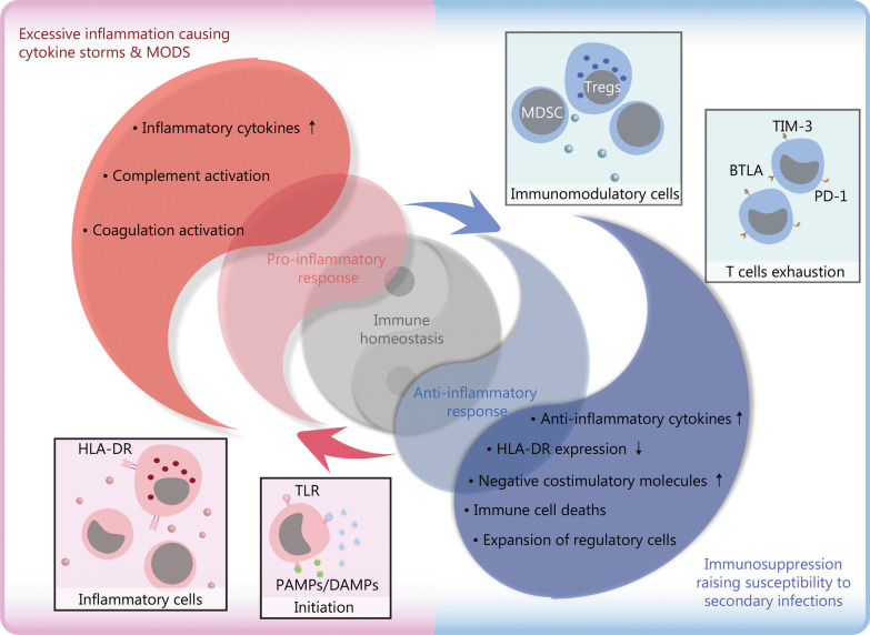
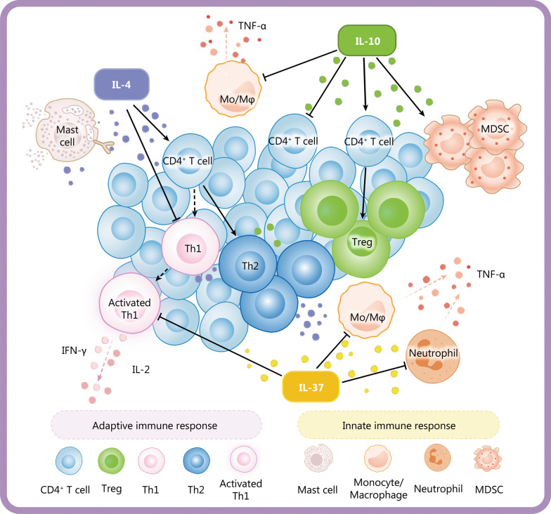
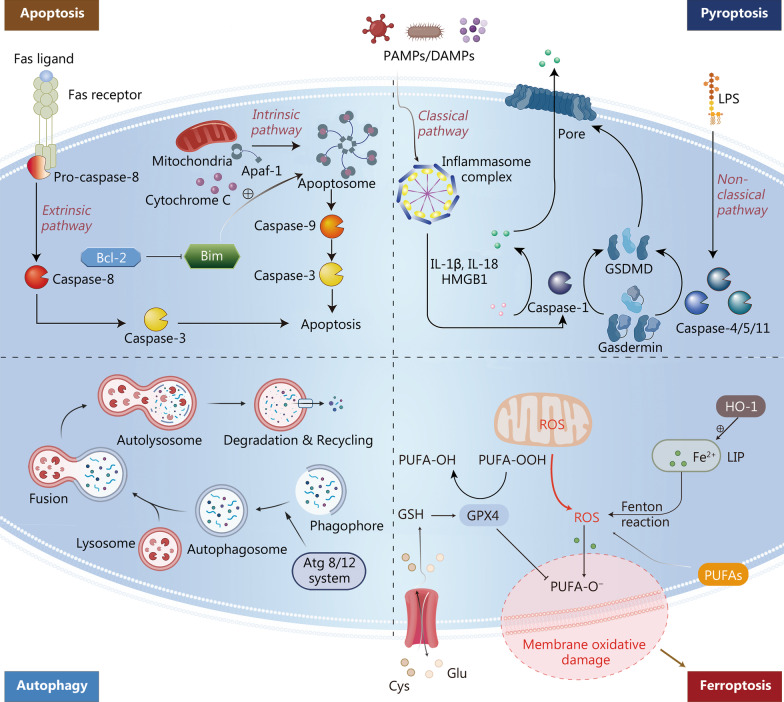
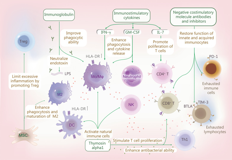
Sepsis is a common complication of combat injuries and trauma, and is defined as a life-threatening organ dysfunction caused by a dysregulated host response to infection. It is also one of the significant causes of death and increased health care costs in modern intensive care units. The use of antibiotics, fluid resuscitation, and organ support therapy have limited prognostic impact in patients with sepsis. Although its pathophysiology remains elusive, immunosuppression is now recognized as one of the major causes of septic death. Sepsis-induced immunosuppression is resulted from disruption of immune homeostasis. It is characterized by the release of anti-inflammatory cytokines, abnormal death of immune effector cells, hyperproliferation of immune suppressor cells, and expression of immune checkpoints. By targeting immunosuppression, especially with immune checkpoint inhibitors, preclinical studies have demonstrated the reversal of immunocyte dysfunctions and established host resistance. Here, we comprehensively discuss recent findings on the mechanisms, regulation and biomarkers of sepsis-induced immunosuppression and highlight their implications for developing effective strategies to treat patients with septic shock.

Keywords: Immune monitoring; Immunomodulatory therapy; Immunosuppression; Sepsis.


Conflict of interest statement

The authors declare that they have no competing interests.

Figures

Fig. 1
Fig. 1
Schematic diagram of immune homeostasis imbalance in sepsis. The immune response is initiated when the host recognizes PAMPs and DAMPs. Inflammatory cells release pro-inflammatory cytokines and cause excessive inflammation in the early stage of inflammation. Under physiological conditions, the dynamic balance between pro-inflammatory and anti-inflammatory responses maintains immune homeostasis. However, after the onset of sepsis, the balance is disrupted. The upregulated expression of pro-inflammatory cytokines released by inflammatory cells and the activation of the complement and coagulation systems, result in excessive inflammation, which further leads to cytokine storms and MODS. Concurrently or subsequently, the increased release of anti-inflammatory cytokines and coinhibitory molecules, decreased expression of HLA-DR, death of immunocytes, and expansion of regulatory cells lead to immunosuppression, increasing the susceptibility to secondary infections, which is the main cause of poor prognosis in septic patients. TLR toll-like receptor, PAMP pathogen-associated molecular pattern, DAMP damage-associated molecular pattern, HLA-DR human leukocyte antigen-DR, MODS multiple organ dysfunction syndromes, Treg regulatory T cell, TIM-3 T-cell immunoglobulin domain and mucin domain-3, BTLA B and T lymphocyte antigens, PD-1 programmed cell death 1
Fig. 2
Fig. 2
Anti-inflammatory cytokines in sepsis. Anti-inflammatory cytokines mainly include IL-4, IL-10 and IL-37. IL-4 can induce CD4+ T cells to differentiate into Th2 cells and promote autocrine signaling of mast cells through positive feedback. It can also stimulate the release of other anti-inflammatory cytokines and inhibit the release of pro-inflammatory cytokines such as IL-2 and IFN-γ by activated Th1. IL-10 may aggravate immunosuppression by decreasing the release of pro-inflammatory cytokines including TNF-α, inhibiting the proliferation of CD4+ T cells and promoting the differentiation of CD4+ T cells into Tregs and the proliferation of MDSCs. IL-37 is closely related to the severity of sepsis-induced immunosuppression by suppressing the pro-inflammatory cytokine release from monocytes and neutrophils. Inflammatory cytokines such as TNF-α, IFN-γ and IL-2 are represented by red dots whereas IL-10, IL-37 and IL-4 are represented by dots in other colors. Th1 T helper 1, Th2 T helper 2, MDSC myeloid-derived suppressor cell, TNF-α tumor necrosis factor-alpha, IFN-γ interferon-γ, IL interleukin, Mo/Mφ monocyte/macrophage
Fig. 3
Fig. 3
Four types of cell death in sepsis. Apoptosis: Apoptosis is activated in either the extrinsic or the intrinsic pathway. The extrinsic pathway is triggered by the Fas/FasL pathway after infection. The death receptor Fas activates caspase-8 by binding to the Fas ligand that is expressed on activated T lymphocytes during cellular immunity followed by activation of caspase-3 to trigger the execution pathway of apoptosis. In the intrinsic pathway, death stimuli including DNA damage and the accumulation of misfolded proteins break the balance between proapoptotic and antiapoptotic signals mediating mitochondrial outer membrane permeabilization after which cytochrome C is released from mitochondria and forms an apoptosome with Apaf-1. The apoptosome activates caspase-9 and finally activates caspase-3. The pro-apoptotic protein Bim accelerates apoptosis while the anti-apoptotic protein Bcl-2 inhibits apoptosis. Pyroptosis: In the classical pathway, the inflammasome complex activates caspase-1 upon simulating PAMPs and DAMPs. Caspase-1 promotes the release of pro-inflammatory cytokines such as IL-1β IL-18 and HMGB1 and then cleaves gasdermin into GSDMD. GSDMD aggregates into a pore on the cell membrane. In the non-classical pathway, LPS activates caspase-4, caspase-5 and caspase-11 which cleave gasdermin into GSDMD to form the pore and drive pyroptosis. Autophagy: Atg 8/12 systems activate the phagophore to form the autophagosome. The autophagosome fuses with the lysosome and further form an autolysosome. Lysosomal enzymes degrade misfolded proteins and damaged organelles in autolysosomes and enter the recycling process. Ferroptosis: Ferroptosis is a ROS-dependent form of cell death defined by iron-dependent accumulation and lipid peroxides that is resulted from an imbalance between the synthesis of oxidants and antioxidants. At the core process, PUFAs and lipids containing PUFAs are particularly sensitive to oxidation by enzymes and nonenzymatic processes such as iron-dependent Fenton reactions to form lipid hydroperoxides that can produce toxic lipid free radicals (e.g., alkoxyl radicals) in the presence of iron. Furthermore, by taking protons from neighboring PUFAs, these free radicals might initiate a new round of lipid oxidation and spread oxidative damage. GPX4 functions as a phospholipid hydroperoxidase in the redox system to reduce phospholipid hydroperoxide production and plays an anti-ferroptosis role. The enzyme HO-1 can accelerate the formation of a labile iron pool and further promote lipid peroxidation. GSDMD gasdermin-D, LPS lipopolysaccharide, Atg autophagy-related gene, ROS reactive oxygen species, PUFA polyunsaturated fatty acid, LIP labile iron pool, GPX4 glutathione peroxidase 4, HMGB1 high mobility group box 1, IL interleukin. Part of the autophagy was created partially utilizing the templates on BioRender.com as a reference
Fig. 4
Fig. 4
Immunity therapies to fight sepsis. Immunomodulatory therapy includes medication to improve immunity and immune stimulation combined with anti-inflammatory approaches can significantly improve the outcome of severe sepsis. IFN-γ, GM-CSF and IL-7 are immunostimulatory cytokines that have been proven to activate early-responding immune cells in sepsis. IFN-γ and GM-CSF can activate innate immune cells to enhance phagocytosis in addition to pro-inflammatory cytokine release and the expression of mHLA-DR on APCs. IL-7 can increase the number of lymphocytes by promoting proliferation and inhibiting apoptosis. Immunoglobulin is a natural protein that neutralizes endotoxins in the body and promotes the phagocytic ability of monocytes and macrophages. Immunoglobulin therapy may be beneficial in improving the prognosis of septic patients with multidrug-resistant bacterial infections. Thymosin alpha1 can activate innate immune cells such as DCs and NK cells, and macrophages stimulate T-cell proliferation and enhance the antibacterial effect of Th1 cells. MSCs can promote the maturation of M2 macrophages and regulatory T cells, thereby promoting bacterial clearance and limiting excessive inflammation, to alleviate organ damage and ultimately reduce sepsis mortality. Coinhibitory molecule antibodies and antagonists targeting TIM-3, PD-1, BTLA, etc., can restore the function of innate and acquired immunocytes, reversing the immune exhaustion state. GM-CSF granulocyte–macrophage colony-stimulating factor, MSC mesenchymal stem cell, BTLA B and T lymphocyte attenuator, Mo/Mφ monocyte/macrophage, DC dendritic cell, NK natural killer cell, HLA-DR human leukocyte antigen-DR, LPS lipopolysaccharide, PD-1 programmed cell death 1, IFN-γ interferon-γ, IL interleukin

References

    1. Singer M, Deutschman CS, Seymour CW, Shankar-Hari M, Annane D, Bauer M, et al. The third international consensus definitions for sepsis and septic shock (sepsis-3) JAMA. 2016;315(8):801–810. doi: 10.1001/jama.2016.0287. - DOI - PMC - PubMed
    1. Seymour CW, Kennedy JN, Wang S, Chang CH, Elliott CF, Xu Z, et al. Derivation, validation, and potential treatment implications of novel clinical phenotypes for sepsis. JAMA. 2019;321(20):2003–2017. doi: 10.1001/jama.2019.5791. - DOI - PMC - PubMed
    1. Gavelli F, Castello LM, Avanzi GC. Management of sepsis and septic shock in the emergency department. Intern Emerg Med. 2021;16(6):1649–1661. doi: 10.1007/s11739-021-02735-7. - DOI - PMC - PubMed
    1. Rudd KE, Johnson SC, Agesa KM, Shackelford KA, Tsoi D, Kievlan DR, et al. Global, regional, and national sepsis incidence and mortality, 1990–2017: analysis for the Global Burden of Disease Study. Lancet. 2020;395(10219):200–211. doi: 10.1016/S0140-6736(19)32989-7. - DOI - PMC - PubMed
    1. McBride MA, Patil TK, Bohannon JK, Hernandez A, Sherwood ER, Patil NK. Immune checkpoints: novel therapeutic targets to attenuate sepsis-induced immunosuppression. Front Immunol. 2020;11:624272. doi: 10.3389/fimmu.2020.624272. - DOI - PMC - PubMed

Publication types