

Small nuclei identification with a hemispherical brain PET
- PMID: 36209191
- PMCID: PMC9547762
- DOI: 10.1186/s40658-022-00498-4
Small nuclei identification with a hemispherical brain PET
Abstract
Background: To confirm the performance of the first hemispherical positron emission tomography (PET) for the brain (Vrain) that we developed to visualise the small nuclei in the deep brain area, we compared 18F-fluorodeoxyglucose (FDG) brain images with whole-body PET images.
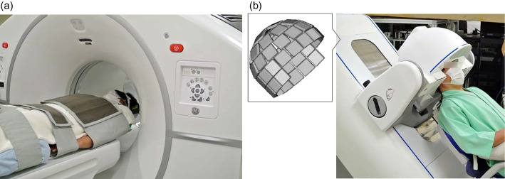
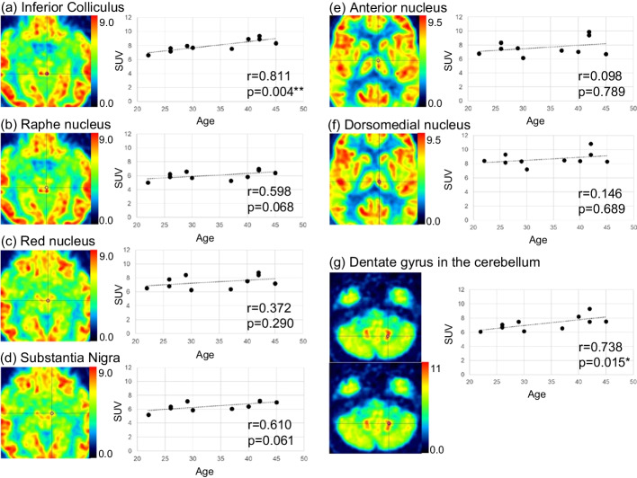
Methods: Ten healthy male volunteers (aged 22-45 years) underwent a representative clinical whole-body PET, followed by Vrain each for 10 min. These two scans were initiated 30 min and 45 min after FDG injection (4.1 ± 0.5 MBq/kg), respectively. First, we visually identified the small nuclei and then compared their standardised uptake values (SUVs) with the participants' age. Next, the SUVs of each brain region, which were determined by applying a volume-of-interest template for anatomically normalised PET images, were compared between the brain images with the Vrain and those with the whole-body PET images.
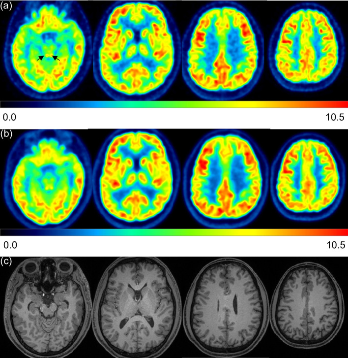
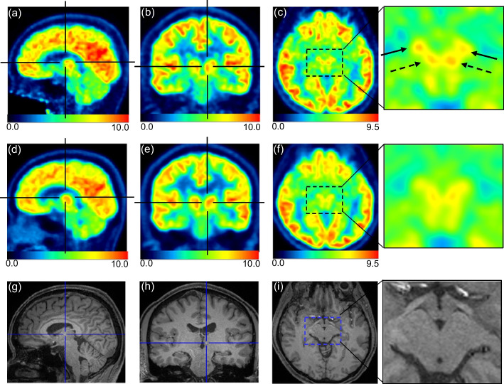
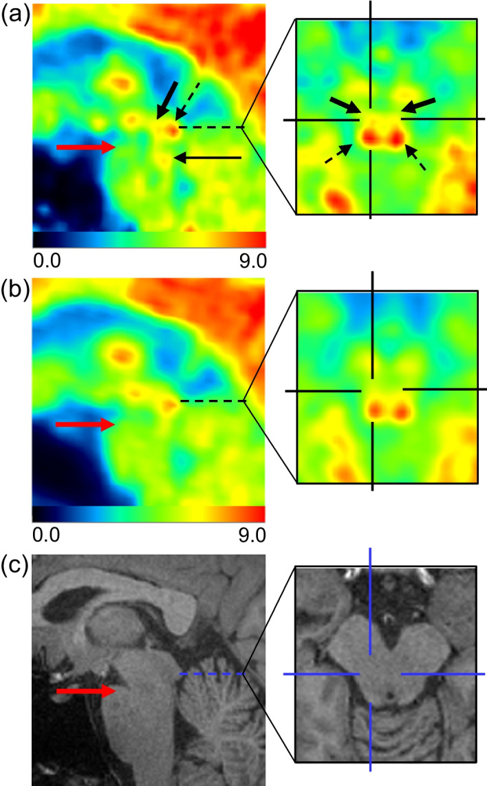
Results: Small nuclei, such as the inferior colliculus, red nucleus, and substantia nigra, were more clearly visualised in Vrain than in whole-body PET. The anterior nucleus and dorsomedial nucleus in the thalamus and raphe nucleus in the brainstem were identified in Vrain but not in whole-body PET. The SUVs of the inferior colliculus and dentate gyrus in the cerebellum positively correlated with age (Spearman's correlation coefficient r = 0.811, p = 0.004; r = 0.738, p = 0.015, respectively). The SUVs of Vrain were slightly higher in the mesial temporal and medial parietal lobes than those in whole-body PET.
Conclusions: This was the first time that the raphe nuclei, anterior nuclei, and dorsomedial nuclei were successfully visualised using the first hemispherical brain PET. TRIAL REGISTRATION : Japan Registry of Clinical Trials, jRCTs032210086, Registered 13 May 2021, https://jrct.niph.go.jp/latest-detail/jRCTs032210086 .
Keywords: Brain PET; FDG; Healthy volunteer; Raphe nucleus; Thalamus.
© 2022. The Author(s).
Conflict of interest statement
This study was financially supported by ATOX Co., Ltd.; G. Akamatsu, H. Tashima, E. Yoshida, and T. Yamaya have applied for patents related to the development of our brain-dedicated PET system; no other potential conflicts of interest relevant to this article exist.
Figures
Similar articles
-
Performance evaluation of VRAIN: a brain-dedicated PET with a hemispherical detector arrangement.Phys Med Biol. 2022 Nov 16;67(22). doi: 10.1088/1361-6560/ac9e87. Phys Med Biol. 2022. PMID: 36317319
-
Individually differentiated serotonergic raphe nuclei measured with brain PET/MR imaging.Radiology. 2014 Aug;272(2):541-8. doi: 10.1148/radiol.14131547. Epub 2014 Mar 21. Radiology. 2014. PMID: 24654972
-
Metabolic rates in small brain nuclei determined by high-resolution PET.J Nucl Med. 2004 Nov;45(11):1811-5. J Nucl Med. 2004. PMID: 15534048 Clinical Trial.
-
Brain PET motion correction using 3D face-shape model: the first clinical study.Ann Nucl Med. 2022 Oct;36(10):904-912. doi: 10.1007/s12149-022-01774-0. Epub 2022 Jul 19. Ann Nucl Med. 2022. PMID: 35854178 Free PMC article.
-
Simultaneous whole-body PET/MR imaging in comparison to PET/CT in pediatric oncology: initial results.Radiology. 2014 Oct;273(1):220-31. doi: 10.1148/radiol.14131732. Epub 2014 May 31. Radiology. 2014. PMID: 24877983
Cited by
-
Visualization of small brain nuclei with a high-spatial resolution, clinically available whole-body PET scanner.Ann Nucl Med. 2024 Feb;38(2):154-161. doi: 10.1007/s12149-023-01886-1. Epub 2023 Nov 21. Ann Nucl Med. 2024. PMID: 37989801 Free PMC article.
-
New Horizons in Brain PET Instrumentation.PET Clin. 2024 Jan;19(1):25-36. doi: 10.1016/j.cpet.2023.08.001. Epub 2023 Oct 6. PET Clin. 2024. PMID: 37806894 Free PMC article. Review.
-
A conformal TOF-DOI Prism-PET prototype scanner for high-resolution quantitative neuroimaging.Med Phys. 2023 Jan 18:10.1002/mp.16223. doi: 10.1002/mp.16223. Online ahead of print. Med Phys. 2023. PMID: 36651630 Free PMC article.
References
LinkOut - more resources
Full Text Sources
