

Quantum tunnelling in the context of SARS-CoV-2 infection
- PMID: 36209224
- PMCID: PMC9547378
- DOI: 10.1038/s41598-022-21321-1
Quantum tunnelling in the context of SARS-CoV-2 infection
Abstract
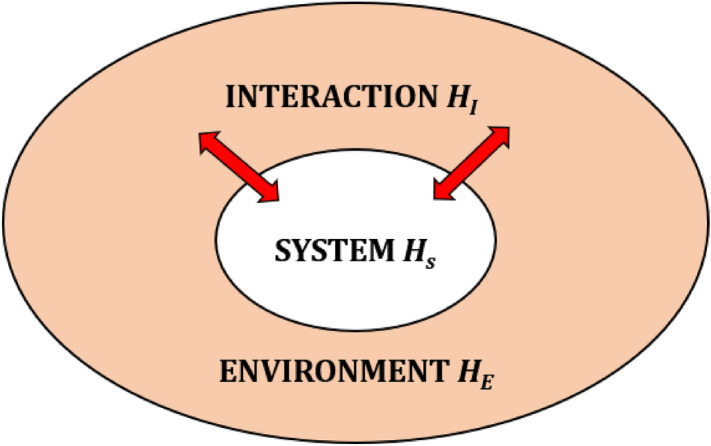
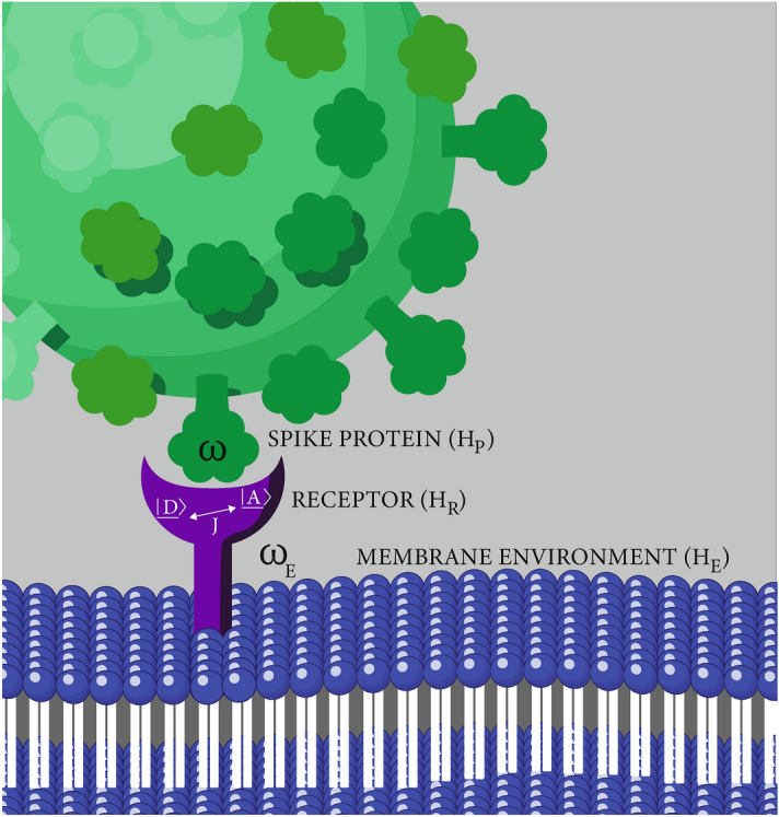
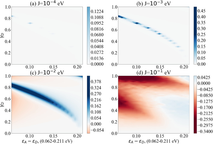
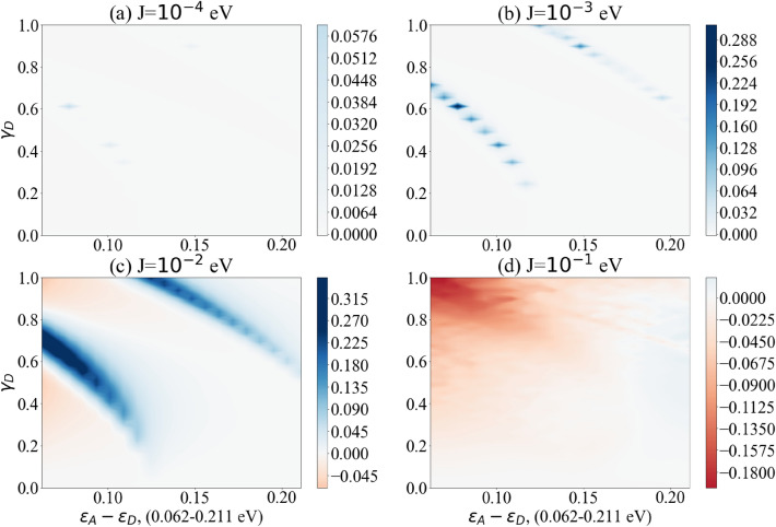
The SARS-CoV-2 pandemic has added new urgency to the study of viral mechanisms of infection. But while vaccines offer a measure of protection against this specific outbreak, a new era of pandemics has been predicted. In addition to this, COVID-19 has drawn attention to post-viral syndromes and the healthcare burden they entail. It seems integral that knowledge of viral mechanisms is increased through as wide a research field as possible. To this end we propose that quantum biology might offer essential new insights into the problem, especially with regards to the important first step of virus-host invasion. Research in quantum biology often centres around energy or charge transfer. While this is predominantly in the context of photosynthesis there has also been some suggestion that cellular receptors such as olfactory or neural receptors might employ vibration assisted electron tunnelling to augment the lock-and-key mechanism. Quantum tunnelling has also been observed in enzyme function. Enzymes are implicated in the invasion of host cells by the SARS-CoV-2 virus. Receptors such as olfactory receptors also appear to be disrupted by COVID-19. Building on these observations we investigate the evidence that quantum tunnelling might be important in the context of infection with SARS-CoV-2. We illustrate this with a simple model relating the vibronic mode of, for example, a viral spike protein to the likelihood of charge transfer in an idealised receptor. Our results show a distinct parameter regime in which the vibronic mode of the spike protein enhances electron transfer. With this in mind, novel therapeutics to prevent SARS-CoV-2 transmission could potentially be identified by their vibrational spectra.
© 2022. The Author(s).
Conflict of interest statement
The authors declare no competing interests.
Figures

Similar articles
-
Vibration assisted electron tunnelling in COVID-19 infection using quantum state diffusion.Sci Rep. 2024 May 27;14(1):12152. doi: 10.1038/s41598-024-62670-3. Sci Rep. 2024. PMID: 38802472 Free PMC article.
-
Coevolution, Dynamics and Allostery Conspire in Shaping Cooperative Binding and Signal Transmission of the SARS-CoV-2 Spike Protein with Human Angiotensin-Converting Enzyme 2.Int J Mol Sci. 2020 Nov 4;21(21):8268. doi: 10.3390/ijms21218268. Int J Mol Sci. 2020. PMID: 33158276 Free PMC article.
-
Hot spot profiles of SARS-CoV-2 and human ACE2 receptor protein protein interaction obtained by density functional tight binding fragment molecular orbital method.Sci Rep. 2020 Oct 8;10(1):16862. doi: 10.1038/s41598-020-73820-8. Sci Rep. 2020. PMID: 33033344 Free PMC article.
-
Angiotensin-Converting Enzyme 2 (ACE2) in the Pathogenesis of ARDS in COVID-19.Front Immunol. 2021 Dec 22;12:732690. doi: 10.3389/fimmu.2021.732690. eCollection 2021. Front Immunol. 2021. PMID: 35003058 Free PMC article. Review.
-
COVID-19 pandemic: Insights into structure, function, and hACE2 receptor recognition by SARS-CoV-2.PLoS Pathog. 2020 Aug 21;16(8):e1008762. doi: 10.1371/journal.ppat.1008762. eCollection 2020 Aug. PLoS Pathog. 2020. PMID: 32822426 Free PMC article. Review.
Cited by
-
Non-Markovian Quantum State Diffusion for the tunnelling in SARS-COVID-19 virus.Comput Struct Biotechnol J. 2025 Jun 9;30:70-79. doi: 10.1016/j.csbj.2025.06.012. eCollection 2025. Comput Struct Biotechnol J. 2025. PMID: 40574961 Free PMC article.
-
Vibration assisted electron tunnelling in COVID-19 infection using quantum state diffusion.Sci Rep. 2024 May 27;14(1):12152. doi: 10.1038/s41598-024-62670-3. Sci Rep. 2024. PMID: 38802472 Free PMC article.
References
-
- Bohr N. Light and life. Nature. 1933;131:421–423. doi: 10.1038/131421a0. - DOI
-
- Schrödinger E. What is life? London. Cambridge***: Cambridge University Press; 1944.
Publication types
MeSH terms
Substances
LinkOut - more resources
Full Text Sources
Medical
Miscellaneous
