Review
. 2022 Oct 8;7(1):358.
doi: 10.1038/s41392-022-01190-w.

Signaling pathways and therapeutic interventions in gastric cancer

Affiliations
Review

Signaling pathways and therapeutic interventions in gastric cancer

Zi-Ning Lei et al. Signal Transduct Target Ther. .

Abstract

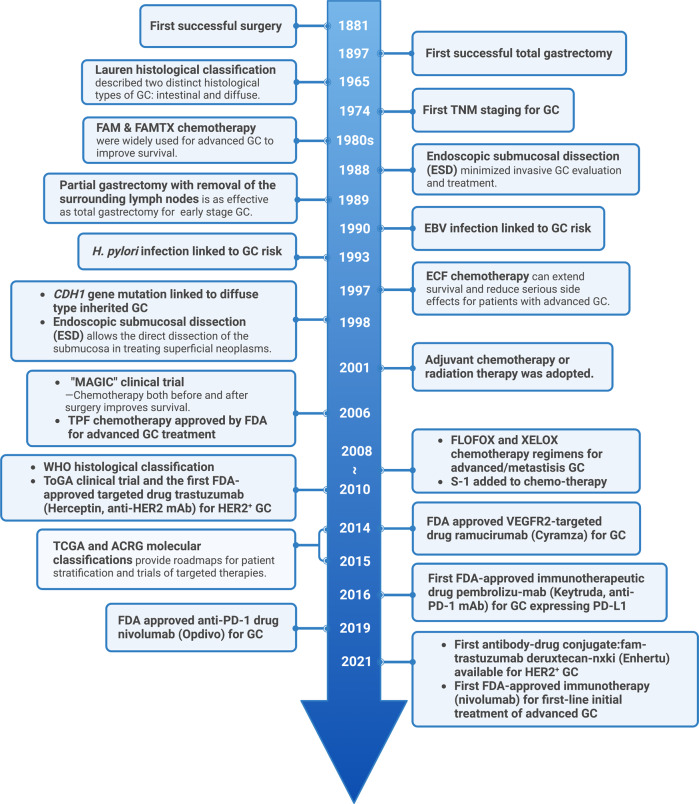
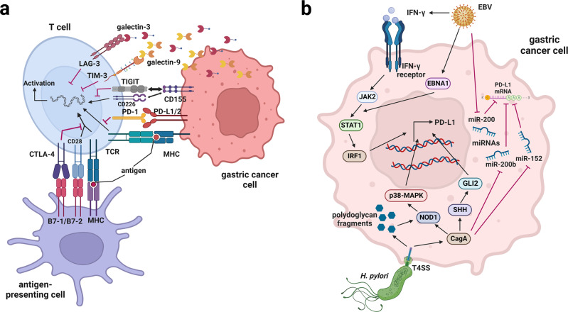
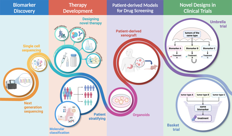
Gastric cancer (GC) ranks fifth in global cancer diagnosis and fourth in cancer-related death. Despite tremendous progress in diagnosis and therapeutic strategies and significant improvements in patient survival, the low malignancy stage is relatively asymptomatic and many GC cases are diagnosed at advanced stages, which leads to unsatisfactory prognosis and high recurrence rates. With the recent advances in genome analysis, biomarkers have been identified that have clinical importance for GC diagnosis, treatment, and prognosis. Modern molecular classifications have uncovered the vital roles that signaling pathways, including EGFR/HER2, p53, PI3K, immune checkpoint pathways, and cell adhesion signaling molecules, play in GC tumorigenesis, progression, metastasis, and therapeutic responsiveness. These biomarkers and molecular classifications open the way for more precise diagnoses and treatments for GC patients. Nevertheless, the relative significance, temporal activation, interaction with GC risk factors, and crosstalk between these signaling pathways in GC are not well understood. Here, we review the regulatory roles of signaling pathways in GC potential biomarkers, and therapeutic targets with an emphasis on recent discoveries. Current therapies, including signaling-based and immunotherapies exploited in the past decade, and the development of treatment for GC, particularly the challenges in developing precision medications, are discussed. These advances provide a direction for the integration of clinical, molecular, and genomic profiles to improve GC diagnosis and treatments.


Conflict of interest statement

The authors declare no competing interests.

Figures

Fig. 1
Fig. 1
Timeline of selected key findings and significant therapy developments in gastric cancer. The major milestones for risk factor identification, classification and staging, and therapy developments for GC are listed. Chemotherapy regimens: FAM: fluorouracil (5-FU) + mitomycin C + doxorubicin; FAMTX: methotrexate + 5-FU + doxorubicin; ECF: epirubicin + cisplatin + 5-FU; TPF: docetaxel + cisplatin + 5-FU; FLOFOX: oxaliplatin + 5-FU + leucovorin; XELOX: capecitabine (Xeloda) + oxaliplatin; S-1: tegafur (5-FU prodrug) + 5-chloro-2,4-dihydroxypyridine (CDHP) + oteracil potassium (Oxo), in a molar ratio of 1:0.4:1. EBV Epstein–Barr virus, TCGA The Cancer Genome Atlas, ACRG Asian Cancer Research Group. This figure was created with Biorender.com
Fig. 2
Fig. 2
Main signaling pathways and fundamental factors in gastric cancer. The major signaling and crosstalk of MAPK, HER2, PI3K/AKT/mTOR, HGF/c-Met, p53, Wnt/β-catenin, and NF-κB pathways, as well as their regulatory roles in cellular processes, are illustrated. GPCRs G-protein-coupled receptors, HGF hepatocyte growth factor, c-MET c-mesenchymal-epithelial transition factor, EGFR epidermal growth factor receptor, HER2/3/4 human epidermal growth factor receptor 2/3/4, MAPKKKs mitogen-activated protein kinase kinase kinases, RTKs receptor tyrosine kinases, RAS rat sarcoma, RAF rapidly accelerated fibrosarcoma, MKK mitogen-activated protein kinase kinase, SAPK/JNK jun amino-terminal kinase, p38-MAPKs p38 group of mitogen-activated protein kinases, MEK mitogen-activated protein kinase kinase, ERK1/2 extracellular signal-related kinase 1/2, PI3K phosphoinositide 3-kinase, AKT protein kinase B, mTORC1/2 mammalian target of rapamycin complex 1/2, PTEN phosphatase and tensin homolog, PDK1 phosphoinositide-dependent protein kinase 1, TSC1/2 tuberous sclerosis complex 1/2, p70S6K1 phosphorylation of ribosomal p70S6 kinase 1, 4E-BP1 eukaryotic translation initiation factor 4E (eIF4E)-binding protein 1, NF-κB nuclear factor kappa-B, GSK3 glycogen synthase kinase 3, BAD Bcl-xl/Bcl-2-asociated death promoter, Casp9 cysteinyl aspartate specific proteinase 9, MDM2 murine double minute 2, p53 tumor protein 53, EMT epithelial-mesenchymal transition, LRP5/6 low-density lipoprotein receptor-related protein 5/6, CKIα casein kinase Iα, APC adenomatous polyposis coli, TCF/LEF T-cell factor/lymphoid enhancer factor, TNFR tumor necrosis factor receptor, TLR toll-like receptors, IKK IκB kinase. This figure was created with Biorender.com
Fig. 3
Fig. 3
The immune checkpoint signaling pathways in gastric cancer and regulations on PD-L1 by H. pylori and EBV. a The immune checkpoint proteins PD-1 on the surface of T cells interact with the ligands PD-L1/PD-L2 on GC cells, or the aberrant CTLA-4 proteins on GC patient T cells interact with B7 on antigen-presenting cells, resulting in an immunosuppressive microenvironment, providing cancer cells with a survival advantage. TIGIT on the T cells membrane competes with the activation of CD226 binding to CD155 from the GC cells. Other immune checkpoint proteins, TIM-3 or LAG-3, interact with galectin-9 or galectin-3 released from GC cells, inhibiting the activation of T cells. b Chronic H. pylori or EBV infection, which are risk factors of GC, can induce upregulation of PD-L1 in GC cells via various signaling pathways and microRNAs, promoting immune escape. EBV Epstein–Barr virus, PD-1 programmed death 1, PD-L1/2 programmed death ligand 1/2, CTLA-4 cytotoxic T-lymphocyte-associated protein 4, TCR T-cell receptor, MHC major histocompatibility complex, TIGIT T cell immunoreceptor with Ig and ITIM domains, TIM-3 T cell immunoglobulin and mucin-domain containing-3, LAG-3 lymphocyte-activation gene 3, IFN-γ interferon gamma, JAK2 Janus kinase 2, STAT1 signal transducer and activator of transcription 1, IRF1 interferon regulatory factor 1, EBNA1 Epstein–Barr nuclear antigen 1, MAPK mitogen-activated protein kinase, NOD1 nucleotide-binding oligomerization domain-containing protein 1, SHH Sonic hedgehog protein, CagA cytotoxin-associated gene A, T4SS type IV secretion system. This figure was created with Biorender.com
Fig. 4
Fig. 4
Current therapies for gastric cancer based on staging. Therapeutic interventions for GC at different stages are illustrated by icons. The majorly used drugs or regimens of chemotherapy, targeted therapy, and immunotherapy are listed. EMR endoscopic mucosal resection, ESD endoscopic submucosal dissection. S-1 is an oral agent that is converted to 5-FU in the body, which contains a 5-FU prodrug called tegafur and the two enzyme inhibitors 5-chloro-2,4-dihydroxypyridine (CDHP) and oteracil potassium (Oxo), in a molar ratio of 1:0.4:1. This figure was adapted and modified from “Gastric Cancer Staging” by Biorender.com (2022). Retrieved from https://app.biorender.com/biorender-templates. Icons were adapted from Adobe Express
Fig. 5
Fig. 5
Overview of targeted therapy and immunotherapy in gastric cancer. The representative therapeutic targets in GC and the corresponding targeted or immunotherapeutic agents that have entered clinical investigations are depicted. EGFR epidermal growth factor receptor, MAPK mitogen-activated protein kinase, HER2 human epidermal growth factor receptor 2, PI3K phosphoinositide 3-kinases, FGFR2 fibroblast growth factor receptor 2, VEGFR2 vascular endothelial growth factor receptor 2, FAK focal adhesion kinase, RhoA Ras homolog family member A, PD-1 programmed death 1, PD-L1/2 programmed death ligand 1/2, ADC antibody-drug conjugate, LRP5/6 low-density lipoprotein receptor-related protein 5/6, DKK Dickkopf, CTLA-4 cytotoxic T-lymphocyte-associated protein 4, CD3 cluster of differentiation 3, TIGIT T cell immunoreceptor with Ig and ITIM domains, LAG-3 lymphocyte-activation gene 3, DNMT DNA methyltransferase, HDAC Histone deacetylases. This figure was created with Biorender.com
Fig. 6
Fig. 6
Essential technologies and processes for elevating biomarker-guided precision medicine. The next-generation sequencing and novel technologies like single cell sequencing for profiling genetic changes enable biomarker identification with higher precision. Biomarkers are the basis for molecular classification and patient stratifying. Meanwhile, biomarker-based novel therapy is developed as the target is selected. New therapeutic agents are developed with lead compound or biologics identified, followed by formulation optimization and possible combination designs. The patient-derived xenograft or organoid research models are useful tools for drug screening and molecular mechanism verifications. Finally, novel clinical trial designs like umbrella trials and basket trials enable precise evaluation of treatment effects under a fine stratification of patients. This figure was drafted with Biorender.com and modified using Adobe Photoshop

References

    1. Sung H, et al. Global cancer statistics 2020: GLOBOCAN estimates of incidence and mortality worldwide for 36 cancers in 185 countries. CA Cancer J. Clin. 2021;71:209–249. doi: 10.3322/caac.21660. - DOI - PubMed
    1. Ajani JA, et al. Gastric cancer, version 2. 2022, NCCN clinical practice guidelines in oncology. J. Natl Compr. Canc. Netw. 2022;20:167–192. doi: 10.6004/jnccn.2022.0008. - DOI - PubMed
    1. Ferlay J, et al. Cancer statistics for the year 2020: an overview. Int. J. Cancer. 2021;149:778–789. doi: 10.1002/ijc.33588. - DOI - PubMed
    1. Ilic M, Ilic I. Epidemiology of stomach cancer. World J. Gastroenterol. 2022;28:1187–1203. doi: 10.3748/wjg.v28.i12.1187. - DOI - PMC - PubMed
    1. Arnold M, et al. Is gastric cancer becoming a rare disease? A global assessment of predicted incidence trends to 2035. Gut. 2020;69:823–829. doi: 10.1136/gutjnl-2019-320234. - DOI - PMC - PubMed

Publication types

MeSH terms