. 2022 Nov;42(11):1141-1161.
doi: 10.1002/cac2.12367. Epub 2022 Oct 9.

SDCBP-AS1 destabilizes β-catenin by regulating ubiquitination and SUMOylation of hnRNP K to suppress gastric tumorigenicity and metastasis

Affiliations

SDCBP-AS1 destabilizes β-catenin by regulating ubiquitination and SUMOylation of hnRNP K to suppress gastric tumorigenicity and metastasis

Jing Han et al. Cancer Commun (Lond). 2022 Nov.

Abstract

Background: Gastric cancer (GC) is among the most malignant tumors, yet the pathogenesis is not fully understood, especially the lack of detailed information about the mechanisms underlying long non-coding RNA (lncRNA)-mediated post-translational modifications. Here, the molecular mechanisms and clinical significance of the novel lncRNA syndecan-binding protein 2-antisense RNA 1 (SDCBP2-AS1) in the tumorigenesis and progression of GC were investigated.

Methods: The expression levels of SDCBP2-AS1 in 132 pairs of GC and adjacent normal tissues were compared, and the biological functions were assessed in vitro and in vivo. RNA pull-down and immunoprecipitation assays were conducted to clarify the interactions of SDCBP2-AS1 and heterogeneous nuclear ribonucleoprotein (hnRNP) K. RNA-sequencing, immunoprecipitation, immunofluorescence, and luciferase analyses were performed to investigate the functions of SDCBP2-AS1.

Results: SDCBP2-AS1 was significantly downregulated in GC tissues and predictive of poor patient prognosis. Silencing of SDCBP2-AS1 promoted the proliferation and migration of GC cells both in vitro and in vivo. Mechanically, SDCBP2-AS1 physically bound to hnRNP K to repress SUMOylation of hnRNP K and facilitated ubiquitination of hnRNP K and β-catenin, thereby promoting the degradation of β-catenin in the cytoplasm. Silencing of SDCBP2-AS1 caused SUMOylation of hnRNP K and stabilized β-catenin activity, which altered transcription of downstream genes, resulting in tumorigenesis and metastasis of GC. Moreover, the knockdown of hnRNP K partially abrogated the effects of SDCBP2-AS1.

Conclusions: SDCBP2-AS1 interacts with hnRNP K to suppress tumorigenesis and metastasis of GC and regulates post-transcriptional modifications of hnRNP K to destabilize β-catenin. These findings suggest SDCBP2-AS1 as a potential target for the treatment of GC.

Keywords: SDCBP2-AS1; gastric cancer; hnRNP K; post-transcriptional modifications; tumorigenesis; β-catenin.


Conflict of interest statement

The authors declare no potential conflicts of interest.

Figures
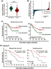
FIGURE 1
FIGURE 1
SDCBP2‐AS1 was downregulated in GC tissues and indicated poor patient outcomes. (A) SDCBP2‐AS1 expression was examined by RT‐qPCR in GC and non‐tumor tissues from 198 patients. (B) Fold‐changes in SDCBP2‐AS1 expression in paired GC tissues (normalized to adjacent non‐normal tissues) from 132 patients. Vertical dotted line represents the median number of samples. (C) Kaplan‐Meier survival analysis of DFS and OS of the 132 patients according to SDCBP2‐AS1 levels in GC tissues. (D) Kaplan‐Meier survival analysis of DFS and OS according to SDCBP2‐AS1 levels in GC tissues by mining a public microarray dataset GSE22377 (n = 43). Data are presented as the mean ± SD of three independent experiments. ***P < 0.001. Abbreviations: SDCBP2‐AS1, syndecan binding protein 2 antisense RNA 1; GC, gastric cancer; RT‐qPCR, quantitative real‐time polymerase chain reaction; DFS, disease‐free survival; OS, overall survival; HR, hazard ratio; SD, standard deviation
FIGURE 2
FIGURE 2
SDCBP2‐AS1 suppressed GC cell proliferation and metastasis in vitro. (A‐B) The proliferation and colony formation assay of stable SDCBP2‐AS1‐knockdown BGC823 and MKN28 cells. (C‐D) The proliferation and colony formation assay of stable SDCBP2‐AS1‐overexpressing SGC7901 cells. (E‐H) The results of wound healing and transwell assay of SDCBP2‐AS1‐knockdown BGC823 and MKN28 cells and SDCBP2‐AS1‐overexpressing SGC7901 cells, respectively. Data are presented as the mean ± SD. *P < 0.05, **P < 0.01, ***P < 0.001. Abbreviations: SDCBP2‐AS1, syndecan binding protein 2 antisense RNA 1; GC, gastric cancer; NT, non‐target; sh, small harpin RNA; Con, empty vector control; SD, standard deviation.
FIGURE 3
FIGURE 3
SDCBP2‐AS1 suppressed growth and metastasis of GC cells in vivo. (A‐D) Representative images (A) and xenograft quantification (B‐D) of nude mice (5 mice/group) injected with stable SDCBP2‐AS1‐knockdown BGC823 cells (sh1) or with NT control cells. (B) Tumor volumes were calculated after injection every 3 days for 21 days. (C) Tumor weights are presented as the mean ± SD. (D) The tumor sections were subjected to staining with H&E and IHC analysis of Ki‐67. (E) Representative images of metastatic sites in the lungs of nude mice injected via the tail vein with BGC823 cells stably transfected with the NT control or sh1 (5 mice/group). (F) Quantification of metastatic foci in lung tissues. (G) Representative images of H&E staining and IHC analysis of Ki‐67 of metastatic sites in lung tissues. The intensity of Ki‐67 staining was quantified. Data are presented as the mean ± SD. *P < 0.05, **P < 0.01, ***P < 0.001. Abbreviations: SDCBP2‐AS1, syndecan binding protein 2 antisense RNA 1; GC, gastric cancer; NT, non‐target; sh, small harpin RNA; H&E, hematoxylin and eosin; IHC, immunohistochemical; SD, standard deviation.
FIGURE 4
FIGURE 4
SDCBP2‐AS1 interacted with hnRNP K in the cytoplasm. (A) RNA‐FISH analysis of SDCBP2‐AS1 in the cytoplasmic (red) and nuclear (blue) fractions of SGC7901 and BGC823 cells. (B) Nuclear‐cytoplasmic RNA fractionation assays showed that SDCBP2‐AS1 was mainly located in the cytoplasm of SGC7901 and BGC823 cells. (C) RNA pull‐down analysis of biotinylated or antisense SDCBP2‐AS1 to identify associated proteins. After silver staining, bands were excised and analyzed by mass spectrometry, which showed that hnRNP K interacts with SDCBP2‐AS1 in BGC823 cells. (D) Western blotting analysis of proteins collected from the SDCBP2‐AS1 pull‐down assay using an antibody against hnRNP K in BGC823 cells. (E) RNA‐IP analysis of BGC823 cells using antibodies against hnRNP K or IgG. The precipitated RNA was used for RT‐qPCR analysis. Enrichment of SDCBP2‐AS1, as determined by RIP analysis of hnRNP K, was relative to the IgG control, showing that SDCBP2‐AS1 interacted with hnRNP K. (F) The predicted secondary structure of SDCBP2‐AS1 RNA. (G) A schematic diagram of full‐length SDCBP2‐AS1 and truncated fragments. (H) In vitro transcribed biotinylated RNA of different constructs of SDCBP2‐AS1 associated with hnRNP K were detected by Western blotting analysis. (I) RIP assays were performed using an anti‐Flag antibody in BGC823 cells stably transfected with Flag‐tagged hnRNP K or deletion mutants. (J) Western blotting analysis was conducted to evaluate the expression of Flag‐tagged hnRNP K or deletion mutants. (K) RT‐qPCR analysis was used to measure enrichment of SDCBP2‐AS1. Data are presented as the mean ± SD. *P < 0.05, **P < 0.01, ***P < 0.001. Abbreviations: SDCBP2‐AS1, syndecan binding protein 2 antisense RNA 1; GC, gastric cancer; FISH, fluorescence in situ hybridization; GAPDH, glyceraldehyde 3‐phosphate dehydrogenase; U6, U6 small nuclear RNA; hnRNP K, heterogeneous nuclear ribonucleoprotein K; Frag, fragment; Pos. Con, positive control; Con, empty vector control; RIP, RNA immunoprecipitation; MFE, minimum free energy; RT‐qPCR, quantitative real‐time polymerase chain reaction; SD, standard deviation.
FIGURE 5
FIGURE 5
Silencing of SDCBP2‐AS1 stabilized β‐catenin and prohibited transcriptional activity. (A) Clustering volcano plot of differentially expressed genes in stable SDCBP2‐AS1‐knockdown BGC823 cells or its NT control. (B) Identification of enriched oncogenic signatures gene sets associated with SDCBP2‐AS1 knockdown by GSEA. (C) β‐catenin and hnRNP K protein levels in whole‐cell lysates were analyzed by Western blotting analysis. (D) The nuclear and cytoplasmic protein levels of hnRNP K and β‐catenin in BGC823 or MKN28 cells stably transfected with the NT control or sh1 or in SGC7901 cells stably overexpressing the empty vector control (Con) or SDCBP2‐AS1 as determined by Western blotting analysis. GAPDH served as the cytosolic control, and histone H3 was used to validate the nuclear content. (E) IF confocal images revealing localization of β‐catenin in SDCBP2‐AS1‐knockdown BGC823 cells and SDCBP2‐AS1‐overexpressing SGC97901 cells. (F) Results of the TOP/FOP‐flash luciferase assays indicating the relative transcriptional activity of β‐catenin in BGC823 or MKN28 cells stably transfected with the NT control or sh1 and in SGC7901 cells stably overexpressing SDCBP2‐AS1. (G‐H) mRNA expression levels of MYC, CCND1, and MMP7 as detected by RT‐qPCR analysis (G) and protein levels of c‐Myc, cyclin D1, MMP7, p‐β‐catenin (Ser33/37/Thr41), Wnt3a, and Wnt5a as detected by Western blotting analysis (H) in BGC823 or MKN28 cells after knockdown of SDCBP2‐AS1 and in SGC7901 cells overexpressing SDCBP2‐AS1. GAPDH served as the endogenous control. **P < 0.01, ***P < 0.001. Abbreviations: SDCBP2‐AS1, syndecan binding protein 2 antisense RNA 1; GC, gastric cancer; GSEA, gene set enrichment analysis; NT, non‐target; sh, small harpin RNA; Con, empty vector control; FDR, false discovery rate; NES, enrichment score; IF, immunofluorescence; RT‐qPCR, quantitative real‐time polymerase chain reaction; hnRNP K, heterogeneous nuclear ribonucleoprotein K; H3, histone 3; GAPDH, glyceraldehyde 3‐phosphate dehydrogenase; MMP7, matrix metallopeptidase 7; CCND1, cyclin D1; p‐β‐catenin, phospho‐β‐catenin.
FIGURE 6
FIGURE 6
SDCBP2‐AS1 destabilized β‐catenin by blocking SUMOylation of hnRNP K. (A) Cells were treated with CHX (20 μg/mL) for 4 h before harvest. (B) Cells were treated with MG132 (10 μmol/L) for 8 h before harvest. The protein levels of β‐catenin were measured in BGC823 cells after the knockdown of SDCBP2‐AS1 or SGC7901 cells overexpressing SDCBP2‐AS1 as determined by Western blotting analysis. (C) Extracts of BGC823 cells after knockdown of SDCBP2‐AS1 or SGC7901 cells overexpressing SDCBP2‐AS1 were used for IP analysis with antibodies against β‐catenin or IgG as a control. Western blotting analysis of β‐catenin, APC, Axin1, GSK3β, and hnRNP K. (D) SDCBP2‐AS1 regulated ubiquitination of β‐catenin. IP assays of endogenous β‐catenin in cell lines with SDCBP2‐AS1 knockdown or overexpression, followed by Western blotting analysis with antibodies against ubiquitin and β‐catenin. (E‐F) BGC823 cells were transfected with Flag‐tagged hnRNP K truncation constructs (E) or HA‐tagged β‐catenin truncation constructs (F) to identify the interaction domains between hnRNP K and β‐catenin. (G) SDCBP2‐AS1 knockdown in BGC823 cells overexpressing HA‐tagged SUMO‐1 and Flag‐tagged wild‐type hnRNP K or the K422R mutant. Cell lysates were subjected to IP analysis followed by Western blotting analysis with antibodies against Flag, HA and β‐catenin. (H) After the knockdown of SDCBP2‐AS1, BGC823 cells overexpressed Flag‐tagged wild‐type hnRNP K or the K422R mutant. Cell lysates were subjected to IP and Western blotting analysis with antibodies against Flag and ubiquitin. Abbreviations: SDCBP2‐AS1, syndecan binding protein 2 antisense RNA 1; CHX, cycloheximide; IP, immunoprecipitation; APC, adenomatous polyposis coli; GSK3β, glycogen synthase kinase 3 beta; hnRNP K, heterogeneous nuclear ribonucleoprotein K; HA, hemagglutinin; GAPDH, glyceraldehyde 3‐phosphate dehydrogenase; Ub, ubiquitin; ARM, armadillo repeats; NT, non‐target; sh, small harpin RNA; Con, empty vector control; EV, empty vector; WT, wildtype; SUMO‐1, small ubiquitin like modifier 1.
FIGURE 7
FIGURE 7
SDCBP2‐AS1 exerted a tumor‐suppressor function partially through interplay with hnRNP K. (A‐C) The results of the cell proliferation (A), colony formation (B), and transwell assays (C) of SGC7901 cells stably transfected with the NT or sh‐hnRNP K (sh1 and sh2) and sequtially transfected with the Con or SDCBP2‐AS1. Knockdown of hnRNP K blocked SDCBP2‐AS1's effects. (D‐E) IF confocal images (D) and Western blotting analysis (E) of the nuclear and cytoplasmic protein levels of hnRNP K and β‐catenin showed that knockdown of hnRNP K reversed the effects of SDCBP2‐AS1 overexpression through localization of β‐catenin in SGC7901 cells. (F) The results of the TOP/FOP‐flash luciferase assays indicating changes in the relative transcriptional activity of β‐catenin in SGC7901 cells after knockdown of hnRNP K or overexpression of SDCBP2‐AS1. (G) RT‐qPCR analysis of the mRNA levels (G) of MYC, CCND1, and MMP7. (H) Western blotting analysis of the protein levels (H) of c‐Myc, cyclin D1, MMP7, p‐β‐catenin (Ser33/37/Thr41), Wnt3a, and Wnt5a. GAPDH served as the endogenous control. (I‐K) Representative images (I), tumor volume growth curves (J), and tumor weight at the end points (K) of xenografts in athymic nude mice formed by subcutaneous injection of SGC7901 cells stably transfected with Con, SDCBP2‐AS1, the NT control, or hnRNP K sh1 (5 mice/group). ***P < 0.001. Abbreviations: SDCBP2‐AS1, syndecan binding protein 2 antisense RNA 1; NT, non‐target; shK, known down hnRNP K with small harpin RNA; Con, empty vector control; IF, immunofluorescence; RT‐qPCR, quantitative real‐time polymerase chain reaction; hnRNP K, heterogeneous nuclear ribonucleoprotein K; H3, histone 3; GAPDH, glyceraldehyde 3‐phosphate dehydrogenase; MMP7, matrix metallopeptidase 7; p‐β‐catenin, phospho‐β‐catenin.
FIGURE 8
FIGURE 8
Schematic illustrating the mechanism of SDCBP2‐AS1‐induced suppression of tumorigenesis in GC. SDCBP2‐AS1 regulates SUMOylation and ubiquitination of hnRNP K. SDCBP2‐AS1 blocks the major SUMOylation site of hnRNP K, which increases ubiquitination of hnRNP K and β‐catenin, and degradation of β‐catenin. Knockout of SDCBP2‐AS1 increases SUMOylation of hnRNP K and translocation of β‐catenin to the nucleus for transcription. Abbreviations: SDCBP2‐AS1, syndecan binding protein 2 antisense RNA 1; APC, adenomatous polyposis coli; β‐TrCP, β‐transducin repeat‐containing protein; CK1, casein kinase1; Dvl, disheveled, dsh homolog; GSK3β, glycogen synthase kinase 3 beta; hnRNP K, heterogeneous nuclear ribonucleoprotein K; LRP, low‐density lipoprotein receptor‐related protein; TCF, T‐cell factor enhancer‐binding factor; SUMO, SUMOylation.

Similar articles

Cited by

References

    1. Sung H, Ferlay J, Siegel RL, Laversanne M, Soerjomataram I, Jemal A, et al. Global Cancer Statistics 2020: GLOBOCAN Estimates of Incidence and Mortality Worldwide for 36 Cancers in 185 Countries. CA Cancer J Clin. 2021;71(3):209‐49. - PubMed
    1. Xia C, Dong X, Li H, Cao M, Sun D, He S, et al. Cancer statistics in China and United States, 2022: profiles, trends, and determinants. Chin Med J (Engl). 2022;135(5):584‐90. - PMC - PubMed
    1. Wang FH, Zhang XT, Li YF, Tang L, Qu XJ, Ying JE, et al. The Chinese Society of Clinical Oncology (CSCO): Clinical guidelines for the diagnosis and treatment of gastric cancer, 2021. Cancer Commun (Lond). 2021;41(8):747‐95. - PMC - PubMed
    1. Chen W, Zheng R, Baade PD, Zhang S, Zeng H, Bray F, et al. Cancer statistics in China, 2015. CA Cancer J Clin. 2016;66(2):115‐32. - PubMed
    1. Yang L, Ying X, Liu S, Lyu G, Xu Z, Zhang X, et al. Gastric cancer: Epidemiology, risk factors and prevention strategies. Chin J Cancer Res. 2020;32(6):695‐704. - PMC - PubMed

Publication types

MeSH terms