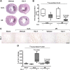
DJ-1 administration exerts cardioprotection in a mouse model of acute myocardial infarction
- PMID: 36210822
- PMCID: PMC9539284
- DOI: 10.3389/fphar.2022.1002755
DJ-1 administration exerts cardioprotection in a mouse model of acute myocardial infarction
Abstract
Cardiovascular diseases, and particularly acute myocardial infarction (MI), are the most common causes of death worldwide. Infarct size is the major predictor of clinical outcomes in MI. The Parkinson's disease associated protein, DJ-1 (also known as PARK7), is a multifunctional protein with chaperone, redox sensing and mitochondrial homeostasis activities. Previously, we provided the evidence for a central role of endogenous DJ-1 in the cardioprotection of post-conditioning. In the present study, we tested the hypothesis that systemic administration of recombinant DJ-1 exerts cardioprotective effects in a mouse model of MI and also explored the associated transcriptional response. We report a significant treatment-induced reduction in infarct size, leukocyte infiltration, apoptosis and oxidative stress. Effects potentially mediated by G-protein-coupled receptor signaling and modulation of the immune response. Collectively, our results indicate a protective role for the exogenously administrated DJ-1 upon MI, and provide the first line of evidence for an extracellular activity of DJ-1 regulating cardiac injury in vivo.
Keywords: DJ-1; PARK7; cardioprotection; ischemia; ischemia/reperfusion injury; myocardial infarction; reperfusion.
Copyright © 2022 Gallinat, Mendieta, Vilahur, Padró and Badimon.
Conflict of interest statement
LB received institutional research grants from AstraZeneca; consultancy fees from Sanofi, Pfizer and Novartis; speaker fees from Amarin, Lilly, Pfizer, and AstraZeneca. TP, GV and LB are shareholders of the academic spin-off companies GlyCardial Diagnostics SL and Ivestatin Therapeutics SL. All unrelated to the present work. LB, GV, and TP are authors of the patents EP3219326A1 and WO2017157958A1 regarding the use of DJ-1-derived polypeptides for the treatment of ischemia/reperfusion injury. AG and GM declare no conflict of interest.
Figures


Similar articles
-
Sphingosine-1-Phosphate Receptor Agonist Fingolimod Increases Myocardial Salvage and Decreases Adverse Postinfarction Left Ventricular Remodeling in a Porcine Model of Ischemia/Reperfusion.Circulation. 2016 Mar 8;133(10):954-66. doi: 10.1161/CIRCULATIONAHA.115.012427. Epub 2016 Jan 29. Circulation. 2016. PMID: 26826180
-
DJ-1 protects against neurodegeneration caused by focal cerebral ischemia and reperfusion in rats.J Cereb Blood Flow Metab. 2008 Mar;28(3):563-78. doi: 10.1038/sj.jcbfm.9600553. Epub 2007 Sep 19. J Cereb Blood Flow Metab. 2008. PMID: 17882163
-
Hyperglycemia-induced inhibition of DJ-1 expression compromised the effectiveness of ischemic postconditioning cardioprotection in rats.Oxid Med Cell Longev. 2013;2013:564902. doi: 10.1155/2013/564902. Epub 2013 Nov 4. Oxid Med Cell Longev. 2013. PMID: 24303086 Free PMC article.
-
The reduction of infarct size--forty years of research.Rev Port Cardiol. 2010 Jun;29(6):1037-53. Rev Port Cardiol. 2010. PMID: 20964114 Review. English, Portuguese.
-
Heat shock protein 70: A promising therapeutic target for myocardial ischemia-reperfusion injury.J Cell Physiol. 2019 Feb;234(2):1190-1207. doi: 10.1002/jcp.27110. Epub 2018 Aug 21. J Cell Physiol. 2019. PMID: 30132875 Review.
Cited by
-
Molecular and Physiological Determinants of Amyotrophic Lateral Sclerosis: What the DJ-1 Protein Teaches Us.Int J Mol Sci. 2023 Apr 21;24(8):7674. doi: 10.3390/ijms24087674. Int J Mol Sci. 2023. PMID: 37108835 Free PMC article. Review.
-
Mitochondrial Dysfunction as a Potential Mechanism Mediating Cardiac Comorbidities in Parkinson's Disease.Int J Mol Sci. 2024 Oct 12;25(20):10973. doi: 10.3390/ijms252010973. Int J Mol Sci. 2024. PMID: 39456761 Free PMC article. Review.
-
DJ-1 as a Novel Therapeutic Target for Mitigating Myocardial Ischemia-Reperfusion Injury.Cardiovasc Ther. 2024 Dec 13;2024:6615720. doi: 10.1155/cdr/6615720. eCollection 2024. Cardiovasc Ther. 2024. PMID: 39742003 Free PMC article. Review.
References
-
- Arac A., Brownell S. E., Rothbard J. B., Chen C., Ko R. M., Pereira M. P., et al. (2011). Systemic augmentation of alphaB-crystallin provides therapeutic benefit twelve hours post-stroke onset via immune modulation. Proc. Natl. Acad. Sci. U. S. A. 108 (32), 13287–13292. 10.1073/pnas.1107368108 - DOI - PMC - PubMed
LinkOut - more resources
Full Text Sources
Miscellaneous

