

Optical coherence tomography systems for evaluation of marginal and internal fit of ceramic reconstructions
- PMID: 36211304
- PMCID: PMC9543067
- DOI: 10.1080/26415275.2022.2122468
Optical coherence tomography systems for evaluation of marginal and internal fit of ceramic reconstructions
Abstract
Purpose: To evaluate the marginal and internal fit of lithium disilicate and zirconia crowns using two optical coherence tomography (OCT) systems in order to estimate inter-system variations.
Materials and methods: Ten lithium disilicate and 10 cubic stabilized zirconia crowns were placed on prepared artificial teeth without cement. Marginal discrepancy and internal cement gap of the crowns were assessed on images obtained using a swept source OCT (SS-OCT) and a spectral domain OCT (SD-OCT). Medians and interquartile ranges were calculated for both materials and OCT systems. Thereafter, Wilcoxon signed rank test was carried out.
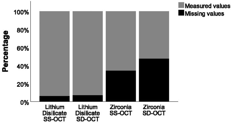
Results: No significant difference was found between the two OCT systems for absolute marginal discrepancy of either lithium disilicate (SS-OCT: 182 µm, SD-OCT: 214 µm; p = .922) or zirconia crowns (SS-OCT: 116 µm, SD-OCT: 121 µm; p = .232). Regarding internal cement gap, no significant difference was found between the two OCT systems for lithium disilicate crowns (SS-OCT: 128 µm, SD-OCT: 128 µm; p = .064). However, for zirconia crowns the SD-OCT showed significantly higher (p = .027) internal cement gap (92 µm) than the SS-OCT (68 µm). Moreover, it was not possible to assess the internal fit of zirconia crowns in 47% and 34% of the sites using SD-OCT and SS-OCT, respectively.
Conclusions: No significant difference was noted in the ability of SS-OCT and SD-OCT to assess the marginal and internal fit of lithium disilicate crowns, nor the marginal fit of zirconia crowns. On the contrary, drawbacks regarding the assessment of internal fit of zirconia crowns using both OCT systems were observed.
Keywords: Optical coherence tomography; lithium disilicate; zirconia.
© 2022 The Author(s). Published by Informa UK Limited, trading as Taylor & Francis Group.
Conflict of interest statement
No potential conflict of interest was reported by the authors.
Figures


Similar articles
-
In vitro Evaluation of the Marginal Fit and Internal Adaptation of Zirconia and Lithium Disilicate Single Crowns: Micro-CT Comparison Between Different Manufacturing Procedures.Open Dent J. 2018 Feb 22;12:160-172. doi: 10.2174/1874210601812010160. eCollection 2018. Open Dent J. 2018. PMID: 29854014 Free PMC article.
-
Evaluation of marginal fit of 2 CAD-CAM anatomic contour zirconia crown systems and lithium disilicate glass-ceramic crown.J Adv Prosthodont. 2015 Aug;7(4):271-7. doi: 10.4047/jap.2015.7.4.271. Epub 2015 Aug 18. J Adv Prosthodont. 2015. PMID: 26330973 Free PMC article.
-
A micro-computed tomography analysis of internal and marginal fits of fixed partial dentures: Effect of preparation finish line designs on monolithic zirconia and heat-pressed zirconia-reinforced lithium disilicate.J Prosthodont. 2023 Jun;32(5):90-99. doi: 10.1111/jopr.13656. Epub 2023 Feb 25. J Prosthodont. 2023. PMID: 36718906
-
Evaluation of marginal and internal fit of lithium disilicate and zirconia all-ceramic CAD-CAM crowns using digital impressions: A systematic review.Prim Dent J. 2023 Mar;12(1):88-95. doi: 10.1177/20501684231154323. Prim Dent J. 2023. PMID: 36916623
-
Effect of cement type on the clinical performance and complications of zirconia and lithium disilicate tooth-supported crowns: A systematic review. Report of the Committee on Research in Fixed Prosthodontics of the American Academy of Fixed Prosthodontics.J Prosthet Dent. 2019 May;121(5):754-765. doi: 10.1016/j.prosdent.2018.10.011. Epub 2019 Mar 15. J Prosthet Dent. 2019. PMID: 30885580
Cited by
-
Microbiological and Imaging-Based Evaluations of Photodynamic Therapy Combined with Er:YAG Laser Therapy in the In Vitro Decontamination of Titanium and Zirconia Surfaces.Microorganisms. 2024 Jun 30;12(7):1345. doi: 10.3390/microorganisms12071345. Microorganisms. 2024. PMID: 39065113 Free PMC article.
References
-
- Clarkson DM. An update on optical coherence tomography in dentistry. Dent Update. 2014;41(2):174–176. - PubMed
-
- Haak R, Siegner J, Ziebolz D, et al. . OCT evaluation of the internal adaptation of ceramic veneers depending on preparation design and ceramic thickness. Dent Mater. 2021;37(3):423–431. - PubMed
LinkOut - more resources
Full Text Sources
Miscellaneous