

Magnetic resonance analysis of deep cerebral venous vasospasm after subarachnoid hemorrhage in rabbits
- PMID: 36211577
- PMCID: PMC9532692
- DOI: 10.3389/fcvm.2022.1013610
Magnetic resonance analysis of deep cerebral venous vasospasm after subarachnoid hemorrhage in rabbits
Abstract
Objective: Arterial spasm is proved to be an inducer of cerebral ischemia and cerebral infarction, while when a venous spasm occurs, cerebral edema is seen to be caused by a disturbance in cerebral blood flow. However, it is unclear and unproven whether venous spasm occurs after subarachnoid hemorrhage (SAH). To provide the theoretical basis for treating cerebral vasospasm after SAH, magnetic resonance imaging (MRI) was employed to observe the changes in the diameter of deep cerebral veins in rabbits after SAH.
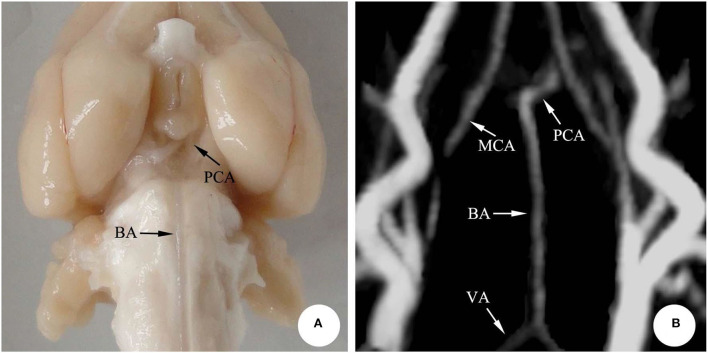
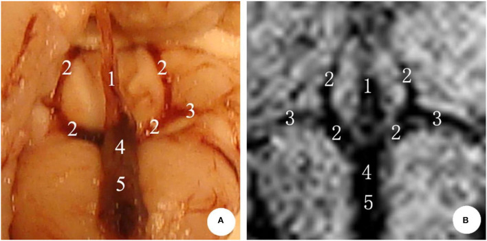
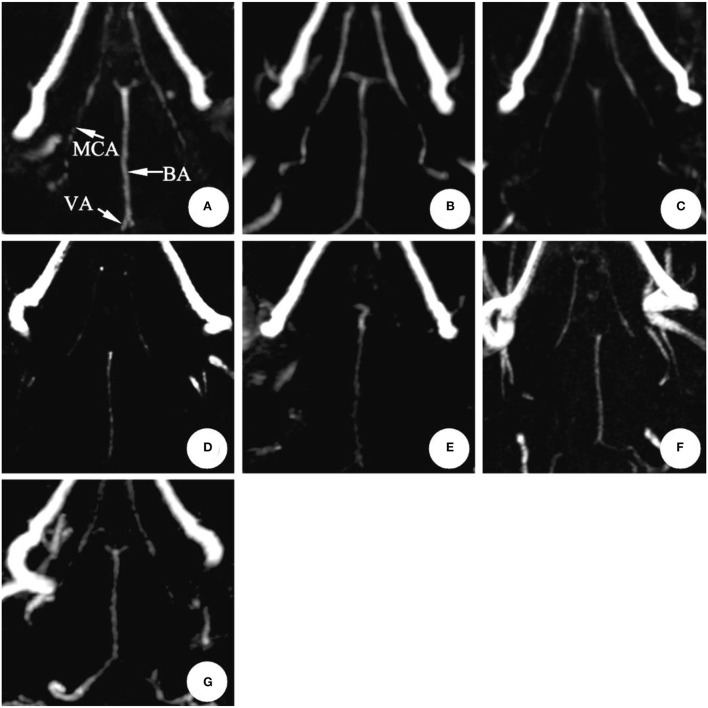
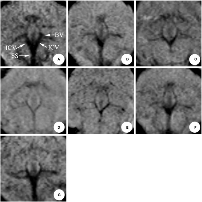
Methods: Fourteen New Zealand rabbits were randomly divided into the SAH group (n = 10) and the normal saline group (NS group, n = 4). Specifically, the SAH models were established by the ultrasound-guided double injections of blood into cisterna magna. Moreover, the MRI was performed to observe the changes in the diameter of deep cerebral veins (internal cerebral vein, basilar vein, and great cerebral vein) and basilar artery before modeling (0 d) and 1, 3, 5, 7, 9, and 11 d after modeling.
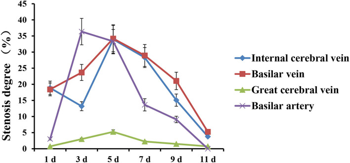
Results: In the SAH group, the diameter of the basilar artery showed no evident change on the 1st d. However, it became narrower obviously on the 3rd d and 5th d, and the stenosis degree was more than 30%. The diameter gradually relieved from 7th to 9th d, and finally returned to normal on the 11th d. Moreover, the diameter of the internal cerebral vein significantly narrowed on the 1st d, the stenosis degree of which was 19%; the stenosis then relieved slightly on the 3rd d (13%), reached the peak (34%) on the 5th d, and gradually relieved from 7th d to 11th d. Moreover, the stenosis degree of the basilar vein was 18% on the 1st d, 24% on the 3rd d, and reached the peak (34%) on the 5th d.
Conclusion: After SAH in rabbits, the cerebral vasospasm was seen to occur in the basilar artery, and likewise, spasmodic changes took place in the deep cerebral vein. Furthermore, the time regularity of spasmodic changes between the cerebral vein and basilar artery was of significant difference, indicating that the venous vasospasm resulted in active contraction.
Keywords: basilar artery; cerebral vasospasm; deep cerebral vein; magnetic resonance imaging; subarachnoid hemorrhage.
Copyright © 2022 Zhang, Fang, Zhang, Zhu, Zhang, Zhu and Deng.
Conflict of interest statement
The authors declare that the research was conducted in the absence of any commercial or financial relationships that could be construed as a potential conflict of interest.
Figures
References
-
- Gupta R, Woodward K, Fiorella D, Woo HH, Liebeskind D, Frei D, et al. Primary results of the Vesalio NeVa VS for the Treatment of Symptomatic Cerebral Vasospasm following Aneurysm Subarachnoid Hemorrhage (VITAL) study. J Neurointerv Surg. (2022) 14:815–9. 10.1136/neurintsurg-2021-017859 - DOI - PubMed
-
- Thompson JC, Chalet FX, Manalastas EJ, Hawkins N, Sarri G, Talbot DA. Economic and humanistic burden of cerebral vasospasm and its related complications after aneurysmal subarachnoid hemorrhage: a systematic literature review. Neurol Therapy. (2022) 11:597–620. 10.1007/s40120-022-00348-6 - DOI - PMC - PubMed
LinkOut - more resources
Full Text Sources
Miscellaneous