. 2022 Oct 5;11(1):2127691.
doi: 10.1080/2162402X.2022.2127691. eCollection 2022.

A newly discovered PD-L1 B-cell epitope peptide vaccine (PDL1-Vaxx) exhibits potent immune responses and effective anti-tumor immunity in multiple syngeneic mice models and (synergizes) in combination with a dual HER-2 B-cell vaccine (B-Vaxx)

Affiliations

A newly discovered PD-L1 B-cell epitope peptide vaccine (PDL1-Vaxx) exhibits potent immune responses and effective anti-tumor immunity in multiple syngeneic mice models and (synergizes) in combination with a dual HER-2 B-cell vaccine (B-Vaxx)

Linlin Guo et al. Oncoimmunology. .

Abstract

Blockade of checkpoint receptors with monoclonal antibodies against CTLA-4, PD-1 and PD-L1 has shown great clinical success in several cancer subtypes, yielding unprecedented responses albeit a significant number of patients develop resistance and remain refractory. Both PD-1/PD-L1 and HER-2 signaling pathway inhibitors have limited efficacy and exhibits significant toxicities that limit their use. Ongoing clinical studies support the need for rationale combination of immuno-oncology agents to make a significant impact in the lives of cancer patients. We introduce the development of a novel chimeric PD-L1 B-cell peptide epitope vaccine (amino acid 130-147) linked to a "promiscuous" T cell measles virus fusion (MVF) peptide (MVF-PD-L1(130); PDL1-Vaxx) or linked to tetanus toxoid (TT3) TT3-PD-L1 (130) via a linker (GPSL). These vaccine constructs are highly immunogenic and antigenic in several syngeneic animal models. The PD-L1 vaccines elicited high titers of polyclonal antibodies that inhibit tumor growth in multiple syngeneic cancer models, eliciting antibodies of different subtypes IgG1, IgG2a, IgG2b and IgG3, induced PD-1/PD-L1 blockade, decreased proliferation, induced apoptosis and caused ADCC of tumor cells. The PDL1-Vaxx induces similar inhibition of tumor growth versus the standard anti-mouse PD-L1 antibody in both syngeneic BALB/c and C57BL/6J mouse models. The combination of PDL1-Vaxx with HER-2 vaccine B-Vaxx demonstrated synergistic tumor inhibition in D2F2/E2 carcinoma cell line. The anti-PDL1-Vaxx block PD-1/PD-L1 interaction and significantly prolonged anti-tumor responses in multiple syngeneic tumor models. The combination of HER-2 vaccine (B-Vaxx) with either PDL1-Vaxx or PD1-Vaxx demonstrated synergistic tumor inhibition. PDL1-Vaxx is a promising novel safe checkpoint inhibitor vaccine.

Keywords: B-cell epitope; CT26; D2F2/E2; PD-L1; Peptide-vaccine; immunotherapy.


Conflict of interest statement

Pravin T.P. Kaumaya is consultant to Imugene, Ltd. Nicholas Ede is CTO at Imugene. All the other authors declare no competing interests. Heather Darby is now at Assurance laboratories, Birmingham, AL. (hdarby@assurancelabs.com).

Figures
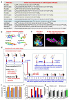
Figure 1.
Figure 1.
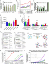
Identification of four B-cell epitope sequences of human PD-L1. (a): Amino acid sequences of human PD-L1. The PD-L1 epitopes of 36–53, 50–67, 95–112 and 130–147 peptides were chosen for investigation. HER-2 or MVF-HER-2 and TT3 sequences are depicted in the table. Briefly, the PD-L1 sequences were subjected to several computerized algorithms that predicts location of epitopes that are surface oriented, have high hydrophilicity, flexibility/mobility, exposure, protrusion and high antigenicity values. The best scoring epitopes are then idealized by correlation with known crystal structures of PD-L1(3BIS) (4Z18), complexes of PD-1:PD-L1(3BIK) 4ZQK, PD-L1:Avelumab PD-L1:/durvalumab (Protein Data Bank http://www.rcsb.org code: 8 M) and PD-L1:/atezolizumab (Protein Data Bank http://www.rcsb.org code: 5X8L, 5XXY)., The PD-L1(130) epitope was synthesized with a Tetanus toxoid (TT3) “promiscuous’ T-cell epitope replacing our usual MVF T-cell epitope. There are two reasons for this change: (i) the MVF T-cell epitope is restricted to the H-2d mice strain and cannot be used in the H-2b mice strain precluding its use as a chimeric vaccines to be used in the MC38/C57BL/6J syngeneic model; (ii) the TT3 epitope is a chimeric vaccine construct works in both H-2d and H-2b strains, thus its inclusion in our epitope repertoire. (b): The secondary structure of the four selected peptide sequences was modeled by using PyMOL 3-D modeling software Version 2.4.0 (Schrodinger, New York, NY, USA). The PyMOL User’s Manual: https://pymol.org. (c): The structure of the PD-1/PD-L1 complex adapted by Zak et al.,, key amino acids involved in the interaction between hPD-1 (light blue ribbon model; navy blue amino acid residues) and hPD-L1(green ribbon model; light green amino acid residues) are illustrated. Amino acids that constitute the central hydrophobic core of the hPD‐1/hPD-L1 interface are indicated in yellow. Red letters indicate strands on both PD-1 and PD-L1. (d): Overview of in vivo studies. Scheme of immunization with MVF-PD-L1 B-cell epitopes in New Zealand White rabbits. Rabbits were intramuscularly (i.m.) immunized with 1 mg of each MVF-peptide immunogen dissolved in ddH2O emulsified (1:1) in Montanide ISA 720 vehicle. The rabbits were boosted with the same doses at 3 weeks interval. Blood was collected via the central auricular artery of each rabbit. The terminal sera were collected at 3Y+3 which is 3 weeks after the last immunization. Scheme of immunization with B-cell epitopes in BALB/c mice (MVF or TT3 linked PD-L1) or C57BL/6J mice (TT3-PD-L1). Mice were subcutaneously immunized with 100 μg of each peptide immunogen dissolved in ddH2O emulsified (1:1) in Montanide ISA 720 vehicle. The rabbits were boosted with the same doses at 3 weeks interval prior to tumor challenge. Blood was collected as indicated and sera tested for antibody titers by ELISA. 2 weeks after the third immunization (3Y), the mice were engrafted subcutaneously with tumor cells as indicated in each set of experiment. Control mice were treated twice weekly with PBS as negative control or with anti-mouse PD-L1 antibody (clone 10F.9G2) as positive control starting 2 days after tumor challenge, mice were treated during the experimental period. Tumor growths were observed and measured by calipers; (e): Immunogenicity of MVF-PD-1 B-cell epitope vaccines were evaluated by ELISA using peptide-coated plates. The 200 ng/well peptides were used to coat the ELISA plates. Titers are defined as the highest dilution of sera with an absorbance value of 0.2 after subtracting the average of blank wells. (f): Antigenicity of PD-L1 peptide vaccines with human PD-L1(PDL1, PD1-H5228, HIS tag) coated EISA plates. (g): In vitro CT26 tumor cells growth inhibition assay. 10% or 5% sera from immunized rabbits were used to culture with CT26 tumor cells, the tumor cells proliferation was tested by MTT. Normal cell growth serves as control. One-way ANOVA statistics analysis was performed, ** indicates p < 0.01, * indicates p < 0.05, (n > 3). Only anti-PD-L1(130) showed statistical difference (p < 0.05) in tumor growth inhibition with 5% of immunized rabbit sera indicating that this epitope was far superior than the others in inhibiting tumor growth.
Figure 2.
Figure 2.
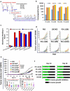
Mechanisms of PD-1/PD-L1 signaling pathway. (a): Antibodies from peptide immunized animals suppress BT474 tumor growth in vitro tested by MTT cell proliferation assay, * indicate p < 0.05, ** indicate p < 0.01 versus with normal cell growth by one-way ANOVA analysis, (n > 3); (b): PD-1/PD-L1 interaction luciferase induction assay. The blockade of PD-1/PD-L1 interaction is directly proportional to increased luciferase signal. Two-way ANOVA was used to analyze the whole curves comparison, *** indicates p < 0.001, * indicates p < 0.05 compared with negative control rabbit IgG; The PD-1 and PD-L1 ligation suppressed TCR activation and attenuated the NFAT responsive luciferase activity. Anti-PD-L1 antibody interrupted the interaction between PD-1 and PD-L1 to release PD-1 signaling and reactive TCR to induce the NFAT luciferase signal. (c): Blockade of the PD-1/PD-L1 interaction by anti-PD-L1 monoclonal antibody atezolizumab and purified anti-MVF-PD-L1 antibodies from immunized rabbit sera. (n = 3); (d): The caspase activity was used to evaluate the apoptosis of MC38 cancer cells after treatment as indicated. Each of antibody was used to treat MC38 cells at the concentration of 500 μg/ml. Atezolizumab was used as the positive control. The luminescence of caspase-9 was measured by using a luminometer. One-way ANOVA was performed to analysis each group versus with control. * indicates p < 0.05, ** indicate p < 0.01 and ns indicate no significance, (n > 3). (e): ADCC assay. INF-gamma induced MC38 colon carcinoma cells served as target cells were incubated with different ratio of effector cells (hPBMC) after treatment with 50 μg purified antibodies as indicated. Avelumab was used as positive control and atezolizumab was served as negative control. One-way ANOVA were performed versus with the experiment control only with effector cells and target cells (E + T), *** indicates p < 0.001, ** indicates p < 0.01 and * indicates p < 0.05, (n ≥ 3); (f) Competitive ELISA was used to determine the relative affinities and specificities of the PD-L1 anti-peptide antibodies, left side. Right side is the normal ELISA activity to determine antibody concentration (50%). In the competitive ELISA, the 100 ng/ml antibody were premix with serial diluted peptide as indicated (panel F1). PD-L1 human recombinant protein (PDL1, PD1-H5228, HIS tag) based competitive ELISA left side, right side is the normal recombinant protein activity, In the competitive ELISA, 20 μg/ml antibody premix with serial diluted peptide as indicated (panel F2). An irrelevant peptide (left panel of Figure 2 F1) was used as control and indicates that no observed inhibition demonstrating the specificity of the PD-L1 peptides (g-h): Mechanisms flow of magnetic microsphere beads-based PD-1/PD-L1 blockade assay, and PD-1/PD-L1 interaction blockade assay; (I): Binding of anti-IgG brilliant violet to antibodies bound to biotin-PD-L1 via PD-1 beads. Leveraging a dual-detection adaptation of the reverse PD-L1 magnetic microsphere-based blocking assay, binding of biotin-PD-L1 to microsphere-coupled PD-1 was detected concurrently with the binding of biotin-PD-L1 to each anti-PD-L1 antibody. MFI was correlated to the dose of anti-PD-L1 antibody in the reaction. In contrast, in this assay there was no detection of positive control atezolizumab on the microspheres. Similarly, no binding of negative control antibody trastuzumab to the PD-1/PD-L1-microsphere complex was detected.
Figure 3.
Figure 3.
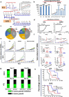
Initial screening of hPD-L1 peptide epitopes in BALB/c mice syngeneic model challenged with CT26 colon carcinoma cell line. (a): Scheme of BALB/c mice vaccination and tumor engraftment. BALB/c mice (PBS, n = 20; PD-L1(130), n = 5 all other groups n = 10) 6–8 weeks old were subcutaneously immunized with MVF-peptide immunogens emulsified in ISA 720 (1:1) with 3 times and three weeks apart. Mice were immunized with 4 MVF-PD-L1 vaccine constructs [PD-L1(36–53), PD-L1(50–67), PD-L1(95–112), PD-L1(130–147)] prior to tumor challenge. Blood was collected as indicated and sera tested for antibody titers by ELISA. 2 weeks after the third immunization (3Y), the mice were engrafted subcutaneously with CT26 tumor cells 105 per mouse. Control mice were treated twice weekly with PBS as negative control or with anti-mouse PD-L1 antibody (clone 10F.9G2) as positive control starting 2 days after tumor challenge, mice were treated during the experimental period. Tumor growths were observed and measured by calipers; (b): Immunogenicity of MVF-PD-L1 peptides in BALB/c mice. Mice immunized with various peptide constructs. Sera were tittered against each individual MVF-PD-L1 peptide immunogen. (c): Antigenicity. Mouse sera tittered against rhPDL1. (d): Individual plots of tumor growths in BALB/c mice immunized with MVF-PD-L1 vaccine constructs as indicated above, anti-mouse PD-L1 antibody (clone 10F.9G2, PD-L1, B7-H1; Cat#BE0101 BioXCell, Lebanon, NH) as positive control. (PBS n = 20; PD-L1(130) n = 5; all other groups n = 10); (e): Two-way ANOVA was used to analyze the whole curves of average tumor volume growing, which shows significant difference between mAb, immunized groups versus PBS group; Plots of tumor volume LWW at day 14 and day 16 for each of group. (f): Percentage of tumor growth inhibition (%TGI) was defined as the difference between median tumor volume (MTV) of treatment group with the positive control group and the value was calculated by formula: %TGI = 100*(MTV control-MTV test)/MTV control which were calculated at day 14 and day 16. The %TGI indicate the mice immunized with MVF-PD-L1(36, 50, 95 & 130) showed higher %TGI versus mAb 10F.9G2, especially MVF-PD-L1(95 & 130) had 59% TGI at day 14, only MVF-PD-L1(130) showed 2% higher TGI compared to mAb 10F.9G2 at day 16. “Two-way ANOVA were used for two parameters between two (versus with PBS) or multiple curves comparison; one-way ANOVA followed by Tukey test were used to compare between two groups; *p < 0.05, **p < 0.01 compared with PBS group”.
Figure 4.
Figure 4.
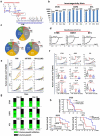
Efficacy of MVF-PD-L1(130) vaccine candidate in BALB/c mice syngeneic model challenged with D2F2 and 4T1 carcinoma cell lines. (a): BALB/c mice (10 mice/gp) 6–8 weeks old were immunized with MVF-PD-L1(130) vaccine emulsified with ISA 720 vehicle with the same scheme as Figure 3a. Four weeks after the last immunization, the mice were challenged with 2 × 105 D2F2 tumor cells. In another set of experiment in a different tumor model, two weeks after the last immunization the mice were subcutaneously injected with 5 × 105 4T1 tumor cells per mouse in each group. The mice in the negative control group were treated with PBS twice weekly whereas mice in the positive control group were ip injected with 100 μl of 2 mg/ml anti-mouse PD-L1 monoclonal antibody (clone 10F.9G2) 2 days after tumor engraftment (n ≥ 9), mice were treated during the experimental period. (b): Immunogenicity of immunized mice was tested by ELISA. (c): Isotypes in BALB/c mice (sera at the time of tumor challenge were used); The isotypes of PDL1-Vaxx antibodies in the mice sera at the time of tumor challenge were analyzed and the predominant isotype was IgG1 (4T1 group 36.2%) and in the D2F2 model it was 43.0% followed by IgG2a 27.0% in the 4T1 model and 25.6 in the D2F2 model followed by IgG2b 17.7% in the 4T1 model versus 16.3% in the D2F2 model. (d): Recombinant activity of MVF-PD-L1(130) immunized mice sera against human PD-L1(PDL1, PD1-H5228, HIS tag); (e): Individual plots of D2F2 and 4T1 tumor growths in BALB/c mice immunized with MVF-PD-L1(130) vaccine. Tumor growth in each individual mouse was monitored in both models 4T1 and D2F2 immunized with PDL1-Vaxx or treated with PBS and anti-mouse PD-L1 antibody (clone, 10F.9G2). (f): Plots of D2F2 and 4T1 tumor volume LWW at day 21 and day 28 of D2F2 tumor or at day 14 and day 16 of 4T1 tumor model of each group. Two-way ANOVA was used to analyze the whole curves of tumor growth. (g): Percentage of tumor growth inhibition (%TGI) was defined as the difference between median tumor volume (MTV) of treatment group with the PBS control group and the value was calculated by formula: %TGI = 100*(MTV control-MTV test)/MTV control which were calculated at different days as indicated in the graphs. (h): The log-rank (Mantel-Cox) test was used to compare the survival curves in multiple groups or between two groups (compared with PBS group), *p < 0.05, **p < 0.01. Two-way ANOVA were used for two parameters between two (versus with PBS) or multiple curves comparison; one-way ANOVA followed by Tukey test were used to compare between two groups (versus with PBS); ns indicates no significance, *p < 0.05, **p < 0.01 compared with PBS group.
Figure 5.
Figure 5.
Efficacy of TT3-PD-L1(130) vaccine candidate in BALB/c mice syngeneic model challenged with CT26, D2F2 and 4T1 carcinoma cell lines. (a): Scheme of BALB/c mice vaccination and tumor engraftment. See Figure 1a for more information on TT3 and MVF epitopes. BALB/c mice (10 mice/gp) 6–8 weeks old were immunized with TT3-PD-L1(130) vaccine emulsified with ISA 720 vehicle. Mice were immunized three times and three weeks apart, 2 weeks after the third immunization (3Y), the mice were engrafted with 1 × 105 CT26 tumor cells per mouse, 2 × 105 D2F2 tumor cells per mouse or 5 × 105 4T1 tumor cells per mouse in each group as designed. Control mice were treated twice weekly with PBS as negative control or with anti-mouse PD-L1 antibody (clone 10F.9G2) as positive control group starting on day 2 after tumor challenge, mice were treated during the experimental period. Tumor growths were observed and measured by calipers; Each of the negative control group mice (10 mice/group) were treated with PBS twice per week and positive control group mice were injected with 100 μl of 2 mg/ml anti-mouse PD-L1 monoclonal antibody (clone 10F.9G2) 2 days after tumor engraftment, twice per week. (b): Immunogenicity of TT3-PD-L1(130) peptide epitope vaccine in BALB/c mice. Mice bleeds were collected as indicated after the primary immunization, and ELISA was used to detect antibody titers in sera. For example, the nomenclature 2Y+1 signifies one week post 2nd injection; 3Y+2 signifies 2 weeks post 3rd injection etc; (c): Isotypes in BALB/c mice (3Y+2) after immunization with TT3-PD-L1(130) and ISA 720. The class of antibodies in mice sera elicited by vaccination with TT3-PD-L1(130) were analyzed by using mouse specific isotyping kit. (d) Antigenicity of TT3-PD-L1(130) peptide epitope vaccine in BALB/c mice using human recombinant protein PD-L1(PDL1, PD1-H5228, HIS tag); (e): Individual plots of tumor growths in BALB/c mice immunized with TT3-PD-L1 vaccine, PBS as negative control and anti-mouse PD-L1 antibody (clone 10F.9G2) as positive control. (f): Plots of tumor volume LWWat day 14 and day 16 for CT26 and 4T1 models or day 21 and day 28 for D2F2 tumor model of each group. Two-way ANOVA was used to analyze the whole curves of tumor growth, which shows significant difference with p < 0.01, one-way ANOVA was used to analyze the multiple groups comparison; (g): Percentage of tumor growth inhibition (%TGI) was defined as the difference between median tumor volume (MTV) of treatment group with the PBS control group and the value was calculated by formula: %TGI = 100*(MTV control-MTV test)/MTV control which were calculated at different days as indicated in the graphs. (h): The Log-rank (Mantel-Cox) test was used to compare the survival curves in multiple groups or between two groups (compared with PBS group), *p < 0.05, **p < 0.01. Two-way ANOVA were used for two parameters between two (versus with PBS) or multiple curves comparison, one-way ANOVA followed by Tukey test were used to compare between two groups (versus with PBS group); ns indicates no significance, * indicates p < 0.05, ** indicate p < 0.01.
Figure 6.
Figure 6.
Efficacy of TT3-PD-L1(130) vaccine candidate in C57BL/6J black mice syngeneic model challenged with MC38 wild type, MC38/HER-2 and B16-F10 carcinoma cell lines. (a): Scheme of C57BL/6J mice vaccination and tumor engraftment. The mice (10 mice/gp) 6–8 weeks old were immunized with TT3-PD-L1(130) vaccine emulsified with ISA 720 vehicle. Mice were immunized three times and three weeks apart, 2 weeks after the third immunization (3Y), the mice were engrafted with 1 × 105 MC38 wild-type tumor cells per mouse, 1 × 105 MC38/HER-2 tumor cells per mouse or 1 × 105 B16-F10 tumor cells per mouse in each group as designed. Control mice were treated twice weekly with PBS as negative control or with anti-mouse PD-L1 antibody (clone 10F.9G2) as positive control group starting on day 2 after tumor challenge, mice were treated during the experimental period. Tumor growths were observed and measured by calipers; (b): Immunogenicity of TT3-PD-L1(130) peptide epitope vaccine in C57BL/6J mice. Mice bleeds were collected as indicated after the primary immunization, and ELISA was used to detect antibody titers in sera. (c): Isotypes in C57BL/6J mice (3Y+2) after immunization with TT3-PD-L1(130) and ISA 720; (d): Antigenicity of TT3-PD-L1(130) in mice using human recombinant protein PD-L1(PDL1, PD1-H5228, HIS tag); (e): Individual plots of tumor growths in C57BL/6J mice immunized with TT3-PD-L1(130) vaccine, PBS as negative control and anti-mouse PD-L1 antibody (clone 10F.9G2) as positive control. (f): Plots of tumor volume LWW at day 14 and day 16 for MC38 wild type, MC38/HER-2 and B16-F10 models of each group. Two-way ANOVA was used to analyze the whole curves of tumor growth, which shows significant difference with p < 0.01, one-way ANOVA was used to analyze the multiple groups comparison; (g): Percentage of tumor growth inhibition (%TGI) was defined as the difference between median tumor volume (MTV) of treatment group with the PBS control group and the value was calculated by formula: %TGI = 100*(MTV control-MTV test)/MTV control which were calculated at different days as indicated in the graphs. (h): The Log-rank (Mantel-Cox) test was used to compare the survival curves in multiple groups or between two groups (compared with PBS group), *p < 0.05, **p < 0.01. Two-way ANOVA were used for two parameters between two (versus with PBS) or multiple curves comparison; one-way ANOVA followed by Tukey test were used to compare between two groups (versus with PBS group); ns indicates no significance, * indicates p < 0.05, ** indicate p < 0.01.
Figure 7.
Figure 7.
Efficacy of combination treatment of MVF-PD-L1(130) vaccine and combo HER-2 vaccines in syngeneic BALB/c mice model challenged with D2F2/E2 mammary tumor cells. (a): Scheme of BALB/c mice vaccination and tumor engraftment. BALB/c mice (10 mice/gp) 6–8 weeks old were immunized with combo HER-2: [MVF-HER-2(266) + MVF-HER-2(597)], triple: [MVF-PD-1(92) + combo HER-2] and triple: [MVF-PD-L1(130) + combo HER-2] vaccine emulsified with ISA 720 vehicle. Mice were immunized at least three times and three weeks apart, and the mice were engrafted with 2 × 105 D2F2/E2 tumor cells per mouse in each group as designed. Control mice were treated twice weekly with PBS as negative control or with anti-mouse PD-1 antibody (clone 29F.1A12) or anti-mouse PD-L1 antibody (clone 10F.9G2) as positive control groups starting on day 2 after tumor challenge, mice were treated during the experimental period. Tumor growths were observed and measured by calipers; D2F2/E2 is generated from a BALB/c mouse background mammary carcinoma D2F2 cell line that can express human HER-2 on the cell surface. This cell line was a kind gift from Wei-Zen Wei (School of Medicine, Way State University, Detroit, MI). (b): Immunogenicity of combo and triple vaccinations in BALB/c mice. Mice bleeds were collected as indicated after the primary immunization, and ELISA was used to detect antibody titers in sera. (c): Isotypes in BALB/c mice (sera at the time of tumor challenge were used) after immunization; (d): Antigenicity of combo and triple vaccinations in BALB/c mice using human recombinant protein PD-L1(PDL1, PD1-H5228, HIS tag) or human recombinant HER-2 (HE2-5225 HIS tag) activity of mice sera at the time of tumor challenge; (e): Individual plots of tumor growths in BALB/c mice immunized with combo or triple vaccines, PBS as negative control and anti-mouse PD-1 antibody (clone 29F.1A12) or anti-mouse PD-L1 antibody (clone 10F.9G2) as positive controls. (f): Plots of tumor volume LWW at day 21 and day 28 of D2F2/E2 tumor model of each group. Two-way ANOVA was used to analyze the whole curves of tumor growth, which shows significant difference with p < 0.01, one-way ANOVA was used to analyze the multiple groups comparison; (g): Percentage of tumor growth inhibition (%TGI) was defined as the difference between median tumor volume (MTV) of treatment group with the PBS control group and the value was calculated by formula: %TGI = 100*(MTV control-MTV test)/MTV control which were calculated at different days as indicated in the graphs. (h): The Log-rank (Mantel-Cox) test was used to compare the survival curves in multiple groups p < 0.001. (i): Scheme of tumor free immunized BALB/c mice re-engraftment. 3 mice in combo 2xHER-2 group (named CM1, CM2, and CM3), 3 mice in triple 2xHER-2+ PD-1(92) group (named TPM1, TMP2, and TMP3), 5 mice in triple 2xHER-2+ PD-L1(130) group (named TLM1, TLM2, TLM3, TLM4, and TLM5) were boosted with the same vaccines at 10-week (70 days) after the primary tumor challenging (named as 5Y, which is 4Y+14), 3 weeks later (5Y+3), each individual mouse bleed were collected and each mouse was challenged (which was 91 days after the primary tumor challenging) with 5 × 105 D2F2/E2 tumor cells. The tumor growth was monitored. (j): Immunogenicity of each mouse at 5Y and 5Y+3 tested by ELISA. (k): Each group of tumor volume (mm3, LWW) showed as line curves, (n ≥ 3), values of mean±SD. Two-way ANOVA followed Dunnett’s multiple comparisons test was performed to analyze the data. **indicates 2xHER-2+ PD-1(92), and 2xHER-2+ PD-L1(130) group showed significantly slower (p < 0.01) tumor growth compared with 2xHER-2 group. Both at day28 and day35, 2xHER-2+ PD-1(92), and 2xHER-2+ PD-L1(130) group mice with significantly smaller tumor versus 2xHER-2 group, #p < 0.05, ##p < 0.01, respectively. (i): The Log-rank (Mantel-Cox) test was used to compare the survival curves, p < 0.01. Two-way ANOVA were used for two parameters between two groups (versus with PBS group) or multiple curves comparison; one-way ANOVA followed by Tukey test were used to compare between two groups (versus with PBS group); the Log-rank (Mantel-Cox) test was used to compare the survival curves in multiple groups or between two groups (for between groups, in (H) versus with PBS group, in (l) versus with 2xHER-2 group). ns indicates no significance, * indicates p < 0.05, ** indicates p < 0.01.

Similar articles

Cited by

References

    1. Ott PA, Hodi FS, Robert C.. CTLA-4 and PD-1/PD-L1 blockade: new immunotherapeutic modalities with durable clinical benefit in melanoma patients. Clin Cancer Res. 2013;19(19):5300–17. doi:10.1158/1078-0432.CCR-13-0143. - DOI - PubMed
    1. Robert C, Thomas L, Bondarenko I, O’Day S, Weber J, Garbe C, Lebbe C, Baurain J-F, Testori A, Grob -J-J, et al. Ipilimumab plus dacarbazine for previously untreated metastatic melanoma. N Engl J Med. 2011;364(26):2517–2526. doi:10.1056/NEJMoa1104621. - DOI - PubMed
    1. Topalian SL, Hodi FS, Brahmer JR, Gettinger SN, Smith DC, McDermott DF, Powderly JD, Carvajal RD, Sosman JA, Atkins MB, et al. Safety, activity, and immune correlates of anti-PD-1 antibody in cancer. N Engl J Med. 2012;366(26):2443–2454. doi:10.1056/NEJMoa1200690. - DOI - PMC - PubMed
    1. Brahmer JR, Tykodi SS, Chow LQ. Safety and activity of anti-PD-L1 antibody in patients with advanced cancer. N Engl J Med. 2012;366(26):2455–2465. doi:10.1056/NEJMoa1200694. - DOI - PMC - PubMed
    1. Hodi FS, O’Day SJ, McDermott DF, Weber RW, Sosman JA, Haanen JB, Gonzalez R, Robert C, Schadendorf D, Hassel JC, et al. Improved survival with ipilimumab in patients with metastatic melanoma. N Engl J Med. 2010;363(8):711–723. doi:10.1056/NEJMoa1003466. - DOI - PMC - PubMed

Publication types