. 2022 Sep 23:12:1020201.
doi: 10.3389/fcimb.2022.1020201. eCollection 2022.

Meningococcal virulence in zebrafish embryos depends on capsule polysaccharide structure

Affiliations

Meningococcal virulence in zebrafish embryos depends on capsule polysaccharide structure

Kim Schipper et al. Front Cell Infect Microbiol. .

Abstract

Neisseria meningitidis or the meningococcus, can cause devasting diseases such as sepsis and meningitis. Its polysaccharide capsule, on which serogrouping is based, is the most important virulence factor. Non-encapsulated meningococci only rarely cause disease, due to their sensitivity to the host complement system. How the capsular polysaccharide structure of N. meningitidis relates to virulence is largely unknown. Meningococcal virulence can be modeled in zebrafish embryos as the innate immune system of the zebrafish embryo resembles that of mammals and is fully functional two days post-fertilization. In contrast, the adaptive immune system does not develop before 4 weeks post-fertilization. We generated isogenic meningococcal serogroup variants to study how the chemical composition of the polysaccharide capsule affects N. meningitidis virulence in the zebrafish embryo model. H44/76 serogroup B killed zebrafish embryos in a dose-dependent manner, whereas the non-encapsulated variant was completely avirulent. Neutrophil depletion was observed after infection with encapsulated H44/76, but not with its non-encapsulated variant HB-1. The survival of embryos infected with isogenic capsule variants of H44/76 was capsule specific. The amount of neutrophil depletion differed accordingly. Both embryo killing capacity and neutrophil depletion after infection correlated with the number of carbons used per repeat unit of the capsule polysaccharide during its biosynthesis (indicative of metabolic cost).

Conclusion: Meningococcal virulence in the zebrafish embryo largely depends on the presence of the polysaccharide capsule but the extent of the contribution is determined by its structure. The observed differences between the meningococcal isogenic capsule variants in zebrafish embryo virulence may depend on differences in metabolic cost.

Keywords: Neisseria meningitidis; innate immunity; isogenic capsule variants; meningococcal polysaccharide capsule; zebrafish embryo infection.


Conflict of interest statement

The authors declare that the research was conducted in the absence of any commercial or financial relationships that could be construed as a potential conflict of interest.

Figures

Figure 1
Figure 1
Dose dependent killing of zebrafish embryos by H44/76_B expressing mCherry (numbers in cfu, 20 embryos in each group). Triplicate with 20 embryos in each group.
Figure 2
Figure 2
Fluorescence microscopy of zebrafish embryos at 28 hpf infected with mCherry expressing H44/76_B meningococci (5000 cfu). (A) Zebrafish embryos infected with red fluorescent meningococci. From top to bottom: No meningococci visible, meningococci in the head, meningococci in the tail and meningococci throughout the body. Images taken at 48 hpi; arrow head shows pericardial edema. Bar represents 750 μm. (B) Distribution of meningococci in infected zebrafish embryos.
Figure 3
Figure 3
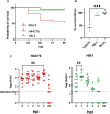
(A) Survival curve of zebrafish embryos infected with capsulated H44/76 (9600 cfu) or non-encapsulated meningococci HB-1 (5200 cfu); 20 embryos in each group. (B) Survival scored at 96 hpi (right panel). Zebrafish embryos were infected with mCherry-expressing H44/76_B (2700 ± 1820 cfu), HB-1 (1960 ± 1420 cfu) or mock-infected. Three independent experiments each with 20 embryos in each group. (C). Survival and multiplication of meningococci (H44/76_B) or non-encapsulated meningococci (HB-1) in zebrafish embryos. Zebrafish embryos were infected with mCherry expressing H44/76_B (3000 cfu) or HB-1 (2800 cfu); 10 embryos in each group. ** P = < 0.01, ***: P = < 0.001.
Figure 4
Figure 4
Fluorescence microscopy of H44/76 meningococci expressing mCherry (red fluorescence) and neutrophils expressing GFP (green fluorescence) in zebrafish embryos. (A). H44/76 infected embryos at 24 hpi, Bar represents 100 μm. (B). HB-1 infected embryo at 24 hpi. (C). Detail of H44/76 infected embryo showing green fluorescent neutrophils and red fluorescent meningococci. (D) Detail of H44/76 infected embryo showing green fluorescent neutrophils and red fluorescent meningococci in the embryo head. Yellow color shows overlap between meningococci and neutrophils.Scale bars represent 100 μm. (E) Number of neutrophils in zebrafish embryos 24hpi of N. meningitidis H44/76 serogroup B (1400 cfu) or non-encapsulated HB-1 (1600 cfu) and mock-infected; 11 embryos infected with H44/76, two groups of 8 embryos infected with HB-1 or mock infected, respectively. IntDen = Integrated density of the green fluorescence signal in the area of interest. * P = < 0.05.
Figure 5
Figure 5
(A) Survival curve of zebrafish embryos infected with H44/76 expressing serogroup B capsule (1560 cfu), no capsule (HB-1) (3600 cfu), serogroup C capsule (H44/76_C) (1200 cfu), serogroup W capsule (H44/76_W) (3600 cfu) or serogroup Y capsule (H44/76_Y) (4800 cfu) all expressing mCherry 20 embryos in each group. (B) Survival of zebrafish embryos infected with H44/76 expressing serogroup B capsule (2700 ± 1820 cfu), no capsule (HB-1) (1960 ± 1420 cfu), serogroup C capsule (H44/76_C) (2160 ± 1660 cfu), serogroup W capsule (H44/76_W) (2760 ± 840 cfu) or serogroup Y capsule (H44/76_Y) (2720 ± 1817 cfu) scored at 96hpi. Three independent experiments, 20 embryos in each group. (C) Quantification of GFP-expressing neutrophils in zebrafish embryos 24 hpi with N. meningitidis H44/76_B (1400 cfu), HB-1 (1600 cfu), H44/76_C (1400 cfu), H44/76_W (4000 cfu), H44/76_Y (3000 cfu) or PBS (Mock); 11, 8, 9, 8, 7 and 8 embryos per group, respectively. IntDen = Integrated density of the green fluorescence signal in the area of interest. * P = < 0.05, ** P = < 0.01, **** P = < 0.0001.
Figure 6
Figure 6
Relation between metabolic cost and meningococcal virulence in zebrafish embryos. Lines represent linear regression. Red: Zebrafish embryo survival after 96 hours post infection with either of the four isogenic capsule variants is the ratio between the number of embryos survived after meningococcal infection and after mock infection (r=0.9090). Blue: Integrated density of the green fluorescence signal as a measure of the number of neutrophils in the area of interest after 24 hours post infection with either of the four isogenic capsule variants divided by that of mock infection; neutrophil count relative to mock-infected zebrafish embryos (r=0.9097).

References

    1. Abramoff M. D., Magalhaes P. J., Ram S. J. (2004). Image processing with ImageJ. Biophotonics Int. 11, 36–42.
    1. Bhattacharjee A. K., Jennings H. J., Kenny C. P., Martin A., Smith I. C. (1975). Structural determination of the sialic acid polysaccharide antigens of Neisseria meningitidis serogroups b and c with carbon 13 nuclear magnetic resonance. J. Biol. Chem. 250, 1926–1932. - PubMed
    1. Bhattacharjee A. K., Jennings H. J., Kenny C. P., Martin A., Smith I. C. (1976). Structural determination of the polysaccharide antigens of Neisseria meningitidis serogroups y, W-135, and BO1. Can. J. Biochem. 54, 1–8. doi: 10.1139/o76-001 - DOI - PubMed
    1. Borrow R., Longworth E., Gray S. J., Kaczmarski E. B. (2000). Prevalence of de-o-acetylated serogroup c meningococci before the introduction of meningococcal serogroup c conjugate vaccines in the united kingdom. FEMS Immunol. Med. Microbiol. 28, 189–191. doi: 10.1111/j.1574-695X.2000.tb01475.x - DOI - PubMed
    1. Bos M. P., Tommassen J. (2005). Viability of a capsule-and lipopolysaccharide-deficient mutant of. Neisseria meningitidis. Infect. Immun. 73, 6194–6197. doi: 10.1128/IAI.73.9.6194-6197.2005 - DOI - PMC - PubMed

Publication types

LinkOut - more resources