

Correlation between retinal vessel rarefaction and psychometric measures in an older Southern Italian population
- PMID: 36212041
- PMCID: PMC9541429
- DOI: 10.3389/fnagi.2022.999796
Correlation between retinal vessel rarefaction and psychometric measures in an older Southern Italian population
Abstract
Objective: To explore the linear association between inner retinal layers thickness and macular capillary density compared to variations of global cognition evaluated by psychometric measures in a cohort of Mediterranean subjects aged 65+ years.
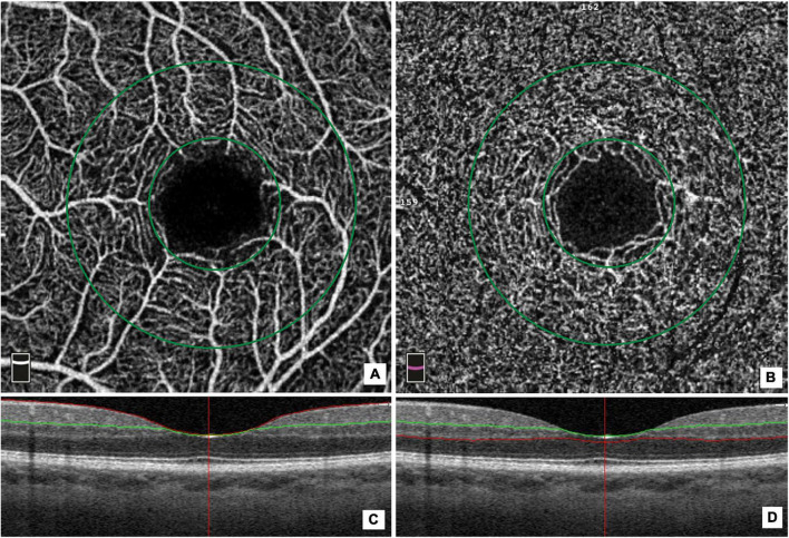
Materials and methods: We performed a cross-sectional analysis of 574 participants aged 65 years+ drawn from a population-based Southern Italian study. All subjects underwent neurological evaluations, including global cognitive screening, the Mini-Mental State Examination (MMSE) and frontal assessment battery (FAB), together with an ophthalmic examination including optical coherence tomography (OCT) and OCT-Angiography. We assessed the average thickness of the ganglion cell complex (GCC) and the retinal nerve fiber layer (RNFL), the foveal avascular zone area, and vascular density (VD) of superficial (SVD) and deep (DVD) capillary plexi at the foveal and parafoveal area. Linear regression was applied to assess associations of ocular measurements with MMSE and FAB scores.
Results: In the linear regression model, foveal DVD (beta = 0.01, 95% CI:0.004-0.052), whole DVD (beta = 0.04, 95% CI:0.02-0.08), and whole SVD (beta = 0.04, 95% CI:0.02-0.07) showed a positive association with MMSE. In addition, foveal SVD (beta = 0.01, 95% CI:0.003-0.05) and whole SVD (beta = 0.03, 95% CI:0.004-0.08) were positively associated with the FAB score. We found no further significant association between the MMSE score or the FAB score and the average thickness of the GCC and RNFL, and FAZ area.
Conclusion: A direct linear association between the VD of the macular capillary plexi with global and frontal cognitive functions was observed in elderly subjects.
Keywords: cognitive function; imaging; older (diseased) population; retina; vessel density.
Copyright © 2022 Giuliani, Sborgia, Niro, Castellana, Lampignano, Puzo, Pascale, Pastore, Buonamassa, Galati, Bordinone, Cassano, Clemente, Landini, Scotti, Gaudiomonte, Guglielmi, Semeraro, Santoro, Alessio, Sardone and Boscia.
Conflict of interest statement
The authors declare that the research was conducted in the absence of any commercial or financial relationships that could be construed as a potential conflict of interest.
Figures
References
LinkOut - more resources
Full Text Sources