Case Reports
. 2022 Sep 20;18(1):68-72.
doi: 10.14797/mdcvj.1067. eCollection 2022.

A Case of Cardiac Arrest Caused by Air Embolism from Routine Root Canal Procedure

Affiliations
Case Reports

A Case of Cardiac Arrest Caused by Air Embolism from Routine Root Canal Procedure

Akarsh Parekh et al. Methodist Debakey Cardiovasc J. .

Abstract

Venous air embolism (VAE) occurs when air is introduced into the venous system and subsequently travels into the right heart and pulmonary circulation. VAE mainly occurs from air that is forced by positive pressure or drawn in by negative pressure. We present a rare case of fatal VAE that occurred during a routine dental root canal procedure. A 69-year-old male was undergoing a root canal procedure at an outpatient dental office under local anesthesia. During the procedure, he went into cardiopulmonary arrest. He was resuscitated, and return of spontaneous circulation was achieved. Thoracic computed tomography was performed and revealed large amounts of air within the right ventricle and portal venous system. VAE should be recognized as a potentially fatal complication resulting from routine dental procedures.

Keywords: air embolism; cardiopulmonary arrest; dental surgery; root canal procedure.


Conflict of interest statement

The authors have no competing interests to declare.

Figures

Electrocardiogram with right bundle branch block
Figure 1
Electrocardiogram.
CT of RV air embolism extending to pulmonary trunk
Figure 2
Computed tomography sagittal view demonstrating right ventricular air embolism with extension into the pulmonary trunk.
CT chest axial view shows air in RV
Figure 3
Computed tomography chest axial view showing presence of air in the right ventricle.
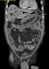
CT of abdomen shows venous air embolism in RV and portal system
Figure 4
Computed tomography of the abdomen showing a venous air embolism in right ventricle and portal system.
CT of head shows air in facial tissue
Figure 5
Computed tomography of the head revealing the presence of air in facial tissue.

References

    1. Palmon SC, Moore LE, Lundberg J, Toung T. Venous air embolism: a review. J Clin Anesth. 1997. May;9(3):251–7. doi: 10.1016/s0952-8180(97)00024-x - DOI - PubMed
    1. Hybels RL. Venous air embolism in head and neck surgery. Laryngoscope. 1980. Jun;90(6 Pt 1):946–54. doi: 10.1002/lary.1980.90.6.946 - DOI - PubMed
    1. Lanfranco J, Romero-Legro I, Freire AX, Nearing K, Ratnakant S. Pulmonary air embolism: an infrequent complication in the radiology suite. Am J Case Rep. 2017. Jan 24;18:80. doi: 10.12659/AJCR.901098 - DOI - PMC - PubMed
    1. Hülsmann M, Hahn W. Complications during root canal irrigation-literature review and case reports. Int Endod J. 2000. May;33(3):186–93. doi: 10.1046/j.1365-2591.2000.00303.x - DOI - PubMed
    1. Frühauf J, Weinke R, Pilger U, Kerl H, Müllegger RR. Soft tissue cervicofacial emphysema after dental treatment: report of 2 cases with emphasis on the differential diagnosis of angioedema. Arch Dermatol. 2005. Nov;141(11):1437–40. doi: 10.1001/archderm.141.11.1437 - DOI - PubMed

Publication types