

Differentiating Glioblastoma Multiforme from Brain Metastases Using Multidimensional Radiomics Features Derived from MRI and Multiple Machine Learning Models
- PMID: 36212721
- PMCID: PMC9534611
- DOI: 10.1155/2022/2016006
Differentiating Glioblastoma Multiforme from Brain Metastases Using Multidimensional Radiomics Features Derived from MRI and Multiple Machine Learning Models
Abstract
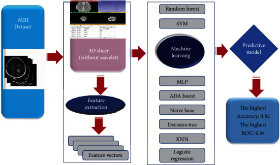
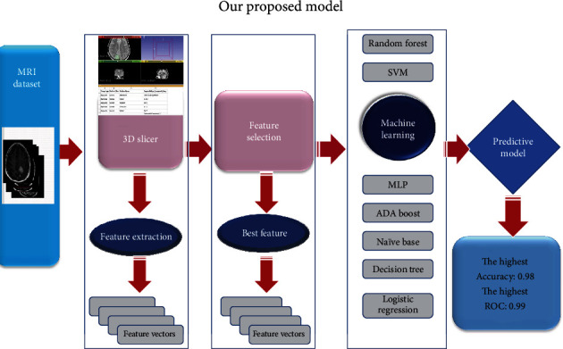
Due to different treatment strategies, it is extremely important to differentiate between glioblastoma multiforme (GBM) and brain metastases (MET). It often proves difficult to distinguish between GBM and MET using MRI due to their similar appearance on the imaging modalities. Surgical methods are still necessary for definitive diagnosis, despite the importance of magnetic resonance imaging in detecting, characterizing, and monitoring brain tumors. We introduced an accurate, convenient, and user-friendly method to differentiate between GBM and MET through routine MRI sequence and radiomics analyses. We collected 91 patients from one institution, including 50 with GBM and 41 with MET, which were proven pathologically. The tumors separately were segmented on all MRI images (T1-weighted imaging (T1WI), contrast-enhanced T1-weighted imaging (T1C), T2-weighted imaging (T2WI), and fluid-attenuated inversion recovery (FLAIR)) to form the volume of interest (VOI). Eight ML models and feature reduction strategies were evaluated using routine MRI sequences (T1W, T2W, T1-CE, and FLAIR) in two methods with (second model) and without wavelet transform (first model) radiomics. The optimal model was selected based on each model's accuracy, AUC-roc, and F1-score values. In this study, we have achieved the result of 0.98, 0.99, and 0.98 percent for accuracy, AUC-roc, and F1-score, respectively, which have yielded a better result than the first model. In most investigated models, there were significant improvements in the multidimensional wavelets model compared to the non-multidimensional wavelets model. Multidimensional discrete wavelet transform can analyze hidden features of the MRI from a different perspective and generate accurate features which are highly correlated with the model accuracy.
Copyright © 2022 Salar Bijari et al.
Conflict of interest statement
The authors declare that they have no competing interests.
Figures
Similar articles
-
Glioblastoma and Solitary Brain Metastasis: Differentiation by Integrating Demographic-MRI and Deep-Learning Radiomics Signatures.J Magn Reson Imaging. 2024 Sep;60(3):909-920. doi: 10.1002/jmri.29123. Epub 2023 Nov 13. J Magn Reson Imaging. 2024. PMID: 37955154
-
An integrative non-invasive malignant brain tumors classification and Ki-67 labeling index prediction pipeline with radiomics approach.Eur J Radiol. 2023 Jan;158:110639. doi: 10.1016/j.ejrad.2022.110639. Epub 2022 Nov 28. Eur J Radiol. 2023. PMID: 36463703
-
Development and validation of a multi-modality fusion deep learning model for differentiating glioblastoma from solitary brain metastases.Zhong Nan Da Xue Xue Bao Yi Xue Ban. 2024 Jan 28;49(1):58-67. doi: 10.11817/j.issn.1672-7347.2024.230248. Zhong Nan Da Xue Xue Bao Yi Xue Ban. 2024. PMID: 38615167 Free PMC article. Chinese, English.
-
Advanced magnetic resonance imaging in glioblastoma: a review.Chin Clin Oncol. 2017 Aug;6(4):40. doi: 10.21037/cco.2017.06.28. Chin Clin Oncol. 2017. PMID: 28841802 Review.
-
Assessment of brain cancer atlas maps with multimodal imaging features.J Transl Med. 2023 Jun 12;21(1):385. doi: 10.1186/s12967-023-04222-3. J Transl Med. 2023. PMID: 37308956 Free PMC article. Review.
Cited by
-
Impact of Wavelet Kernels on Predictive Capability of Radiomic Features: A Case Study on COVID-19 Chest X-ray Images.J Imaging. 2023 Jan 30;9(2):32. doi: 10.3390/jimaging9020032. J Imaging. 2023. PMID: 36826951 Free PMC article.
-
One Step Forward-The Current Role of Artificial Intelligence in Glioblastoma Imaging.Life (Basel). 2023 Jul 14;13(7):1561. doi: 10.3390/life13071561. Life (Basel). 2023. PMID: 37511936 Free PMC article. Review.
-
The first case of glioma detected by an artificial intelligence algorithm running on real-time data in neurosurgery: illustrative case.J Neurosurg Case Lessons. 2023 May 8;5(19):CASE22536. doi: 10.3171/CASE22536. Print 2023 May 8. J Neurosurg Case Lessons. 2023. PMID: 37158388 Free PMC article.
-
Radiomics and Deep Features: Robust Classification of Brain Hemorrhages and Reproducibility Analysis Using a 3D Autoencoder Neural Network.Bioengineering (Basel). 2024 Jun 24;11(7):643. doi: 10.3390/bioengineering11070643. Bioengineering (Basel). 2024. PMID: 39061725 Free PMC article.
-
Development and validation of a preoperative magnetic resonance imaging-based and machine learning model for the noninvasive differentiation of intracranial glioblastoma, primary central nervous system lymphoma and brain metastases: a retrospective analysis.Front Oncol. 2025 Apr 22;15:1541350. doi: 10.3389/fonc.2025.1541350. eCollection 2025. Front Oncol. 2025. PMID: 40330833 Free PMC article.
References
-
- Tateishi M., Nakaura T., Kitajima M., et al. An initial experience of machine learning based on multi-sequence texture parameters in magnetic resonance imaging to differentiate glioblastoma from brain metastases. Journal of the Neurological Sciences . 2020;410, article 116514 doi: 10.1016/j.jns.2019.116514. - DOI - PubMed
MeSH terms
LinkOut - more resources
Full Text Sources
Medical
Miscellaneous
