. 2022 Sep 23:10:987669.
doi: 10.3389/fbioe.2022.987669. eCollection 2022.

Microfluidics combined with fluorescence in situ hybridization (FISH) for Candida spp. detection

Affiliations

Microfluidics combined with fluorescence in situ hybridization (FISH) for Candida spp. detection

Violina Baranauskaite Barbosa et al. Front Bioeng Biotechnol. .

Abstract

One of the most prevalent healthcare-associated infection is the urinary tract infection (UTI), caused by opportunistic pathogens such as Candida albicans or non-albicans Candida species (NACS). Urine culture methods are routinely used for UTI diagnostics due to their specificity, sensitivity and low-cost. However, these methods are also laborious, time- and reagent-consuming. Therefore, diagnostic methods relying on nucleic acids have been suggested as alternatives. Nucleic acid-based methods can provide results within 24 h and can be adapted to point-of-care (POC) detection. Here, we propose to combine fluorescence in situ hybridization (FISH) with a microfluidic platform for the detection of Candida spp. As a case study we used C. tropicalis, which is reported as the second most common NACS urine isolate obtained from patients suspected with UTI. The microfluidic platform proposed in this study relies on hydrodynamic trapping, and uses physical barriers (e.g., microposts) for the separation of target cells from the suspension. Using a specific peptide nucleic acid (PNA) probe, the FISH procedure was applied onto previously trapped C. tropicalis cells present inside the microfluidic platform. Fluorescence signal intensity of hybridized cells was captured directly under the epifluorescence microscope. Overall, the PNA probe successfully detected C. tropicalis in pure culture and artificial urine (AU) using FISH combined with the microfluidic platform. Our findings reveal that FISH using nucleic acid mimics (PNA) in combination with microfluidics is a reliable method for the detection of microorganisms such as C. tropicalis. As such, this work provides the basis for the development of a POC detection platform in the future.

Keywords: C. tropicalis; FISH; UTI; detection; microfluidics.


Conflict of interest statement

The authors declare that the research was conducted in the absence of any commercial or financial relationships that could be construed as a potential conflict of interest.

Figures

FIGURE 1
FIGURE 1
Schematic representation of microfluidic channel layout with single inlet and outlet (A). Enlarged view representing pre-filters (B) and the detection region (C) containing microposts of different geometries: front (Micropost I), lateral (Micropost II) and support (Micropost III and IV). Rectangles 1–5 enlarged microchannel layout sections.
FIGURE 2
FIGURE 2
Schematic representation of the device computational domain (A) and detailed view of the mesh (inset) (B).
FIGURE 3
FIGURE 3
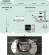
Schematic representation of gravity- and pressure-driven fluid handling systems applied in FISH integration with microfluidics experiments and microfluidic device illustration (A).
FIGURE 4
FIGURE 4
Representative examples of microfluidic channel detection region at cross-section (A) and horizontal view (B). White dashed line represents micropost height. Black line micropost contour; Black dashed line micropost contact with glass substrate. Original magnification 400 X (A), 100 X (B).
FIGURE 5
FIGURE 5
Contours of the velocity magnitude (m/s) for proposed microfluidic devices along the horizontal plane of symmetry (Symmetry plane II, Figure 2A) predicted by the CFD simulations (A), maximum velocity along the lateral gaps (B) and detail of the contours of the velocity magnitude in the lateral gaps (C).
FIGURE 6
FIGURE 6
Representative example of C. tropicalis subjected to FISH in microchannel (A,B) Previously trapped C. tropicalis (≈1 × 10⁸ cells/mL) after fixation/permeabilization, (C,D) hybridization and washing steps of FISH protocol. The microscope parameters maintained the same. Black dashed arrow represents cells at lateral microposts; Rectangle enlarged microchannel section; Black arrow trapped cell cells at front microposts; White dashed arrow fluorescence cells; White arrow fluorescence cells in different focus plane. Original magnification 400 X (A,B), 1,000 X (C,D).
FIGURE 7
FIGURE 7
Morphological growth forms of C. tropicalis: (A) yeast, (B) pseudohyphae, (C) hyphae. The microscope parameters maintained the same. Black arrow represents trapped cell. Original magnification 400 X (A–C).
FIGURE 8
FIGURE 8
Representative example of artificial urine (AU) contaminated with C. tropicalis subjected to microchannel integrated FISH method (A,C). Previously trapped C. tropicalis (≈1 × 10⁵ cells/mL) after fixation/permeabilization step. And subjected to hybridization solution (HS) alone (control, dashed circles) (B) or (D) PNA probe suspended in HS (200 nM). The microscope parameters were kept the same. Black arrow represents trapped cell; Dashed circle hybridized cell contour. Original magnification 400 X (A,C), 1,000 X (B,D) (E) The fluorescence signal quantification of C. tropicalis. The data is shown as mean fluorescence intensity (arbitrary units a. u.) ± SEM. p>0.05: * vs HS alone [Neg], HS + PNA probe [Pos].

References

    1. Almeida C., Azevedo N. F., Bento J. C., Cerca N., Ramos H., Vieira M. J., et al. (2013a). Rapid detection of urinary tract infections caused by Proteus spp. using PNA-FISH. Eur. J. Clin. Microbiol. Infect. Dis. 32, 781–786. 10.1007/s10096-012-1808-2 - DOI - PubMed
    1. Almeida C., Cerqueira L., Azevedo N. F., Vieira M. J. (2013b). Detection of Salmonella enterica serovar Enteritidis using real time PCR, immunocapture assay, PNA FISH and standard culture methods in different types of food samples. Int. J. Food Microbiol. 161, 16–22. 10.1016/j.ijfoodmicro.2012.11.014 - DOI - PubMed
    1. Almeida C., Sousa J. M., Rocha R., Cerqueira L., Fanning S., Azevedo N. F., et al. (2013c). Detection of Escherichia coli O157 by peptide nucleic acid fluorescence in situ hybridization (PNA-FISH) and comparison to a standard culture method. Appl. Environ. Microbiol. 79, 6293–6300. 10.1128/AEM.01009-13 - DOI - PMC - PubMed
    1. Alvarez-Lerma F., Nolla-Salas J., Leon C., Palomar M., Jorda R., Carrasco N., et al. (2003). Candiduria in critically ill patients admitted to intensive care medical units. Intensive Care Med. 29, 1069–1076. 10.1007/s00134-003-1807-y - DOI - PubMed
    1. Anwer A. G., Sandeep P. M., Goldys E. M., Vemulpad S. (2009). Distinctive autofluorescence of urine samples from individuals with bacteriuria compared with normals. Clin. Chim. Acta 401, 73–75. 10.1016/j.cca.2008.11.021 - DOI - PubMed