. 2022 Oct 4:32:101357.
doi: 10.1016/j.bbrep.2022.101357. eCollection 2022 Dec.

Induced resistance to ifosfamide in osteosarcoma cells suggests a more aggressive tumor profile

Affiliations

Induced resistance to ifosfamide in osteosarcoma cells suggests a more aggressive tumor profile

Maria Tereza de Oliveira Rodrigues et al. Biochem Biophys Rep. .

Abstract

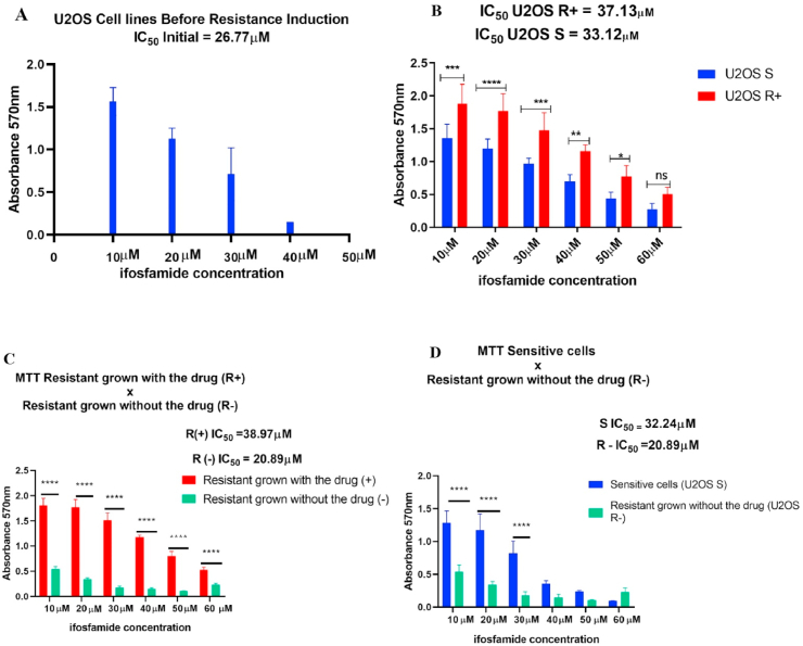
Aims: Osteosarcoma (OS) is the most common primary malignant bone sarcoma among children and adolescents. Treatment is based on neo-adjuvant and adjuvant chemotherapy, using the standard drugs cisplatin, methotrexate, doxorubicin, and ifosfamide (IFO). Due to the high capacity of tumor resistance, the current work aimed to analyze genes related to cycle control and cell differentiation in OS cells sensitive to and with induced resistance to IFO. This was to assess whether the differentiated expression of these genes may affect resistance to the drug IFO used in OS treatment, and thus establish possible biomarkers of disease progression.

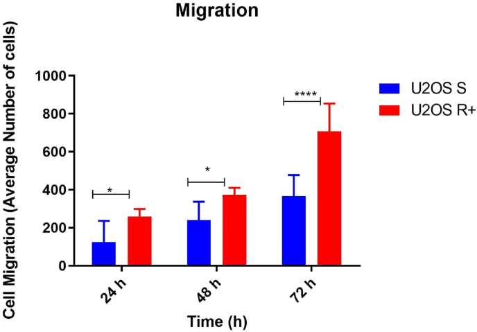
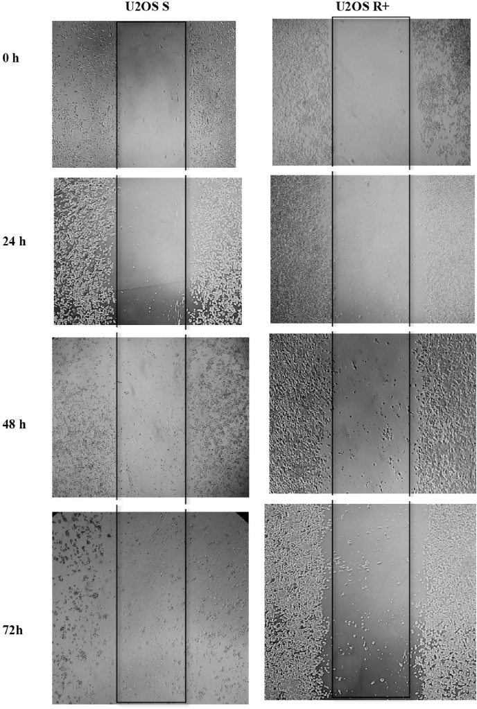
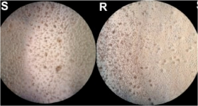
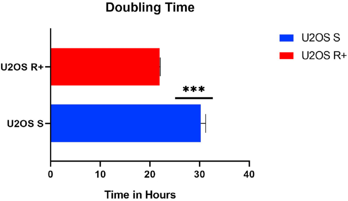
Materials and methods: In this work, the treatment-sensitive OS U2OS lineage was used, and the same lineage was submitted to the process of induction of IFO resistance. These cells were evaluated by MTT, migration and proliferation assays and submitted to gene expression analysis.

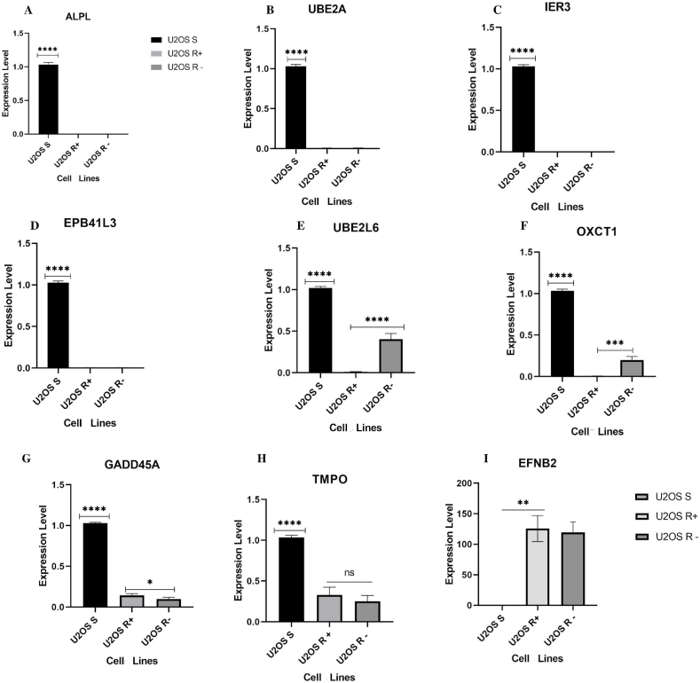
Key findings: The results demonstrate that after induction of resistance to IFO, resistant U2OS cells show a more aggressive tumor behavior, with greater capacity for cell migration, proliferation, and invasion compared to sensitive cells. Gene analysis indicates that resistance-induced cells have differentiated expression of the genes EPB41L3, GADD45A, IER3, OXCT1, UBE2L6, UBE2A ALPL, and EFNB2. Our results suggest new perspectives on possible resistance biomarkers, especially the genes EFNB2 and EPB41L3, given that these genes have rarely been studied their expression linked to osteosarcoma. They show how the resistance induction model can be useful for studies on tumor cell behavior.

Keywords: Ifosfamide; Osteosarcoma; Resistance-induced.


Conflict of interest statement

The authors declare that they have no known competing financial interests or personal relationships that could have appeared to influence the work reported in this paper.

Figures

Fig. 1
Fig. 1
Induction of resistance to IFO. In A the MTT assay demonstrated that 26.77 μM of IFO inhibited approximately 50% of osteosarcoma U2OS cells. B indicates that after the resistance induction protocol, the IFO value necessary to inhibit 50% of the cells became 37.13 μM. In addition, the U2OS R+ group showed a significant difference in the rate of metabolic activity in relation to the U2OS S cells with *p < 0.05 and p < 0.0001 (****) at different drug concentrations. C- Significant difference between U2OS R+ and U2OS R-cells at all drug concentrations with p < 0.0001 (****). D – U2OS R- and U2OS S cells with significant differences in concentrations of 10 μM, 20 μM, and 30 μM (****); however, there was no difference in the higher concentrations of IFO, indicating the sensitivity to treatment in the two cell groups.
Fig. 2
Fig. 2
Migration of the U2OS S and U2OS R+ lineages. The x-axis corresponds to the time in hours and the y-axis is the average of the number of cells that migrated. At 24 and 48 h, U2OS R+ cells had greater migration capacity compared to U2OS S cells (*), and at 72 h the difference in migration between the sensitive and resistant groups was of p < 0.0001 (****).
Fig. 3
Fig. 3
In vitro cell migration of U2OS S and U2OS R+ groups at 0 h, 24 h, 48 h, and 72 h intervals.
Fig. 4
Fig. 4
Non-adherence survival test. Analysis of cells not adhered to the LB agar-coated bottom plate. U2OS S (S) represents the group of sensitive cells, U2OS R+ (R) the group of resistant cells. Optical microscopy view. 10x magnification.
Fig. 5
Fig. 5
Cell proliferation assay – Doubling Time, indicating that U2OS S cells have an average time of 30 h to proliferate, while U2OS R+ cells have a shorter proliferation time, corresponding to 21 h.
Fig. 6
Fig. 6
Expression of tumor resistance genes in human osteosarcoma U2OS lineage groups. Expression levels were evaluated according to the mean of experimental and biological triplicates. The black bar indicates group U2OS S; the light gray bar U2OS R+ and the dark gray bar U2OS R-. The x-axis represents the group of cells and the y-axis the expression level. Graphs A – D represent the genes that were up-regulated in lineage U2OS S compared to groups U2OS R+ and U2OS R- (****p < 0.0001). Graph E represents the expression of UBE2L6 up-regulated in U2OS S (****p < 0.0001), and U2OS R-had greater expression (****p < 0.0001) compared to U2OS R+. Graph FOXCT1 with high expression in U2OS S (****p < 0.0001) and U2OS R- (***p = 0.0004) with greater expression in relation to U2OS R+. Graph GGADD45A up-regulated in U2OS S (****P < 0.0001), and in group U2OS R+ the expression was higher than in U2OS R- (*p = 0.0442). Graph H shows high expression of TMPO in U2OS S (****P < 0.0001) and there was no significant difference in expression between groups U2OS R+ and U2OS R-; Graph IEFNB2 up-regulated in U2OS R+ and U2OS R- (****p < 0.0001) in comparison to group U2OS S, which was down-regulated. The results were validated using one-way ANOVA.

References

    1. Van Driel M., Van Leeuwen J.P.T.M. Cancer and bone: a complex complex. Arch. Biochem. Biophys. 2014;561:159–166. doi: 10.1016/j.abb.2014.07.013. - DOI - PubMed
    1. Meazza C., Scanagatta P. Metastatic osteosarcoma: a challenging multidisciplinary treatment. Expert Rev. Anticancer Ther. 2016;16:543–556. doi: 10.1586/14737140.2016.1168697. - DOI - PubMed
    1. Moreno F., Cacciavillano W., Cipolla M., Coirini M., Streitenberger P., López Martí J., Palladino M., Morici M., Onoratelli M., Drago G., Schifino A., Cores M., Rose A., Jotomliansky J., Varel M., García Lombardi M. Vol. 64. 2017. (Childhood Osteosarcoma: Incidence and Survival in Argentina. Report from the National Pediatric Cancer Registry, ROHA Network 2000–2013, Pediatr. Blood Cancer). - DOI - PubMed
    1. Varshney J., Scott M., Largaespada D., Subramanian S. Understanding the osteosarcoma pathobiology: a comparative oncology approach. Vet. Sci. 2016;3:3. doi: 10.3390/vetsci3010003. - DOI - PMC - PubMed
    1. Sarbay H., Demir Ü.F., Yılmaz G., Atay A.A., Malbora B. Ifosfamide induced encephalopathy in a child with osteosarcoma. J. Oncol. Pharm. Pract. 2020 doi: 10.1177/1078155220963545. - DOI - PubMed