. 2022 Sep 23:16:940989.
doi: 10.3389/fncir.2022.940989. eCollection 2022.

An implantable neurophysiology platform: Broadening research capabilities in free-living and non-traditional animals

Affiliations

An implantable neurophysiology platform: Broadening research capabilities in free-living and non-traditional animals

Matt Gaidica et al. Front Neural Circuits. .

Abstract

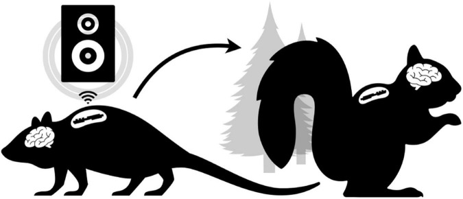
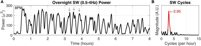
Animal-borne sensors that can record and transmit data ("biologgers") are becoming smaller and more capable at a rapid pace. Biologgers have provided enormous insight into the covert lives of many free-ranging animals by characterizing behavioral motifs, estimating energy expenditure, and tracking movement over vast distances, thereby serving both scientific and conservational endpoints. However, given that biologgers are usually attached externally, access to the brain and neurophysiological data has been largely unexplored outside of the laboratory, limiting our understanding of how the brain adapts to, interacts with, or addresses challenges of the natural world. For example, there are only a handful of studies in free-living animals examining the role of sleep, resulting in a wake-centric view of behavior despite the fact that sleep often encompasses a large portion of an animal's day and plays a vital role in maintaining homeostasis. The growing need to understand sleep from a mechanistic viewpoint and probe its function led us to design an implantable neurophysiology platform that can record brain activity and inertial data, while utilizing a wireless link to enable a suite of forward-looking capabilities. Here, we describe our design approach and demonstrate our device's capability in a standard laboratory rat as well as a captive fox squirrel. We also discuss the methodological and ethical implications of deploying this new class of device "into the wild" to fill outstanding knowledge gaps.

Keywords: accelerometer; closed-loop; implantable; physiology; sleep; wireless.


Conflict of interest statement

The authors declare that the research was conducted in the absence of any commercial or financial relationships that could be construed as a potential conflict of interest.

Figures

FIGURE 1
FIGURE 1
Conceptual Overview. The laboratory setting (left) does not have tools to enable freely behaving neurophysiology in novel experimental paradigms (e.g., low-latency wireless cuing of an external speaker to modulate neural rhythms in real-time). Ideally, the same “biologger” platform can translate to ethical free-ranging experiments (right) utilizing alternative device modes that support autonomous deployment.
FIGURE 2
FIGURE 2
Biologger hardware. (A) High-level overview of the power, digital, and analog systems. (B) Component placement relative to the four-layer PCB. (C) The 3D-printed case with the PCB and electrodes encapsulated in silicone.
FIGURE 3
FIGURE 3
Biologger iOS app configuration utility and data dump module. (A) The app is shown in a connected state with a biologger. These settings represent a recording schedule mode where two channels of biopotentials (EEG2 and EEG3) will record with a 20% duty cycle and accelerometer (Axy) data will record at 10 Hz. (B) The data dump module is shown below the biologger interface. The bottom of the biologger is displayed here to appreciate how the programming port pins interface with the pogo connector (circled in red) in the custom clamp module. Data from the biologger memory is transferred serially through the main data controller board on the micro-SD card.
FIGURE 4
FIGURE 4
Closed-loop slow-wave activity detection and audio stimulation. (A) Peri-detection EEG data are shown (black) with a 0.5–4 Hz bandpass filter applied to better visualize slow-wave (SW) activity. The signal estimate (red) is based on the center frequency and phase estimation of the biologger which is relayed to the base station at t = 0. The base station subsequently estimates a phase delay time to play 50 ms audio stimulus at the up-going phase of the ongoing SW activity. (B) Session-wide (n = 202 trials) values for center frequency (Fc, left) and phase delay time (right). Sham trial distributions (10% probability) are shown in red.
FIGURE 5
FIGURE 5
Biologging overnight in a freely behaving captive fox squirrel. (A) Eight hours of biologger data beginning at 8 p.m. showing EEG data (black) and 3D accelerometer data from the x-axis (blue), y-axis (orange), and z-axis (yellow). Representative sleep and wake epochs are marked along the top and the same data is shown in where (B) time has been restricted to a 10-s window. (C) The EEG spectrogram showing the relative power for each frequency (1–20 Hz) across time (red colors indicate high power).
FIGURE 6
FIGURE 6
Squirrel SW cycle duration. (A) Power in the SW band (0.5–4 Hz) was calculated from overnight recording data (8 h). Arrows indicate high SW power and also demonstrate characteristic SW cycle frequency in a 1-h window. (B) The SW time-frequency relationship was calculated to determine fundamental frequencies that may occur in the SW activity. Highly significant (P < 0.001) values and the peak magnitude were calculated (both in red).

References

    1. Anafi R. C., Kayser M. S., Raizen D. M. (2019). Exploring phylogeny to find the function of sleep. Nat. Rev. Neurosci. 20 109–116. 10.1038/s41583-018-0098-9 - DOI - PubMed
    1. Aulehner K., Bray J., Koska I., Pace C., Palme R., Kreuzer M., et al. (2022). The impact of tethered recording techniques on activity and sleep patterns in rats. Sci. Rep. 12:3179. 10.1038/s41598-022-06307-3 - DOI - PMC - PubMed
    1. Aulsebrook A. E., Jones T. M., Rattenborg N. C., Roth T. C., Lesku J. A. (2016). Sleep ecophysiology: integrating neuroscience and ecology. Trends Ecol. Evol. 31 590–599. 10.1016/j.tree.2016.05.004 - DOI - PubMed
    1. Bakker J., Klomp R., Rijnbeek M. W., Arndt S. S., Philippens I. H., Langermans J. A. (2014). Recovery time after intra-abdominal transmitter placement for telemetric (neuro) physiological measurement in freely moving common marmosets (Callitrix jacchus). Anim. Biotelem. 2:10. 10.1186/2050-3385-2-10 - DOI
    1. Barron D. G., Brawn J. D., Weatherhead P. J. (2010). Meta-analysis of transmitter effects on avian behaviour and ecology. Methods Ecol. Evol. 1 180–187. 10.1111/j.2041-210X.2010.00013.x - DOI

Publication types

LinkOut - more resources