. 2022 Sep 21:13:983924.
doi: 10.3389/fendo.2022.983924. eCollection 2022.

15-Deoxy-Delta-12,14-prostaglandin J2 modulates pro-labour and pro-inflammatory responses in human myocytes, vaginal and amnion epithelial cells

Affiliations

15-Deoxy-Delta-12,14-prostaglandin J2 modulates pro-labour and pro-inflammatory responses in human myocytes, vaginal and amnion epithelial cells

Zahirrah Bm Rasheed et al. Front Endocrinol (Lausanne). .

Abstract

Background: Prematurity is the leading cause of childhood death under the age of five. The aetiology of preterm birth is multifactorial; however, inflammation and infection are the most common causal factors, supporting a potential role for immunomodulation as a therapeutic strategy. 15-Deoxy-Delta-12,14-prostaglandin J2 (15dPGJ2) is an anti-inflammatory prostaglandin and has been shown to delay lipopolysaccharide (LPS) induced preterm labour in mice and improve pup survival. This study explores the immunomodulatory effect of 15dPGJ2 on the transcription factors NF-κB and AP-1, pro-inflammatory cytokines, and contraction associated proteins in human cultured myocytes, vaginal epithelial cell line (VECs) and primary amnion epithelial cells (AECs).

Methods: Cells were pre-incubated with 32µM of 15dPGJ2 and stimulated with 1ng/mL of IL-1β as an in vitro model of inflammation. Western immunoblotting was used to detect phosphorylated p-65 and phosphorylated c-Jun as markers of NF-κB and AP-1 activation, respectively. mRNA expression of the pro-inflammatory cytokines IL-6, IL-8, and TNF-α was examined, and protein expression of COX-2 and PGE2 were detected by western immunoblotting and ELISA respectively. Myometrial contractility was examined ex-vivo using a myograph.

Results: 15dPGJ2 inhibited IL-1β-induced activation of NF-κB and AP-1, and expression of IL-6, IL-8, TNF-α, COX-2 and PGE2 in myocytes, with no effect on myometrial contractility or cell viability. Despite inhibiting IL-1β-induced activation of NF-κB, expression of IL-6, TNF-α, and COX-2, 15dPGJ2 led to activation of AP-1, increased production of PGE2 and increased cell death in VECs and AECs.

Conclusion: We conclude that 15dPGJ2 has differential effects on inflammatory modulation depending on cell type and is therefore unlikely to be a useful therapeutic agent for the prevention of preterm birth.

Keywords: 15dPGJ2; activator protein (AP)-1; cytokines; inflammation; nuclear factor - kappa B (NF - κB); preterm labour (PTL); prostaglandins.


Conflict of interest statement

The authors declare that the research was conducted in the absence of any commercial or financial relationships that could be construed as a potential conflict of interest.

Figures

Figure 1
Figure 1
15dPGJ2 inhibits IL-1β-induced NF-κB and AP-1 activation in myometrial cells. Myometrial cells were pre-incubated for 2 hours with 32µM 15dPGJ2 or vehicle control before stimulation with 1ng/ml of IL-1β for 15 minutes. Whole cell lysates were prepared and assessed by Western immunoblotting for serine 536-phosphorylated p65 (Ser536-p-p65) as a marker of NF-κB activation, and serine 73-phosphorylated c-Jun (Ser73-p-c-Jun) as a marker of AP-1 activation. Representative immunoblots are shown for p-p65 (A) and p-c-Jun (B) with β-actin as a loading control. Densitometry analysis of the immunoblots showed a significant increase in p-p65 and p-c-Jun with IL-1β stimulation after 15 minutes (p<0.01). Pre-incubation with 15dPGJ2 significantly inhibited IL-1β-induced p-p65 and p-c-Jun (p<0.05) activation. n=5-6. *p<0.05, **p<0.01. Data are presented as mean ± SEM.
Figure 2
Figure 2
15dPGJ2 inhibits IL-1β-induced IL-6, IL-8, and TNF-α cytokines mRNA production in myometrial cells. Myometrial cells were pre-incubated for 2 hours with 32µM 15dPGJ2 or vehicle prior to 1ng/ml of IL-1β stimulation. Cells were harvested at -2, 0, 4, 8, 12 and 24 hours post IL-1β stimulation. Quantitative RT-qPCR was used to determine the effect of 15dPGJ2 on IL-6, IL-8, and TNF-α mRNA. Fold change took into account the reference gene β actin. A significant increase in IL-6 was observed with IL-1β at 8 and 12 hours (p < 0.01, p < 0.05), and inhibition of this effect was seen when cells were pre-incubated with 15dPGJ2 at 8 hours (p < 0.05) (A). A significant increase in IL-8 was seen at 8 hours (p < 0.01), and inhibition of this effect was seen when cells were pre-incubated with 15dPGJ2 at 8 hours (p < 0.05) (B). TNF-α mRNA was upregulated at 4 hours with IL-1β stimulation (p < 0.001), the effect of which was inhibited on pre-incubation with 15dPGJ2 (p < 0.001) (C). n = 3. The effect of IL-1β was compared to -2 non-stimulated (vehicle) time point. *p < 0.05, **p < 0.01, ***p < 0.001. The effect of 15dPGJ2 was determined by comparing the IL-1β plus 15dPGJ2 treatment to IL-1β alone p < 0.05, †††p < 0.001. Data are presented as mean ± SEM.
Figure 3
Figure 3
15dPGJ2 inhibits IL-1β-induced contraction associated genes and proteins in myometrial cells. Myometrial cells were pre-incubated for 2 hours with 32µM 15dPGJ2 or vehicle before stimulation with 1ng/ml of IL-1β. For cPLA2-α mRNA, cells were harvested at -2, 0, 4, 8, 12 and 24 post 1ng/ml of IL-1β stimulation. COX-2 mRNA and protein were harvested after 4 hours. mRNA level was quantified using RT-qPCR and whole cells lysates were prepared and assessed by RT-qPCR and western immunoblotting used β-actin as the loading controland reference gene. PGE2 was quantified using ELISA after 24 hours stimulation of 1ng/ml IL-1β. IL-1β-induced cPLA2-α mRNA at 8 and 12 hours and was significantly inhibited by 15dPGJ2 (p < 0.001) (A). IL-1β stimulation significantly increased COX-2 production (p < 0.01) and pre-incubation with 15dPGJ2 significantly decreased mRNA (p < 0.01) (B) and protein expression p < 0.05). (C). IL-1β stimulation increased PGE2 production and pre-incubating the myocytes with 15dPGJ2 significantly inhibited the production (p < 0.05) (D). n = 3. *p < 0.05, **p < 0.01, ***p < 0.001, †††p < 0.001. Data are presented as mean ± SEM.
Figure 4
Figure 4
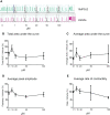
15dPGJ2 has no effect on myometrial contractility. Myometrial tissues biopsies were dissected and mounted on the myograph machine and stretched to 4g tension to achieve spontaneous contractions. After spontaneous contractions were established, the tissues were treated with 1, 10, 20, 50 and 100µM of 15dPGJ2 or vehicle control. A representative trace is shown for two 15dPGJ2 treated strips and a vehicle control strip (A). A cumulative dose response with 15dPGJ2 or vehicle was performed and the effect on total and average area under the curve (B, C), peak amplitude (D) and rate of contractility (E) was determined. 15dPGJ2 had no significant effect on myometrial contractility. V = vehicle. n = 3 biological replicates, each containing at least two 15dPGJ2 treated strips per biological replicate. Data are presented as mean ± SEM.
Figure 5
Figure 5
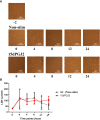
15dPGJ2 effects on myometrial cells integrity. Myometrial cells were pre-incubated with 32µM of 15dPGJ2 or vehicle for 2 hours and assessed for 24 hours. Morphology of the cells were assessed by brightfield microscopy (A) and LDH release into supernatant was detected at -2, 0, 4, 8, 12 and 24 hours (B). No difference in LDH release was seen between timepoints or between vehicle control and 15dPGJ2 treated cells. n = 3. Data are presented as mean ± SEM. NS, Non-stim.
Figure 6
Figure 6
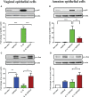
Effect of 15dPGJ2 on NF-κB and AP-1 in vaginal and amnion epithelial cells. Vaginal (VEC) and amnion epithelial cells (AECs) were pre-incubated with 32μM of 15dPGJ2 for 2 hours then stimulated with 1ng/ml of IL-1β. Whole cell lysate was used to determine serine 536-phosphorylated p65 (Ser536-p-p65) as a marker of NF-κB activation and serine 73-phosphorylated c-Jun (Ser73-p-c-Jun) as a marker of AP-1 via western immunoblotting. A representative blot was shown above each graph with β-actin as a loading control. Pre-incubation with 15dPGJ2 significantly inhibited IL-1β stimulated NF-κB in VECs (p < 0.001) (A) and AECs (p < 0.01) (B). AP-1 however significantly increased after incubation with 15dPGJ2 alone as well as with 15dPGJ2 and IL-1β in VECs (p < 0.01) (C) and also in 15dPGJ2 and IL-1β treated AECs (p<0.01) (D). *p < 0.05, **p < 0.01, ***p < 0.001. n = 3-6. Data are presented as mean ± SEM.
Figure 7
Figure 7
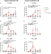
Effect of 15dPGJ2 on IL-6, IL-8 and TNF-α mRNA in vaginal and amnion epithelial cells. VEC and AECs were pre-incubated with 32μM of 15dPGJ2 for 2 hours then stimulated with 1ng/ml of IL-1β. Cells were harvested, and total RNA was quantified after -2, 0, 4, 8, 12 and 24 hours. Quantitative PCR was performed looking at IL-6, IL-8 and TNF-α mRNA level. Fold change took into account the reference gene β actin IL-1β stimulation increased IL-6 mRNA expression in VEC at 4 (**) and 24 (***) hours but no statistically significant inhibition was achieved with 15dPGJ2 pre-incubation (A). IL-8 mRNA expression increased with IL-1β stimulation and this effect was inhibited by 15dPGJ2 at 4, 8, 12 (***, †††) and 24 (**, †) hours (B). TNF-α mRNA also increased with IL-1β stimulation and this effect was inhibited by 15dPGJ2 at 4, 8, 12 (***, †††) and 24 (*, †) hours (C). In amniocytes, IL-1β stimulation significantly increased IL-6 expression in which this effect was inhibited at 8 (***, †††), 12 (***, ††) and 24 (***, †††) hours (D). Pre-incubation of 15dPGJ2 before IL-1β stimulation increased IL-8 mRNA compared to IL-1β alone (††) at 8 hours (E). TNF-α mRNA expression increased at 4 hours with IL-1β stimulation (***) but the inhibition by 15dPGJ2 did not reach statistical significance (F). n = 3. *p < 0.05, **p < 0.01, ***p < 0.001 = effect of IL-1β compared to -2 non-stimulated time point. p < 0.05, †† p < 0.01, ††† p < 0.001 = effect of IL-1β 15dPGJ2 compared to IL-1β treatment alone. Data are presented as mean ± SEM.
Figure 8
Figure 8
Effect of 15dPGJ2 on COX-2 and PGE2 in vaginal and amnion epithelial cells. VECs and AECs were pre-incubated with 32μM of 15dPGJ2 for 2 hours then stimulated with 1ng/ml of IL-1β for 4 hours to examine COX-2 protein expression and 24 hours to examine PGE2 production. Whole cell lysate was used to determine COX-2 protein expression and an ELISA was used to determine PGE2 concentration in cell culture supernatant. 15dPGJ2 pre-incubation significantly inhibited IL-1β stimulated COX-2 protein expression in VECs (p < 0.05) (A) and AECs (B) (p < 0.05) at 4 hours. PGE2 concentration was increased in VEC culture supernatant when treated with 15dPGJ2 (p < 0.001) and 15dPGJ2 plus IL-1β (p < 0.01) (C) and in supernatant of 15dPGJ2 treated AECs (p < 0.05) (D). n=3-7. *p < 0.05, **p < 0.01, ***p < 0.001. Data are presented as mean ± SEM.
Figure 9
Figure 9
Effect of 15dPGJ2 MAPKs in vaginal and amnion epithelial cells. VECs and AECs were pre-incubated with 32μM of 15dPGJ2 for 2 hours then stimulated with 1ng/ml of IL-1β for 15 minutes. Whole cell lysate was used to determine, phosphorylated-JNK1/JNK2(Thr183/Tyr185), phosphorylated-p38(Thr180/Tyr182) and phosphorylated-ERK (Thr202/Tyr204) via Western immunoblotting. A representative blot was shown above each graph with β-actin as a loading control. In VECs, incubation with 15dPGJ2 alone for 2 hours significantly increased the basal level p-JNK (p < 0.01) (A) and p-p38 (p < 0.01) (B) but had no effect on p-ERK (C). Treatment with 1ng/ml of IL-1β increased p-ERK, and this was attenuated by pre-incubation with 15dPGJ2 (p < 0.01) in VECs (C). In AECs, no effect was seen on p-JNK with IL-1β and/or 15dPGJ2 (D). IL-1β stimulation of AECs led to an increase in p-p-38 (p < 0.001) (E) and p-ERK (p < 0.001) (F), the response of which were attenuated upon pre-incubation with 15dPGJ2. *p < 0.05, **p < 0.01, ***p < 0.001, NS non significant. n = 3-6.​ Data are presented as mean ± SEM.
Figure 10
Figure 10
Effect of 15dPGJ2 on cell viability in vaginal and amnion epithelial cells. VECs and AECs were incubated with or without 32μM of 15dPGJ2 and the morphology of the cells were assessed by brightfield light microscopy at -2, 0, 2, 4, 6, 18 and 24 hours. 15dPGJ2 or vehicle control was inserted in the corresponding wells at -2 timepoint following image and supernatant capture. The culture medium at each time point was used to detect cell viability using the MTT assay. The morphology of VECs changed from a healthy polygonal shape to an abnormal round shape compared to the control (A). The effect of 15dPGJ2 on cell viability was detected as early as 2 hours (p < 0.05), and a steady change was seen at 4, 6, 18 and 24 hours (p < 0.001) (B). In AECs, the morphological shape of the cells started to change from an ovoid, slab stone- and cobblestone-like pattern to a stretched star-like pattern at 18 hours compared to the control (C). The cell viability of amnion epithelial cells was significantly changed by 18 hours in 15dPGJ2 treated cells (p < 0.05) (D). *p < 0.05, ***p < 0.001, NS non significant. n = 3.​ Data are presented as mean ± SEM.
Figure 11
Figure 11
Illustration on the effect of IL-1β and 15dPGJ2 on myocytes, vaginal and amnion epithelial cells. Pre-incubation of 15dPGJ2 in myocytes for 2 hours inhibited IL-1β induced activation of the transcription factors NF-κB and AP-1. This led to inhibition of cytokine expression and PGE2 production. In VECs and AECs, 15dPGJ2 inhibited IL-1β-stimulated NF-κB but led to activation of AP-1. A downstream inhibition of the pro-labour cytokines IL-6, IL-8, and TNF-α was seen in VECs. In contrast upregulation of IL-8 was seen in AECs. PGE2 production was increased both VECs and AECs following treatment with 15dPGJ2, and this may be due to AP-1 activation. In addition, increased cell death was seen in VECs and AECs with 15dPGJ2 treatment. We conclude that 15dPGJ2 has differential effects depending on cell type, and due to its ability to increase the production of pro-labour and pro- inflammatory mediators, we would not advocate its development as a novel therapeutic strategy for preterm birth prevention. Created with BioRender.com

Similar articles

Cited by

References

    1. Howson CP, Kinney MV, Lawn JE. Born too soon: The global action report on preterm birth. In: March of dimes, PMNCH, save the children. Geneva: World Health Organisation; (2012).
    1. Liu L, Oza S, Hogan D, Perin J, Rudan I, Lawn JE, et al. . Global, regional, and national causes of child mortality in 2000-13, with projections to inform post-2015 priorities: an updated systematic analysis. Lancet (2015) 385(9966):430–40. doi: 10.1016/S0140-6736(14)61698-6 - DOI - PubMed
    1. UnitedNations . (2022). Available at: https://sdgs.un.org/goals/goal3.
    1. Romero R, Espinoza J, Kusanovic JP, Gotsch F, Hassan S, Erez O, et al. . The preterm parturition syndrome. BJOG. (2006) 113 Suppl 3:17–42. doi: 10.1111/j.1471-0528.2006.01120.x. - DOI - PMC - PubMed
    1. Agrawal V, Hirsch E. Intrauterine infection and preterm labor. Semin Fetal. Neonatal. Med (2012) 17(1):12–9. doi: 10.1016/j.siny.2011.09.001 - DOI - PMC - PubMed

Publication types

MeSH terms