. 2022 Sep 23:16:983151.
doi: 10.3389/fnana.2022.983151. eCollection 2022.

Scribble basal polarity acquisition in RPE cells and its mislocalization in a pathological AMD-like model

Affiliations

Scribble basal polarity acquisition in RPE cells and its mislocalization in a pathological AMD-like model

Alicia Segurado et al. Front Neuroanat. .

Abstract

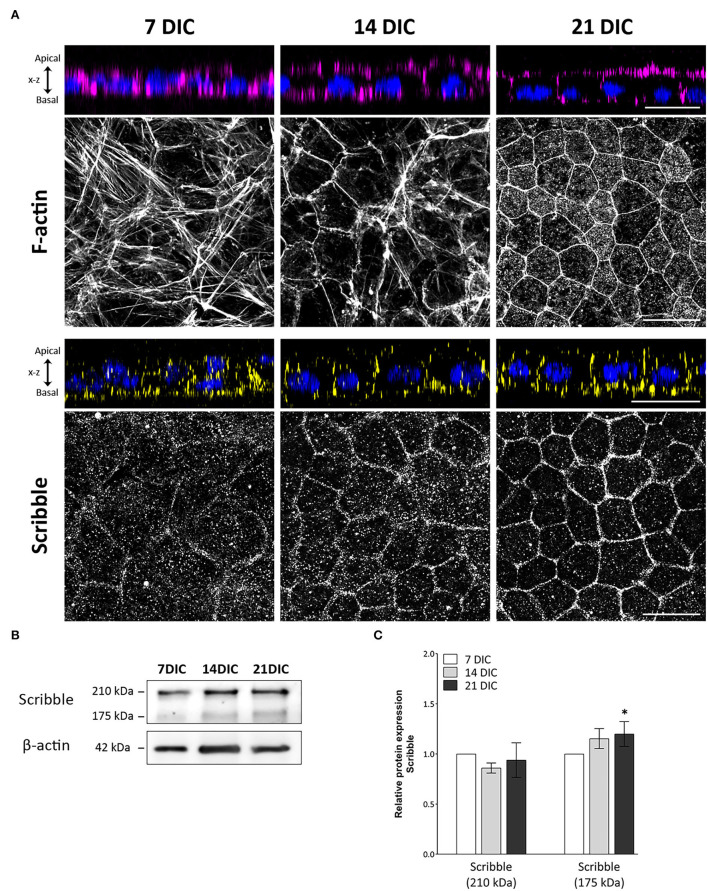
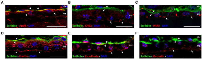
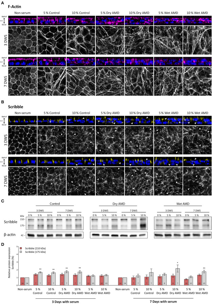
Apicobasal polarity is a hallmark of retinal pigment epithelium cells and is required to perform their functions; however, the precise roles of the different proteins that execute polarity are still poorly understood. Here, we have studied the expression and location of Scribble, the core member of the polarity basal protein complex in epithelial-derived cells, in human and mouse RPE cells in both control and pathological conditions. We found that Scribble specifically localizes at the basolateral membrane of mouse and human RPE cells. In addition, we observed an increase in the expression of Scribble during human RPE development in culture, while it acquires a well-defined basolateral pattern as this process is completed. Finally, the expression and location of Scribble were analyzed in human RPE cells in experimental conditions that mimic the toxic environment suffered by these cells during AMD development and found an increase in Scribble expression in cells that develop a pathological phenotype, suggesting that the protein could be altered in cells under stress conditions, as occurs in AMD. Together, our results demonstrate, for the first time, that Scribble is expressed in both human and mouse RPE and is localized at the basolateral membrane in mature cells. Furthermore, Scribble shows impaired expression and location in RPE cells in pathological conditions, suggesting a possible role for this protein in the development of pathologies, such as AMD.

Keywords: Scribble; Scribble complex; age-related macular degeneration (AMD); cell polarity; differentiation; epithelium; retina; retinal pigment epithelium (RPE).


Conflict of interest statement

The authors declare that the research was conducted in the absence of any commercial or financial relationships that could be construed as a potential conflict of interest.

Figures

Figure 1
Figure 1
Time-course expression pattern of actin and Scribble protein during hRPE cell development in culture. (A) Orthogonal views and corresponding z-stack maximum projections showing actin (magenta) and Scribble protein (yellow) localization as hRPE cells gradually form the mature epithelial tissue. DAPI (blue): nuclear labeling. Scale bars: 20 μm. (B) Western blot analyses showing expression of Scribble (210 kDa) and Scribble (175 kDa) from 7 to 21 DIC. β-actin was used as loading control. (C) Graphical representation of the relative expression of Scribble (210 kDa) and Scribble (175 kDa) during the development of hRPE cells in culture. In both cases, 7 DIC (n = 9), 14 DIC (n = 9), and 21 DIC (n = 9). DIC in (A–C):Days in culture. Data are presented as mean ± sd. Statistical information: The Kolmogorov-Smirnov and Levene tests were used to evaluate the normality and homoscedasticity of the sample distribution, respectively. Data were determined to be non-parametrically distributed. The potential significant statistical differences were analyzed by the Kruskal-Wallis test, followed by Dunn–Bonferroni's post-hoc test. Asterisks indicate statistical differences. *p-value < 0.05 vs. 7 DIC.
Figure 2
Figure 2
Localization of Scribble protein in wild-type adult mouse RPE. Double immunolabeling for Scribble (green, arrowheads) and other proteins (red, arrows): carrier protein for lipids, ApoE (A); apical polarity proteins CRB2 (B) and PAR3 (C); F-actin (D); and adherens and tight junctional protein E-cadherin (E) and occludin (F), respectively. DAPI (in blue): nuclear labeling. Scale bars: 20 μm. Ch, Choroid; RPE, retinal pigment epithelium.
Figure 3
Figure 3
Expression pattern of actin and Scribble proteins in hRPE cells after serum exposure. (A) Orthogonal views and corresponding z-stack maximum projections showing actin (magenta) in cells without serum and after the serum exposure of both control patients and patients with AMD patients. (B) Orthogonal views and corresponding z-stack maximum projections showing Scribble (yellow) in cells without serum and after serum exposure of control patients and patients with AMD. DAPI (blue): nuclear labeling. Scale bars in (A,B): 20 μm. (C) Western blot analyses showing expression of Scribble (210 kDa) and Scribble (175 kDa) in cells without serum and after control and AMD patients serum exposure. β-actin was used as loading control. DWS in (A–C): Days with serum. (D) Graphical representation of the relative expression of Scribble (210 kDa) and Scribble (175 kDa) in cells without serum and after serum exposure of control patients and patients with AMD. Data are presented as mean ± SD. Arrows in (B) point out to areas where Scribble has translocated from the plasma membrane to the cytoplasm. The number within each column corresponds to the number of samples per data. Statistical information: The Kolmogorov-Smirnov and Levene tests were used to assess the normality and homoscedasticity of the sample distribution, respectively. Data were determined to be non-parametrically distributed. The potential significant statistical differences were analyzed by the Kruskal–Walli's test followed by Dunn–Bonferroni's post-hoc test. Asterisks indicate statistical differences. *p-value < 0.05; **p-value < 0.01 vs. non-serum group.

Similar articles

References

    1. Ach T., Tolstik E., Messinger J. D., Zarubina A. V., Heintzmann R., Curcio C. A. (2015). Lipofuscin redistribution and loss accompanied by cytoskeletal stress in retinal pigment epithelium of eyes with age-related macular degeneration. Invest. Ophthalmol. Visual Sci. 56, 3242–3252. 10.1167/iovs.14-16274 - DOI - PMC - PubMed
    1. Assémat E., Bazellières E., Pallesi-Pocachard E., Le Bivic A., Massey-Harroche D. (2008). Polarity complex proteins. Biochim. Biophys. Acta Biomembranes 1778, 614–630. 10.1016/j.bbamem.2007.08.029 - DOI - PubMed
    1. Barreda D., Gutiérrez-González L. H., Martínez-Cordero E., Cabello-Gutiérrez C., Chacón-Salinas R., Santos-Mendoza T. (2020). The scribble complex PDZ proteins in immune cell polarities. J. Immunol. Res. 2020, 5649790. 10.1155/2020/5649790 - DOI - PMC - PubMed
    1. Bhutto I., Lutty G. (2012). Understanding age-related macular degeneration (AMD): relationships between the photoreceptor/retinal pigment epithelium/Bruch's membrane/choriocapillaris complex. Mol. Aspects Med. 33, 295–317. 10.1016/j.mam.2012.04.005 - DOI - PMC - PubMed
    1. Bilder D., Li M., Perrimon N. (2000). Cooperative regulation of cell polarity and growth by Drosophila tumor suppressors. Science 289, 113–116. 10.1126/science.289.5476.113 - DOI - PubMed

LinkOut - more resources