. 2022 Sep 27:2022:7432842.
doi: 10.1155/2022/7432842. eCollection 2022.

Progression in Time of Dentate Gyrus Granule Cell Layer Widening due to Excitotoxicity Occurs along In Vivo LTP Reinstatement and Contextual Fear Memory Recovery

Affiliations

Progression in Time of Dentate Gyrus Granule Cell Layer Widening due to Excitotoxicity Occurs along In Vivo LTP Reinstatement and Contextual Fear Memory Recovery

Karina Hernández Mercado et al. Neural Plast. .

Abstract

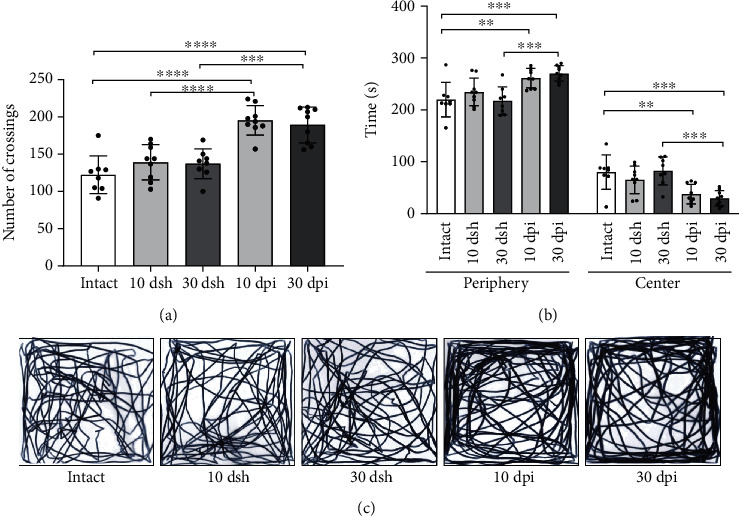
The dentate gyrus (DG) is the gateway of sensory information arriving from the perforant pathway (PP) to the hippocampus. The adequate integration of incoming information into the DG is paramount in the execution of hippocampal-dependent cognitive functions. An abnormal DG granule cell layer (GCL) widening due to granule cell dispersion has been reported under hyperexcitation conditions in animal models as well as in patients with mesial temporal lobe epilepsy, but also in patients with no apparent relation to epilepsy. Strikingly, it is unclear whether the presence and severity of GCL widening along time affect synaptic processing arising from the PP and alter the performance in hippocampal-mediated behaviors. To evaluate the above, we injected excitotoxic kainic acid (KA) unilaterally into the DG of mice and analyzed the evolution of GCL widening at 10 and 30 days post injection (dpi), while analyzing if KA-induced GCL widening affected in vivo long-term potentiation (LTP) in the PP-DG pathway, as well as the performance in learning and memory through contextual fear conditioning. Our results show that at 10 dpi, when a subtle GCL widening was observed, LTP induction, as well as contextual fear memory, were impaired. However, at 30 dpi when a pronounced increase in GCL widening was found, LTP induction and contextual fear memory were already reestablished. These results highlight the plastic potential of the DG to recover some of its functions despite a major structural alteration such as abnormal GCL widening.


Conflict of interest statement

The authors declare no competing financial or nonfinancial interests.

Figures

Figure 1
Figure 1
Experimental design. Kainic acid injections or sham surgeries were performed on day 0. Mice were handled and habituated to the experimenter for three consecutive days before the onset of behavioral procedures. On days 8 or 28 after surgery, mice performed the open field task. The next day (day 9 or 29), mice underwent contextual fear conditioning (CFC), and 24 h later, contextual fear memory (CFM) was evaluated. After memory retrieval, subjects were sacrificed and brains were extracted for histological analysis. A different group of animals, not exposed to behavioral evaluation, was used for in vivo LTP recordings.
Figure 2
Figure 2
Morphological changes in the DG over time after KA injection. Left images show representative Nissl-stained coronal sections of the hippocampus from (a) 10 dsh, (b) 10 dpi, (e) 30 dsh, and (f) 30 dpi. (a) At 10 dsh, sections display a mechanical lesion in the upper blade of DG as result of the cannula insertion (head arrow); scale bars: 300 μm. (e) At 30 dsh, the mechanical lesion is no longer evident (head arrow). (b) At 10 dpi, a focal lesion zone in the upper blade characterized by a thinning of the granule cell layer is observed (white arrow). Outside this lesion area, the upper blade shows GCL widening. (f) At 30 dpi, both DG blades display a more severe widening compared to 10 dpi. The black arrows indicate the prominent widening in the lower blade. The white circle and the asterisk symbols indicate cell loss in CA1 and in the hilus, respectively. (c–g) The site where KA spreads in the upper blade as revealed by the fluorescent tracer; insets show 40x magnifications. (d–h) A visual model of the DG based on Nissl-stained coronal sections: upper blade in gray, lesion site in blue, and lower blade in green. (i) Volume of the DG based on Cavalieri analysis (intact n = 3; 10 dsh n = 5; 30 dsh n = 5; 10 dpi n = 6; 30 dpi n = 5; one-way ANOVA followed by Bonferroni's post hoc test, F(14, 60) = 123.7, p < 0.0001). (j) Volume from the lesion site in the upper blade from KA-injected mice (10 dpi n = 6; 30 dpi n = 5; unpaired t-test with Welch's correction, F(4, 5) = 18.31, p < 0.0069). (k) GCL width from each DG section analyzed in KA-injected mice (10 dpi n = 29 sections from 6 mice; 30 dpi n = 24 sections from 5 mice; unpaired t-test with Welch's correction; upper blade, F(23, 28) = 3.625, p = 0.0015; lower blade, F(24, 28) = 6.493, p < 0.0001; upper blade focal lesion, F(23, 28) = 3.962, p = 0.0007). (l) Average volume and Gundersen's coefficient after a stereological Cavalieri analysis from the same mice used in histograms (i, j); statistical significances are indicated in histograms (i, j). A Gundersen's coefficient less than 0.1 means statistically valid volume of data. The columns represent the mean SD from each group. Dots superimposed in the graph represent individual values. Asterisks indicate statistically significant differences (p < 0.05; ∗∗p < 0.01; ∗∗∗p < 0.001; ∗∗∗∗p < 0.0001).
Figure 3
Figure 3
KA-induced alterations in the DG modify in vivo perforant pathway LTP along time. (a) Schematic representation of experimental procedures. Conditioning stimulus trains began after 20 min of baseline recording and were maintained for 120 min. (b) Representative traces of the excitatory postsynaptic potential (EPSP) obtained during baseline (dotted line) and 140 min after stimulation (full line). (c) Plot of the EPSP slope for the last 20 min after high-frequency stimulation. Intact and 30 dsh groups displayed similar slope percentages of EPSP (intact 171.1%; 30 dsh 172.3%). The EPSP slope percentage in the 10 dsh group was 139.7%, that is, less than those in intact and 30 dsh groups. In the 10 dpi group, LTP induction was impaired, as opposed to the 30 dpi group where LTP induction was recovered. Notably, LTP from mice at 30 dpi took longer time to reach its maximal response compared to all other groups. All these findings are more evident in the population spike slope percentage. (d) Inner numbers show the mean ± SEM of slope (percent of baseline) from the last 20 min of recording. Application of conditioning stimulus trains is indicated (arrow). Repeated measures ANOVA followed by post hoc Fisher's test; intact n = 4; 10 dsh n = 4; 30 dsh n = 4; 10 dpi n = 4; 30 dpi n = 4.
Figure 4
Figure 4
Mice at 10 dpi and 30 dpi show increased motility and anxiety-like behavior. (a) Motor behavior was evaluated through the number of crossings. KA-injected mice at 10 and 30 dpi show a higher number of crossings than intact and sham mice (one-way ANOVA followed by a Bonferroni's post hoc test, F(4, 38) = 18.58, p < 0.0001). (b) Anxiety-like behavior was evaluated through the time spent in the periphery and center zones of the field. KA-injected mice at 10 and 30 dpi spent more time on periphery and less time on center zones than intact and sham mice (two-way ANOVA followed by a Bonferroni's post hoc test, interaction F(4, 76) = 16.44, p < 0.0001). (c) Representative path length tracks from a mouse from each group are shown. The columns represent the mean with SD from each group. Dots superimposed in the graph represent individual values. Asterisks indicate statistically significant differences (p ≤ 0.05; ∗∗p < 0.01; ∗∗∗p < 0.001; ∗∗∗∗p < 0.0001). Intact n = 8; 10 dsh n = 9; 30 dsh n = 8; 10 dpi n = 9; 30 dpi n = 9.
Figure 5
Figure 5
Contextual fear memory was impaired at 10 dpi but restored at 30 dpi. (a) Learning curve for contextual fear conditioning. All groups, except mice at 10 dpi, showed progressive learning reflected by the increase in the percentage of freezing time across trials. Mice at 10 dpi show lower freezing percentage time than intact mice on trials 3 and 4. Points represent the mean with SD for each group across trials (asterisks denote differences from intact vs 10 dpi; two-way ANOVA followed by a Bonferroni's post hoc test, (F(4,192) = 43.07, p < 0.0001). (b) Contextual fear memory retrieval. Mice at 10 dsh show lower freezing than intact mice, but more freezing than mice at 10 dpi. At 30 dpi, mice show similar freezing as intact and 30 dsh mice. The columns represent mean and SD for each group. Dots superimposed on the graph represent individual values (one-way ANOVA followed by Bonferroni's post hoc test, F(4, 38) = 28.29, p < 0.0001). (c, d) Graphs show the positive correlation between the volume of the (c) lower blade (Spearman correlation coefficient, r = 0.8167; 95% confidence interval (0.5552 to 0.9313); n = 18) and the volume of (d) the upper blade (Spearman correlation coefficient, r = 0.6594; 95% confidence interval (0.2644 to 0.8650); n = 18) with the percentage of freezing in lesioned mice. Light-blue circles correspond to 10 dpi mice (n = 9) and navy-blue circles to 30 dpi mice (n = 9). For (a, b), intact n = 8, 10 dsh n = 9, 30 dsh n = 8, 10 dpi n = 9, 30 dpi n = 9. Asterisks indicate statistically significant differences, p ≤ 0.05; ∗∗p ≤ 0.01; ∗∗∗p ≤ 0.001.

Similar articles

Cited by

References

    1. Bird C. M., Burgess N. The hippocampus and memory: insights from spatial processing. Nature Reviews. Neuroscience . 2008;9(3):182–194. doi: 10.1038/nrn2335. - DOI - PubMed
    1. Knierim J. J., Neunuebel J. P., Deshmukh S. S. Functional correlates of the lateral and medial entorhinal cortex: objects, path integration and local–global reference frames. Philos Trans R Soc B Biol Sci . 2014;369(1635):p. 20130369. doi: 10.1098/rstb.2013.0369. - DOI - PMC - PubMed
    1. Witter M. P. The perforant path: projections from the entorhinal cortex to the dentate gyrus. Progress in Brain Research . 2007:43–61. doi: 10.1016/S0079-6123(07)63003-9. - DOI - PubMed
    1. Leutgeb J. K., Leutgeb S., Moser M.-B., Moser E. I. Pattern separation in the dentate gyrus and CA3 of the hippocampus. Science (80) . 2007;315(5814):961–966. doi: 10.1126/science.1135801. - DOI - PubMed
    1. Bouilleret V., Ridoux V., Depaulis A., Marescaux C., Nehlig A., Le Gal La Salle G. Recurrent seizures and hippocampal sclerosis following intrahippocampal kainate injection in adult mice: electroencephalography, histopathology and synaptic reorganization similar to mesial temporal lobe epilepsy. Neuroscience . 1999;89(3):717–729. doi: 10.1016/S0306-4522(98)00401-1. - DOI - PubMed

Publication types

LinkOut - more resources