

Kimura disease, a rare cause of inguinal lymphadenopathy: A case report
- PMID: 36213635
- PMCID: PMC9537629
- DOI: 10.3389/fmed.2022.1023804
Kimura disease, a rare cause of inguinal lymphadenopathy: A case report
Abstract
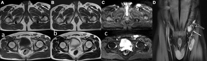
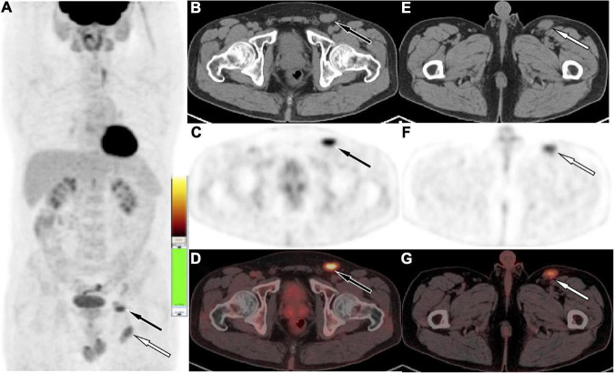
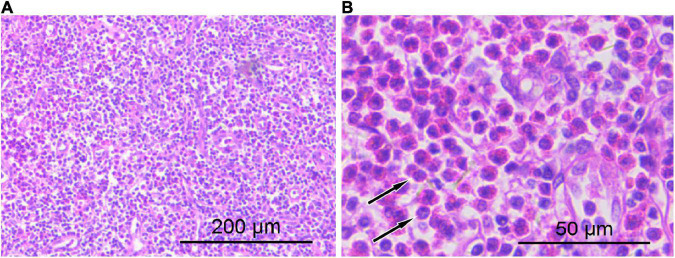
Kimura's disease (KD) is a rare chronic granulomatous disease of unknown etiology that mainly involves damage to lymph nodes, soft tissues, and salivary glands. The clinical symptoms are mainly painless subcutaneous soft tissue masses, often involving head and neck lymph nodes and salivary glands, and are mainly characterized by diffuse eosinophilic infiltration, lymphocyte, and vascular proliferation. There are few reports in the literature that KD affects only inguinal lymph nodes. We report in this study a 41-year-old male patient who presented to the hospital for medical help with soft tissue masses in the groin. Magnetic resonance imaging (MRI) showed multiple abnormal soft tissue nodules around the iliac vessels in the left groin, and a contrast-enhanced scan showed obvious homogeneous enhancement. Diffusion-weighted imaging showed limited movement of water molecules and showed an obvious high signal. Fluoro18-labeled deoxyglucose positron emission tomography/computed tomography (18F-FDG PET/CT) was recommended for further evaluation of the patient's general condition, and the results showed that except for the radioactive uptake in the lesions in the left groin region, no obvious abnormality was found in the rest of the body. Based on these imaging findings, the patient was first suspected to have malignant lesions, and then the patient underwent histopathological examination, which was confirmed to be KD. Our case study suggests that KD affects only the inguinal lymph nodes is rare and should be considered as one of the imaging differential diagnoses for lymphadenopathy such as lymphoma, metastases, and Castleman's disease.
Keywords: Kimura’s disease; PET/CT; flfluoro18-labeled deoxyglucose; inguinal lymphadenopathy; magnetic resonance imaging.
Copyright © 2022 Hu, Li, Yang, Li, Cai and Wang.
Conflict of interest statement
The authors declare that the research was conducted in the absence of any commercial or financial relationships that could be construed as a potential conflict of interest.
Figures
References
-
- Deshpande AH, Nayak S, Munshi MM, Bobhate SK. Kimura’s disease, diagnosis by aspiration cytology. Acta Cytol. (2002) 46:357–63. - PubMed
Publication types
LinkOut - more resources
Full Text Sources