Review
. 2023 Jan;68(1):13-22.
doi: 10.1165/rcmb.2022-0318PS.

Pathological Roles for Endothelial Colony-Forming Cells in Neonatal and Adult Lung Disease

Affiliations
Review

Pathological Roles for Endothelial Colony-Forming Cells in Neonatal and Adult Lung Disease

Jason O Robertson et al. Am J Respir Cell Mol Biol. 2023 Jan.

Abstract

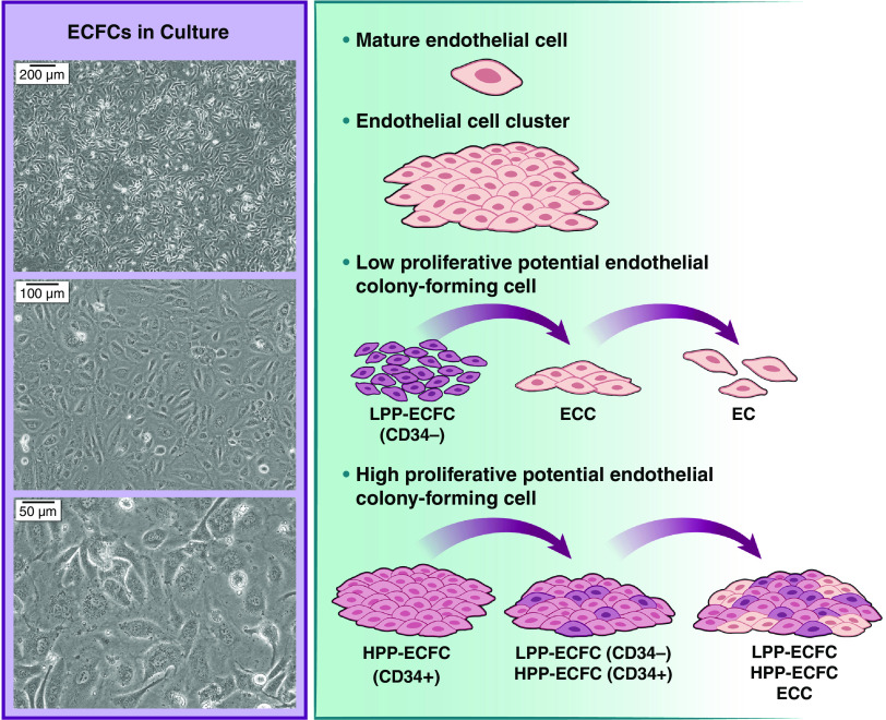
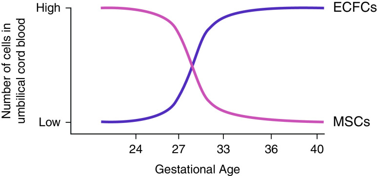
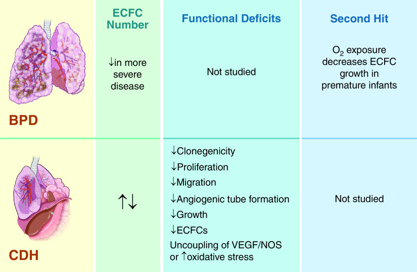
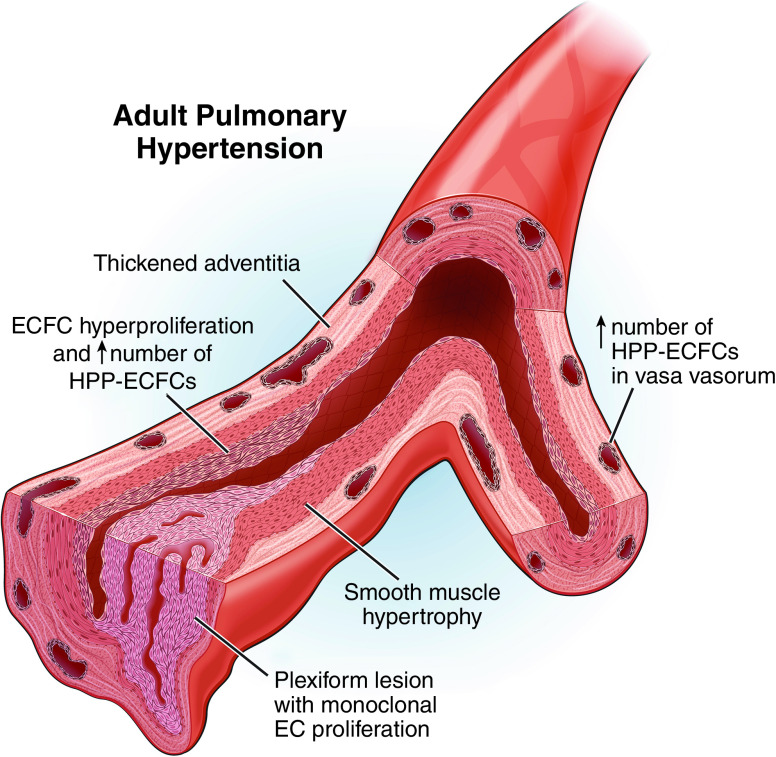
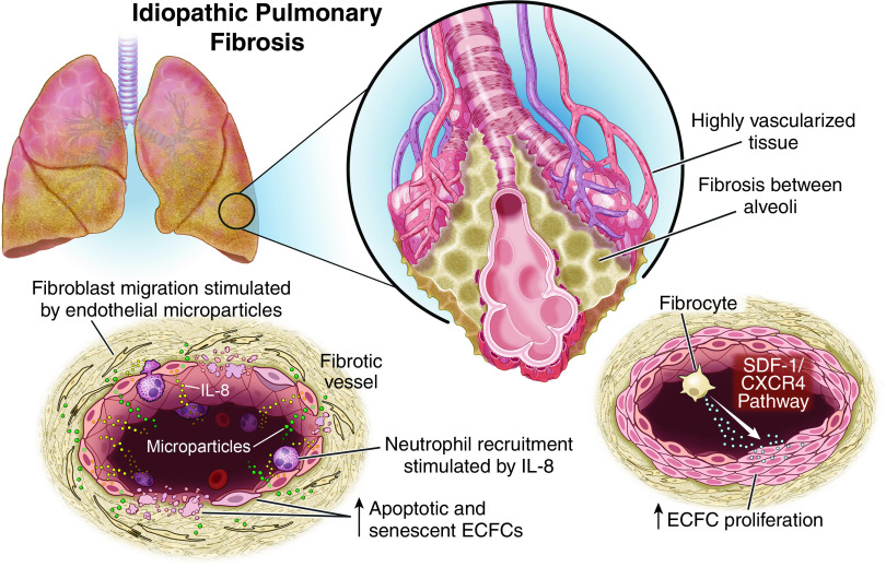
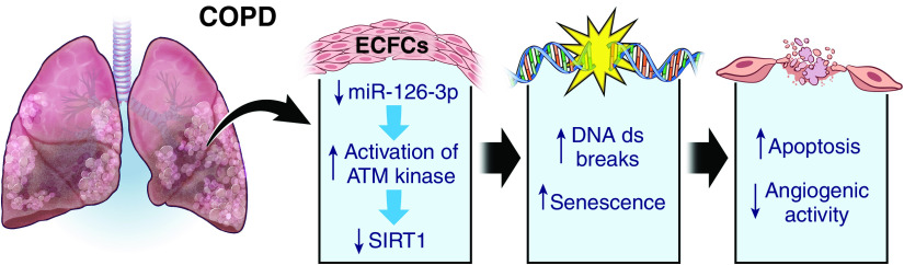
Endothelial colony-forming cells (ECFCs) are vascular resident and circulating endothelial cell subtypes with potent angiogenic capacity, a hierarchy of single-cell clonogenic potentials, and the ability to participate in de novo blood vessel formation and endothelial repair. Existing literature regarding ECFCs in neonatal and adult pulmonary diseases is confounded by the study of ambiguously defined "endothelial progenitor cells," which are often not true ECFCs. This review contrasts adult and fetal ECFCs, discusses the effect of prematurity on ECFCs, and examines their different pathological roles in neonatal and adult pulmonary diseases, such as bronchopulmonary dysplasia, congenital diaphragmatic hernia, pulmonary artery hypertension, pulmonary fibrosis, and chronic obstructive pulmonary disease. Therapeutic potential is also discussed in light of available preclinical data.

Keywords: ECFCs; bronchopulmonary dysplasia; congenital diaphragmatic hernia; pulmonary artery hypertension; pulmonary fibrosis.


Figures

Figure 1.
Figure 1.
Clonogenic hierarchy of endothelial colony-forming cells (ECFCs) in culture. Despite appearing homogenous in culture, ECFCs have well-defined hierarchies on the basis of proliferative potential. CD34+  HPP ECFCs are the only cells capable of self-renewal in stringent single-cell culture. EC = endothelial cell; ECC = endothelial cell cluster; HPP = high proliferative potential; LPP = low proliferative potential.
Figure 2.
Figure 2.
Time course for appearance of ECFCs and mesenchymal stem cells (MSCs) in umbilical cord blood. The number of ECFC colonies enumerated from 33- to 36-week gestational age infants is comparable to that from term infants and roughly three times as many as from 24- to 28-week infants. Rather, early gestational age cord blood has been shown to be enriched in MSCs, which are not as numerous later in gestation.
Figure 3.
Figure 3.
Summary of available data regarding ECFCs in neonatal lung diseases, including bronchopulmonary dysplasia (BPD) and congenital diaphragmatic hernia (CDH). Top panels: BPD lungs are characterized by decreased alveolarization and vascular growth; abnormal vascular remodeling, tone, and reactivity; and emphysema, fibrosis, and increased arteriolar medial thickness. ECFC number has been shown to be reduced in more severe cases of BPD, but functional studies have not been performed with ECFCs from infants with BPD. Bottom panels: Lungs in patients with CDH are characterized by bilateral pulmonary hypoplasia (worst on the side ipsilateral to the diaphragmatic hernia), decreased airway branching and alveolarization, and vascular bed hypoplasia with decreased arborization, altered vasoreactivity, and adventitial thickening. Two human studies that looked at ECFC numbers in infants with CDH came to opposite conclusions. A fetal lamb study supports a reduction in ECFC number in CDH and is in accordance with the human study that shows reduced angiogenic functions of these cells. NOS = nitric oxide synthase; VEGF = vascular endothelial growth factor.
Figure 4.
Figure 4.
Pathogenic contribution of ECFCs to adult pulmonary artery hypertension (PAH). ECFCs in the pulmonary vasculature of patients with PAH are hyperproliferative and comprised of more HPP-ECFCs than healthy individuals. In total, ECFCs represent between 5% and 30% of endothelial cells in PAH. It is thought that ECFCs could be responsible for the development of plexiform lesions, given the fact that these lesions are monoclonal endothelial cell proliferations. HPP-ECFC numbers are also increased in the vasa vasorum. This may contribute to the hypoxia-induced angiogenic expansion of the vasa vasorum, which is an important contributor to pulmonary vascular remodeling.
Figure 5.
Figure 5.
Pathogenic contribution of ECFCs to idiopathic pulmonary fibrosis (IPF). Pulmonary fibrosis is a heterogeneous lung disease in which fibrotic areas have decreased vascular density and adjacent, nonfibrotic areas are highly vascularized. In humans with IPF, ECFCs were increased in those with worse gas exchange, and they were more proliferative in patients undergoing disease exacerbations. It remains unclear whether this is an adaptive response or a contributor to the pathogenesis. Two different models of ECFC function in IPF have been experimentally demonstrated. First, fibrocytes increase ECFC proliferation and differentiation in vitro through the SDF-1/CXCR4 functional pathway, perhaps implying a role for fibrocytes during disease exacerbations. In the second, ECFCs are senescent or apoptotic, leading to increased IL-8 secretion, which may contribute to lung neutrophil invasion. In addition, endothelial microparticles released from IPF ECFCs stimulate fibroblast migration and are more numerous in patients with more significantly impaired gas exchange. It is unclear if ECFCs have different roles in IPF lungs depending on their location within the remodeled lung, the severity of the disease, or the presence of a disease exacerbation. CXCR4 = C-X-C chemokine receptor type 4; IL-8 = interleuken-8; SDF-1 = stromal-cell derived factor-1.
Figure 6.
Figure 6.
Pathogenic contribution of ECFCs to chronic obstructive pulmonary disease (COPD). ECFCs from smokers and patients with moderate COPD display increased DNA double-strand breaks and senescence, leading to decreased in vivo angiogenic activity and increased apoptosis. ATM = ataxia telangiectasia-mutated; ds = double strand; SIRT1 = sirtuin-1.

References

    1. Medina RJ, Barber CL, Sabatier F, Dignat-George F, Melero-Martin JM, Khosrotehrani K, et al. Endothelial progenitors: a consensus statement on nomenclature. Stem Cells Transl Med . 2017;6:1316–1320. - PMC - PubMed
    1. Dight J, Zhao J, Styke C, Khosrotehrani K, Patel J. Resident vascular endothelial progenitor definition and function: the age of reckoning. Angiogenesis . 2022;25:15–33. - PMC - PubMed
    1. Gumina DL, Su EJ. Endothelial progenitor cells of the human placenta and fetoplacental circulation: a potential link to fetal, neonatal, and long-term health. Front Pediatr . 2017;5:41. - PMC - PubMed
    1. Yoon CH, Hur J, Park KW, Kim JH, Lee CS, Oh IY, et al. Synergistic neovascularization by mixed transplantation of early endothelial progenitor cells and late outgrowth endothelial cells: the role of angiogenic cytokines and matrix metalloproteinases. Circulation . 2005;112:1618–1627. - PubMed
    1. Yoder MC, Mead LE, Prater D, Krier TR, Mroueh KN, Li F, et al. Redefining endothelial progenitor cells via clonal analysis and hematopoietic stem/progenitor cell principals. Blood . 2007;109:1801–1809. - PMC - PubMed

Publication types