. 2022 Oct 18;119(42):e2117467119.
doi: 10.1073/pnas.2117467119. Epub 2022 Oct 10.

Acoustic force spectroscopy reveals subtle differences in cellulose unbinding behavior of carbohydrate-binding modules

Affiliations

Acoustic force spectroscopy reveals subtle differences in cellulose unbinding behavior of carbohydrate-binding modules

Markus Hackl et al. Proc Natl Acad Sci U S A. .

Abstract

Protein adsorption to solid carbohydrate interfaces is critical to many biological processes, particularly in biomass deconstruction. To engineer more-efficient enzymes for biomass deconstruction into sugars, it is necessary to characterize the complex protein-carbohydrate interfacial interactions. A carbohydrate-binding module (CBM) is often associated with microbial surface-tethered cellulosomes or secreted cellulase enzymes to enhance substrate accessibility. However, it is not well known how CBMs recognize, bind, and dissociate from polysaccharides to facilitate efficient cellulolytic activity, due to the lack of mechanistic understanding and a suitable toolkit to study CBM-substrate interactions. Our work outlines a general approach to study the unbinding behavior of CBMs from polysaccharide surfaces using a highly multiplexed single-molecule force spectroscopy assay. Here, we apply acoustic force spectroscopy (AFS) to probe a Clostridium thermocellum cellulosomal scaffoldin protein (CBM3a) and measure its dissociation from nanocellulose surfaces at physiologically relevant, low force loading rates. An automated microfluidic setup and method for uniform deposition of insoluble polysaccharides on the AFS chip surfaces are demonstrated. The rupture forces of wild-type CBM3a, and its Y67A mutant, unbinding from nanocellulose surfaces suggests distinct multimodal CBM binding conformations, with structural mechanisms further explored using molecular dynamics simulations. Applying classical dynamic force spectroscopy theory, the single-molecule unbinding rate at zero force is extrapolated and found to agree with bulk equilibrium unbinding rates estimated independently using quartz crystal microbalance with dissipation monitoring. However, our results also highlight critical limitations of applying classical theory to explain the highly multivalent binding interactions for cellulose-CBM bond rupture forces exceeding 15 pN.

Keywords: acoustic force spectroscopy; biofuels; carbohydrate-binding module; nanocellulose; single-molecule force spectroscopy.


Conflict of interest statement

The authors declare no competing interest.

Figures

Fig. 1.
Fig. 1.
Schematic of a generic cellulosome and AFS experimental setup to characterize single-molecule model protein–ligand and CBM3a–polysaccharide unbinding forces (not to scale). (A) Generic bacterial cell surface-anchored cellulosome is shown adhering to a single cellulose fiber. The CBM binds to cellulose and directs the CDs to the cellulose surface. Shear forces due to the substrate or cell movement are exerted on the cellulosome scaffold. (B) Side and bottom view of CBM3a structure with key aromatic residues involved in binding to cellulose (PDB ID: 1NBC). The aromatic residues W118, H57, and Y67 form a flat binding surface complementary to the cellulose surface. (C) Schematic outlining the measurement of the unbinding force of model DIG ligand from surface-bound aDIG antibody to validate the bead preparation method as well as analysis procedure of AFS traces. (D) Schematic outlining the measurement of the unbinding force of His-GFP tagged CBM3a from an NCC surface using the AFS assay.
Fig. 2.
Fig. 2.
Multilayer deposition of NCC within the AFS chip enables the characterization of a uniform and reproducible surface. (A) Flowchart and (B) process flow diagram of the NCC deposition method, (C) Fluorescence image of the NCC-modified AFS chip. GFP-CBM3a-wt was used to bind to and visualize the deposited NCC film. The arrow indicates a representative area where a bubble was stuck at some point during the NCC deposition, resulting in less NCC bound and hence a lesser amount of GFP-CBM3a bound to that area as well. (Scale bar, 500 µm.) (D) AFM image (3 × 3 µm) of the NCC film deposited on a glass slide showing a densely covered surface. The red line represents the area used to obtain the average height profile trace shown below. Despite minor aggregation of NCC crystals during layer-by-layer deposition, height differences are less than 20 nm.
Fig. 3.
Fig. 3.
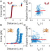
No correlation was observed between tether length and rupture force for DIG–aDIG and CBM3a–cellulose interactions. (A) FD curves of DNA anchored to the chip surface by the DIG–aDIG bond (n = 7). The extension at ∼65 pN is characteristic for a single DNA tether and indicates overstretching of DNA. (B) Example of FD curves for DNA anchored to the chip surface by the NCC–CBM bond. The red line shows the WLC fit with lp = 42 nm and S = 1,300 pN. Despite following the WLC model, the tethers show a reduction in length of 25% on average. No overstretching was observed, since CBMs detach from the surface well below 65 pN. (C) Scatterplot and linear fit (red line) of rupture force and dimensionless length during force calibration (lfc) for DIG–aDIG (n = 156). (D) Scatterplot and linear fit (red line) of rupture force and lfc for CBM3a-wt at 1 pN/s (n = 259). No significant correlation is found between the measured rupture force and lfc (SI Appendix, Tables S1 and S2). The insets in A, C and B, D symbolize the aDIG antibody and CBM3a, respectively.
Fig. 4.
Fig. 4.
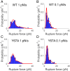
AFS reveals distinct multimodal CBM–cellulose rupture force distribution at lower loading rates. (A and B) Obtained rupture force histograms and fit to a double normal distribution for CBM3a-wt at a loading rate of 1 pN/s (n = 259) and 0.1 pN/s (n = 161), respectively. (C and D) Rupture force histograms and fit to a double normal distribution for CBM3a Y67A at a loading rate of 1 pN/s (n = 138) and 0.1 pN/s (n = 159). The fit parameters are summarized in SI Appendix, Table S3. The tail toward higher rupture forces is observed in all cases; however, the Y67A mutant displays only a single peak at both loading rates, whereas CBM3a-wt shows no clear single peak, but rather two or more rupture force peaks.
Fig. 5.
Fig. 5.
Application of the DHS model to obtained CBM3a–cellulose rupture forces highlights limitations of classical theory to study multivalent protein–polysaccharide unbinding interactions. (A and B) Force-dependent bond lifetime obtained from transforming rupture force distributions at 0.1 pN/s (o) and 1 pN/s (Δ) using Eq. 1 for WT and Y67A, respectively. The fit of Eq. 2 is shown for ν = 2/3 (red, solid line) and ν = 1/2 (green, dashed line). (CF) Rupture force distributions at 0.1 and 1 pN/s with the fit of Eq. 3 for WT and Y67A, respectively, using the parameters obtained from fitting Eq. 2 to data in A and B for ν = 2/3 (red, solid line) and ν = 1/2 (green, dashed line). While both shape factors yield a qualitatively similar fit, only ν = 2/3 results in p(f)df=1.
Fig. 6.
Fig. 6.
MD simulations provide structural insights into the multiple interactions of CBM3a residues with the cellulose surface. (A) Representative configuration of CBM3a-wt interacting with the cellulose crystal as obtained from unbiased MD simulations. The vector formed between the Cα of W118 (left green sphere) and Cα of Y67 (right green sphere) indicates the horizontal alignment of the CBM toward the reducing end of the crystal. The angle between this vector and the normal vector of the surface is defined as θ. (B) Close-up top view of the planar binding motif residues of CBM3a-wt identified to be in close contact with cellulose during MD simulations. Backbone and hydrogen atoms were omitted for clarity. Select interresidual H bonds are indicated by the dotted green line. (C) Comparison of average number of H bonds with cellulose for CBM3a-wt and Y67A mutant residues\. Reduction in S9 seems to be compensated by increased stabilization for N10, N16, and D56 in the mutant. (D) Average intramolecular H-bond formation between pairs of amino acids in both the WT and Y67A mutant. The Y67A mutation leads to the total bond rupture between the H57 and A67 pair; however, significantly greater interactions are observed between D56 and R112. Error bars in C represent the average deviation of all trajectories of two independent simulations, and error bars in D are SEM.
Fig. 7.
Fig. 7.
Summary of hypothesized origin of multimodal rupture force distribution observed for CBM3a-wt bound to cellulose. The planar binding motif may be grouped into two regions, as highlighted in blue and red oval regions shown here. For CBM3a-wt, both regions are intact and interacting with the surface in a multimodal manner. Hence, pulling on the protein yields a bimodal rupture force distribution, depending on which region ruptures from the surface first. The Y67A mutant binds to the cellulose crystal slightly tilted, reducing the interactions between the red region and the substrate. Applying a force on the mutant may therefore result in a unimodal rupture force distribution, since only the blue binding region highlighted is fully engaged with the substrate at any given point in time.

Similar articles

Cited by

References

    1. Georgelis N., Yennawar N. H., Cosgrove D. J., Structural basis for entropy-driven cellulose binding by a type-A cellulose-binding module (CBM) and bacterial expansin. Proc. Natl. Acad. Sci. U.S.A. 109, 14830–14835 (2012). - PMC - PubMed
    1. Cosgrove D. J., Loosening of plant cell walls by expansins. Nature 407, 321–326 (2000). - PubMed
    1. Qin L., et al. , Plant degradation: A nematode expansin acting on plants. Nature 427, 30 (2004). - PubMed
    1. Suits M. D. L., et al. , Conformational analysis of the Streptococcus pneumoniae hyaluronate lyase and characterization of its hyaluronan-specific carbohydrate-binding module. J. Biol. Chem. 289, 27264–27277 (2014). - PMC - PubMed
    1. Jedrzejas M. J., Pneumococcal virulence factors: Structure and function. Microbiol. Mol. Biol. Rev. 65, 187–207 (2001). - PMC - PubMed

Publication types

LinkOut - more resources