. 2022 Oct 18;119(42):e2211568119.
doi: 10.1073/pnas.2211568119. Epub 2022 Oct 10.

Origin recognition complex harbors an intrinsic nucleosome remodeling activity

Affiliations

Origin recognition complex harbors an intrinsic nucleosome remodeling activity

Sai Li et al. Proc Natl Acad Sci U S A. .

Abstract

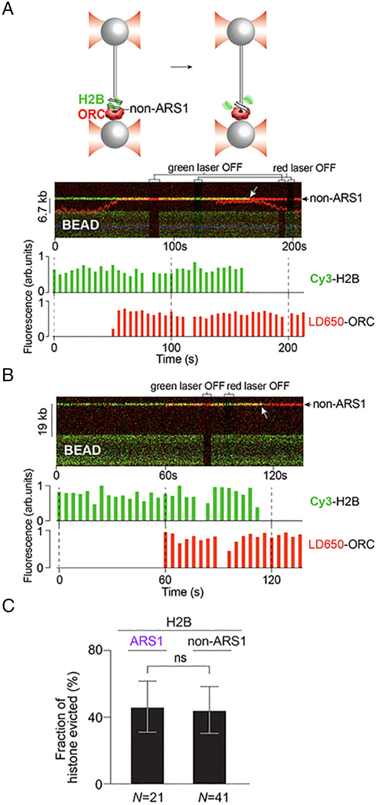
Eukaryotic DNA replication is initiated at multiple chromosomal sites known as origins of replication that are specifically recognized by the origin recognition complex (ORC) containing multiple ATPase sites. In budding yeast, ORC binds to specific DNA sequences known as autonomously replicating sequences (ARSs) that are mostly nucleosome depleted. However, nucleosomes may still inhibit the licensing of some origins by occluding ORC binding and subsequent MCM helicase loading. Using purified proteins and single-molecule visualization, we find here that the ORC can eject histones from a nucleosome in an ATP-dependent manner. The ORC selectively evicts H2A-H2B dimers but leaves the (H3-H4)2 tetramer on DNA. It also discriminates canonical H2A from the H2A.Z variant, evicting the former while retaining the latter. Finally, the bromo-adjacent homology (BAH) domain of the Orc1 subunit is essential for ORC-mediated histone eviction. These findings suggest that the ORC is a bona fide nucleosome remodeler that functions to create a local chromatin environment optimal for origin activity.

Keywords: DNA replication; nucleosome; nucleosome remodeling; origin of replication; origin recognition complex.


Conflict of interest statement

The authors declare no competing interest.

Figures

Fig. 1.
Fig. 1.
ORC-Cdc6 stably binds to nucleosomes at the ARS1 site and non-ARS1 sites. (A) Schematic of the single-molecule experimental setup and the biotinylated λ601-ARS1 DNA template for ORC-nucleosome binding assays. The flow cell contains 3 laminar-flow channels. Two streptavidin-coated beads were optically trapped in channel 1 and then moved into channel 2 that contained biotinylated λ DNA. The tethered λ DNA was then moved to channel 3, containing only buffer to confirm single-tether force-extension characteristics. Cy3-labeled nucleosomes were then assembled onto the DNA in channel 4 and LD650-labeled ORC binding assessed in channel 5. (B) Representative kymograph showing Cy3-H2B-nucleosomes (green) on λ601-ARS1 DNA between two optically trapped beads (DNA was not directly visualized in this case since there was no DNA-staining dye present). Note that nucleosomes assemble at numerous locations on DNA, not just at the 601 site. Cy3-H2B-nucleosomes (green) and LD650-ORC (red) colocalize, yielding a yellow color. At the indicated region, the green laser is turned off, revealing the presence of the red LD650-ORC signal. (C) Quantitation of stably bound ORC-Cdc6 at ARS1 versus non-ARS1 DNA in the absence or presence of nucleosomes. Significance was obtained using an unpaired two-tailed t test (ns, not significant; ***P < 0.001). Error bars represent SDs.
Fig. 2.
Fig. 2.
ORC ejects H2B from nucleosomes assembled adjacent to ARS1. (A) Schematic and a representative kymograph showing LD650-ORC (red) in the presence of Cdc6 encountering a Cy3-H2B nucleosome located at the 601-ARS1 site and evicting H2B from the nucleosome (white arrow). The corresponding fluorescence intensities of Cy3-H2B and LD650-ORC at the nucleosome position are plotted below the kymograph. (B) Two additional kymographs and the corresponding fluorescence intensity plots showing H2B eviction (loss of the Cy3 signal) from an ARS1-adjacent nucleosome after ORC-nucleosome interaction.
Fig. 3.
Fig. 3.
ORC ejects H2B from nucleosomes at non-ARS sites. (A and B) Two representative kymographs showing LD650-ORC encountering a Cy3-H2B nucleosome located at a non-ARS1 site via one-dimensional search (A) or three-dimensional search (B). White arrows indicate the H2B eviction events. The corresponding fluorescence intensity plots are shown below each kymograph. (C) Fraction of H2B evicted by ORC at ARS1 versus non-ARS1 sites. Significance was obtained using an unpaired two-tailed t test (ns, not significant). Error bars represent SDs.
Fig. 4.
Fig. 4.
ORC also ejects H2A but not H3. (A) A representative kymograph showing LD650-ORC encountering a nucleosome at a non-ARS1 site and evicting Cy3-H2A from it. The corresponding fluorescence intensity plots are shown below. (B and C) Normalized average intensity of H2A (B) and H2B (C) fluorescence signal at nucleosome sites as a function of time in the absence or presence of ORC. N indicates the number of nucleosomes analyzed for each condition. (D) Two representative kymographs of Cy3-ORC encountering a Cy5-H3 nucleosome at an ARS1 (Top) or non-ARS1 (Bottom) site. The corresponding fluorescence intensity plots are shown below each kymograph. (E) Normalized average intensity of H3 fluorescence signal at nucleosome sites as a function of time in the absence or presence of ORC. Error bars in (B), (C), and (E) represent SEMs. Significance was obtained using an unpaired two-tailed t test at the 400-s time point (ns, not significant; *P < 0.05; **P < 0.01). (F) Fraction of nucleosome-bound ORC that remained immobile after H2A/H2B eviction at ARS1 versus non-ARS1 sites. Error bars in (F) represent SDs. N indicates the number of analyzed events. Significance was obtained using an unpaired two-tailed t test (ns, not significant).
Fig. 5.
Fig. 5.
Requirement of ATP hydrolysis for the nucleosome remodeling activity of ORC. Fraction of histones (H2A, H2B, or H3) evicted versus retained upon ORC–nucleosome interaction in the presence of ATP or AMP-PNP. Error bars represent SDs.
Fig. 6.
Fig. 6.
ORC differentially remodels H2A and H2A.Z nucleosomes. (A) Schematic and a representative kymograph of LD650-ORC (red) encountering a Cy3-H2A.Z nucleosome (green). The corresponding fluorescence intensity plots are shown below the kymograph. (B) Fraction of ORC that remained stably bound to H2A versus H2A.Z nucleosomes. (C) Fraction of H2A versus H2A.Z evicted upon ORC–nucleosome interaction. N indicates the number of analyzed events. Significance was obtained using an unpaired two-tailed t test (ns, not significant; ***P < 0.001). Error bars represent SDs.
Fig. 7.
Fig. 7.
The nucleosome remodeling activity of ORC depends on the BAH domain of Orc1. (A) Schematic of the full-length Orc1 and Orc1ΔBAH. (B) Two representative kymographs of LD650-ORCΔBAH (red) encountering a Cy3-H2A or Cy3-H2B nucleosome (green). The corresponding fluorescence intensity plots are shown below each kymograph. (C) Fraction of ORC or ORCΔBAH that stably bound to Cy3-H2B or Cy3-H2A nucleosomes for at least 400 s. (D) Fraction of H2B or H2A evicted upon ORC or ORCΔBAH interaction with a nucleosome. N indicates the number of analyzed events. Significance was obtained using an unpaired two-tailed t test (ns, not significant; **P < 0.01; ***P < 0.001). Error bars represent SDs.
Fig. 8.
Fig. 8.
H2B is depleted at strong ORC binding sites genome-wide. (A) Workflow of analyzing specific histone content in the S. cerevisiae genome. The diagram shows the situation in which H2B is hemagglutinin (HA) tagged. The same procedure applies to HA-tagged H4. (B) Heatmap of the Orc1 ChIP-seq data (19) aligned at the center of Orc1 peaks and sorted based on the peak intensity (ranked 1 to 1,000 from Top to Bottom). The two panels represent biological replicates. (C) Heatmap of the H4/H2B MNase-ChIP-seq data (41) at the top 1,000 Orc1 sites. The heatmaps are aligned at the center of Orc1 peaks with the same rank order as in (B). Different panels represent replicates of MNase-seq (input) and MNase-H4/H2B ChIP-seq (IP) datasets. (D) Histograms of the normalized H2B and H4 content (estimated by IP/input) over strong (Left; ranked 1 to 400) and weak (Right; ranked 401 to 1,000) Orc1 binding sites.

Similar articles

Cited by

References

    1. Bell S. P., Labib K., Chromosome duplication in Saccharomyces cerevisiae. Genetics 203, 1027–1067 (2016). - PMC - PubMed
    1. Bleichert F., Botchan M. R., Berger J. M., Mechanisms for initiating cellular DNA replication. Science 355, eaah6317 (2017). - PubMed
    1. O’Donnell M., Langston L., Stillman B., “Principles and concepts of DNA replication in bacteria, archaea, and eukarya” in DNA Replication, Bell S. D., Mechali M., DePhamphilis M. L., Eds. (Cold Spring Harbor Laboratory Press, 2013), chap. 1, pp. 1–13. - PMC - PubMed
    1. Bell S. P., Stillman B., ATP-dependent recognition of eukaryotic origins of DNA replication by a multiprotein complex. Nature 357, 128–134 (1992). - PubMed
    1. MacAlpine D. M., Rodríguez H. K., Bell S. P., Coordination of replication and transcription along a Drosophila chromosome. Genes Dev. 18, 3094–3105 (2004). - PMC - PubMed

Publication types

LinkOut - more resources