. 2023 Dec;21(6):1163-1181.
doi: 10.1016/j.gpb.2022.09.010. Epub 2022 Oct 7.

Protein Lactylation and Metabolic Regulation of the Zoonotic Parasite Toxoplasma gondii

Affiliations

Protein Lactylation and Metabolic Regulation of the Zoonotic Parasite Toxoplasma gondii

Deqi Yin et al. Genomics Proteomics Bioinformatics. 2023 Dec.

Abstract

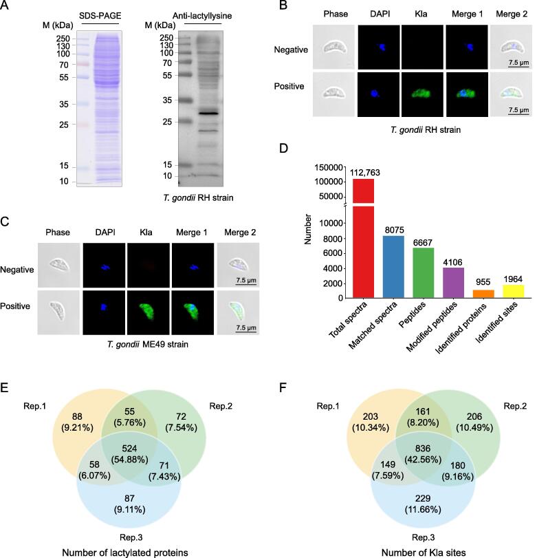
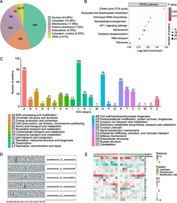
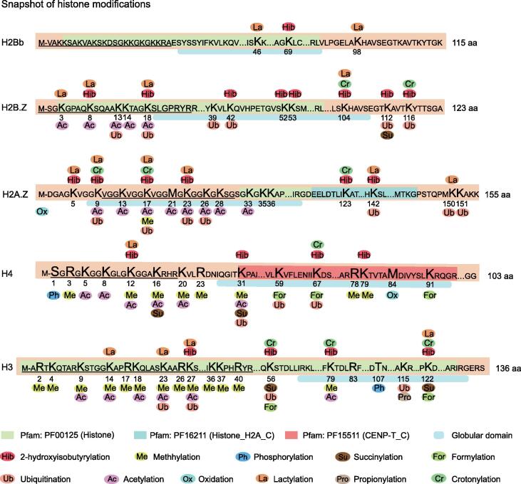
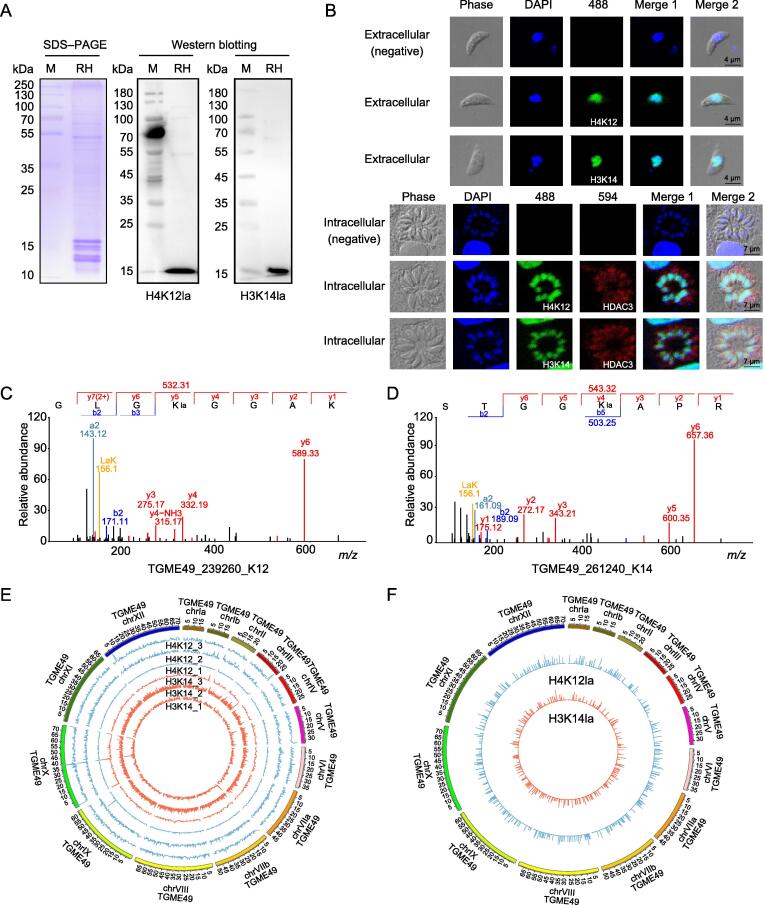
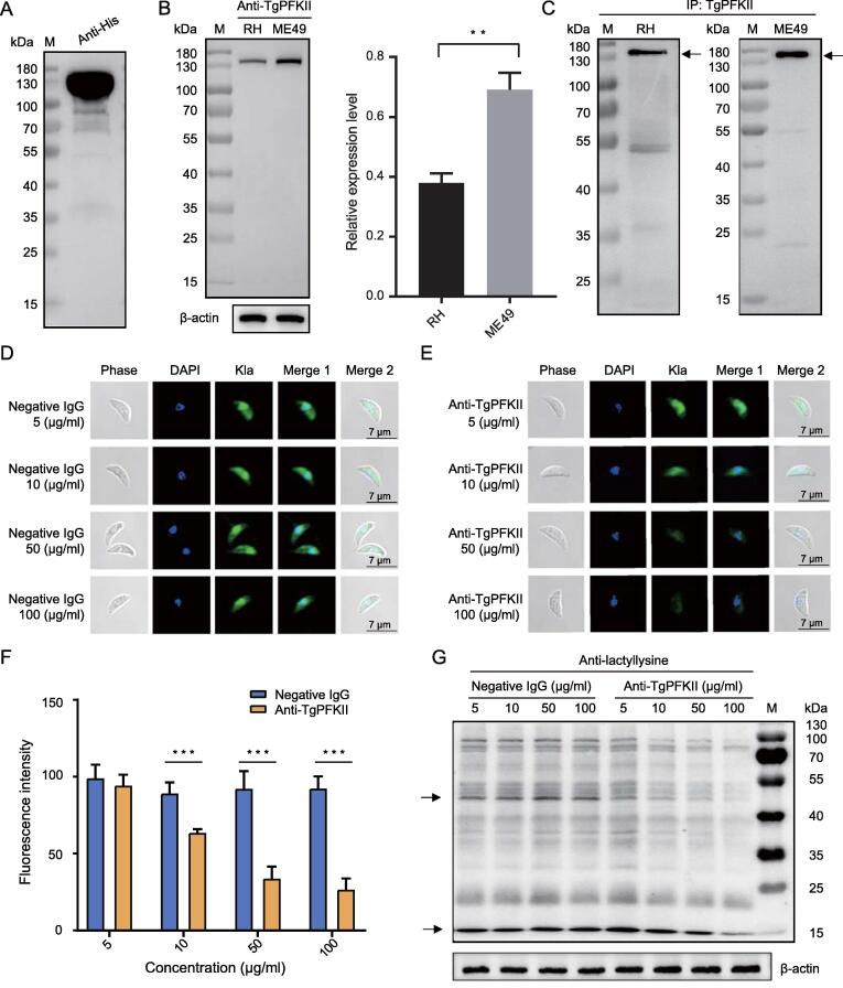
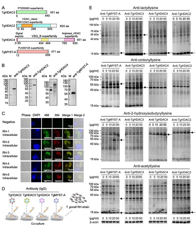
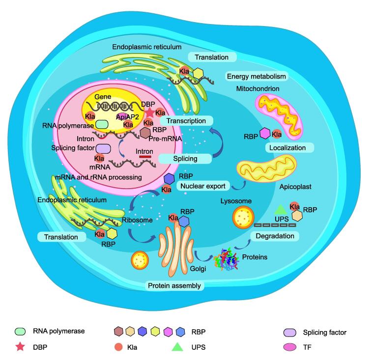
The biology of Toxoplasma gondii, the causative pathogen of one of the most widespread parasitic diseases (toxoplasmosis), remains poorly understood. Lactate, which is derived from glucose metabolism, is not only an energy source in a variety of organisms, including T. gondii, but also a regulatory molecule that participates in gene activation and protein function. Lysine lactylation (Kla) is a type of post-translational modifications (PTMs) that has been recently associated with chromatin remodeling; however, Kla of histone and non-histone proteins has not yet been studied in T. gondii. To examine the prevalence and function of lactylation in T. gondii parasites, we mapped the lactylome of proliferating tachyzoite cells and identified 1964 Kla sites on 955 proteins in the T. gondii RH strain. Lactylated proteins were distributed in multiple subcellular compartments and were closely related to a wide variety of biological processes, including mRNA splicing, glycolysis, aminoacyl-tRNA biosynthesis, RNA transport, and many signaling pathways. We also performed a chromatin immunoprecipitation sequencing (ChIP-seq) analysis using a lactylation-specific antibody and found that the histones H4K12la and H3K14la were enriched in the promoter and exon regions of T. gondii associated with microtubule-based movement and cell invasion. We further confirmed the delactylase activity of histone deacetylases TgHDAC2-4, and found that treatment with anti-histone acetyltransferase (TgMYST-A) antibodies profoundly reduced protein lactylation in T. gondii. This study offers the first dataset of the global lactylation proteome and provides a basis for further dissecting the functional biology of T. gondii.

Keywords: ChIP-seq; Lactylation; Metabolism; Protein post-translational modification; Toxoplasma gondii.


Conflict of interest statement

The authors have declared no competing interests.

Figures

Figure 1
Figure 1
Lactylation is widespread inT. gondiiillustrated by Western blotting, IFA, and LC–MS/MS A. SDS–PAGE and Western blotting analysis of tachyzoite lysate (20 μg) probed with an anti-lactyllysine antibody. B. IFA of tachyzoites of T. gondii RH with an anti-lactyllysine antibody (green). The negative control antibody is a normal rabbit IgG. Nuclei were stained with DAPI (blue). C. IFA of tachyzoites of T. gondii ME49 with an anti-lactyllysine antibody (green). The negative control antibody is a normal rabbit IgG. Nuclei were stained with DAPI (blue). D. LC–MS/MS data of lysine lactylation. E. Venn diagram showing the overlapping lactylated proteins obtained from triplicates. F. Venn diagram showing the overlapping Kla sites obtained from triplicate replications. M, marker; Kla, lysine lactylation; LC–MS/MS, liquid chromatography–tandem mass spectrometry; IFA, immunofluorescence assay; T. gondii, Toxoplasma gondii; SDS–PAGE, sodium dodecyl sulfate–polyacrylamide gel electrophoresis; IgG, immunoglobulin G; DAPI, 4′,6-diamidino-2-phenylindole.
Figure 2
Figure 2
Lactylated proteins are distributed in multiple subcellular compartments and involved in various biological functions A. Subcellular distribution of the identified lactylated proteins. B. KEGG enrichment analysis of the lactylated proteins (Fisher’s exact test, P < 0.05). C. Functional classification of lactylated proteins based on the KOG database. D. Probability sequence motifs of lactylation sites consisting of 20 residues surrounding the targeted lysine residue were produced using Motif-x. The size of each letter corresponds to the frequency of that amino acid residue occurring in that position. E. A heatmap shows the high (red) or low (green) occurrence frequency of the amino acids surrounding the lactylated lysines. The red box indicates the amino acid occurring more frequently in that position, while the green box indicates the amino acid occurring less frequently  in that position. KEGG, Kyoto Encyclopedia of Genes and Genomes; DC, difference score; KOG, EuKaryotic Orthologous Groups.
Figure 3
Figure 3
Multiple PTMs occur at the same histone sites The numbers indicate the specific locations of the modification sites on histones. Ellipses with different colors represent different types of PTMs. The different colored boxes represent the different domains. The horizontal light blue line describes the characteristics of amino acids that mediate PPI or biological processes (specific information from the Universal Protein Resource database, https://www.uniprot.org/uniport). PTM, post-translational modification; PPI, protein–protein interaction.
Figure 4
Figure 4
H4K12 and H3K14 ofT. gondiiare identified as histone lactylation sites A. Western blotting of histones H4K12la and H3K14la. B. Indirect IFA of histones H4K12la and H3K14la in the intracellular and extracellular stages of the parasite. Rabbit IgG was used as the negative control. C. and D. MS/MS spectra of histone H4K12la (C) and H3K14la (D) obtained from T. gondii. The a and b ions refer to the N-terminal part of the peptide, and the y ion refers to the C-terminal part of the peptide. E. Distribution of the binding regions of histone lactylation sites (H4K12la and H3K14la) on the chromosome shown in circos map. The outermost circle shows the chromosomes and the inner circles show the distribution trend for each sample. Data represent three independent experiments. F. Distribution of overlapping H4K12la and H3K14la peaks in the whole genome. The length of the circled bars represents the degree of enrichment and the position within the circle corresponds to the position of the peaks. ChIP-seq, chromatin immunoprecipitation sequencing.
Figure 5
Figure 5
Anti-TgPFKII antibodies reduced protein lactylation A. The purified His-tagged TgPFKII was verified by Western blotting using the anti-His antibody. B. Western blotting analysis of TgPFKII expression levels (131 kDa) in T. gondii RH and ME49 using the anti-TgPFKII antibody. β-actin was used for normalization. The data are presented as mean ± standard deviation of three independent experiments (*, P < 0.05; **, P < 0.01; Student’s t-test). C. The soluble proteins derived from T. gondii RH and ME49 were immunoprecipitated with an anti-TgPFKII antibody, and the TgPFKII protein (arrow-headed) precipitated was detected by the anti-TgPFKII antibody. D. No effect on protein lactylation (green) with different concentrations of negative IgG (mouse IgG) analyzed by IFA. E. The anti-TgPFKII antibodies inhibited protein lactylation (green) in a concentration-dependent manner. F. Immunofluorescence intensity was statistically analyzed for the groups treated with the anti-TgPFKII antibody and the negative control antibody, respectively. Statistical significance analysis was performed using Student’s t test. All data are shown as mean ± standard deviation (n > 10). ***, P < 0.001. G. Western blotting assay was used to test the effect of different concentrations of anti-TgPFKII antibodies on the Kla level of T. gondii RH. β-actin was used for normalization. IP, immunoprecipitation; PFKII, phosphofructokinase II.
Figure 6
Figure 6
Preliminary analysis of the function of TgHDAC2, TgHDAC3, TgHDAC4, and TgMYST-A in associated with protein lactylation A. Sequence characteristics of TgHDAC2, TgHDAC3, TgHDAC4, and TgMYST-A. B. Western blotting analysis of the expression of native TgHDAC2 (71 kDa), TgHDAC3 (53 kDa), TgHDAC4 (101 kDa), and TgMYST-A (56 kDa) with protein-specific antibodies. C. Indirect IFA (co-localization) of TgHDAC2, TgHDAC3, TgHDAC4, and TgMYST-A. D. Flow chart on the effect of specific antibodies on PTMs. E. Modulation of anti-TgHDAC2, anti-TgHDAC3, anti-TgHDAC4, and anti-TgMYST-A antibodies on lactylation, crotonylation, 2-hydroxyisobutyrylation, and acetylation of T. gondii. Proteins with variation in modification level was pointed with an arrow. β-actin was used for normalization. HDAC2, HADC3, and HDAC4 are histone deacetylases; MYST-A is a kind of histone lysine acetyltransferase.
Figure 7
Figure 7
A schematic diagram of the involvement of lactylation in gene regulation The schematic diagram shows that lactylated proteins, such as TFs, RNA polymerases, DBPs, and RBPs, are involved in transcriptional and translational regulatory processes. The font inside the blue box indicates that lactylated RBPs may play a wide range of roles in various post-transcriptional processes at different cellular locations. Lactylated RBPs and splice factors are major players in splicing pre-mRNA into mature mRNA in the nucleus. In addition, lactylated RBPs can assist in the translation of mRNA into proteins in the ribosome, and some RBPs may be degraded by the UPS. TF, transcription factor; DBP, DNA-binding protein; UPS, ubiquitin–proteasome system; RBP, RNA-binding protein.

References

    1. Elmore S.A., Jones J.L., Conrad P.A., Patton S., Lindsay D.S., Dubey J.P. Toxoplasma gondii: epidemiology, feline clinical aspects, and prevention. Trends Parasitol. 2010;26:190–196. - PubMed
    1. Matta S.K., Rinkenberger N., Dunay I.R., Sibley L.D. Toxoplasma gondii infection and its implications within the central nervous system. Nat Rev Microbiol. 2021;19:467–480. - PubMed
    1. Xia N., Yang J., Ye S., Zhang L., Zhou Y., Zhao J., et al. Functional analysis of Toxoplasma lactate dehydrogenases suggests critical roles of lactate fermentation for parasite growth in vivo. Cell Microbiol. 2018;20 - PubMed
    1. Bougdour A., Braun L., Cannella D., Hakimi M.A. Chromatin modifications: implications in the regulation of gene expression in Toxoplasma gondii. Cell Microbiol. 2010;12:413–423. - PubMed
    1. Seo H.H., Han H.W., Lee S.E., Hong S.H., Cho S.H., Kim S.C., et al. Modelling Toxoplasma gondii infection in human cerebral organoids. Emerg Microbes Infect. 2020;9:1943–1954. - PMC - PubMed

Publication types