. 2022 Oct 10;5(12):e202101308.
doi: 10.26508/lsa.202101308.

VEGFR3 modulates brain microvessel branching in a mouse model of 22q11.2 deletion syndrome

Affiliations

VEGFR3 modulates brain microvessel branching in a mouse model of 22q11.2 deletion syndrome

Sara Cioffi et al. Life Sci Alliance. .

Abstract

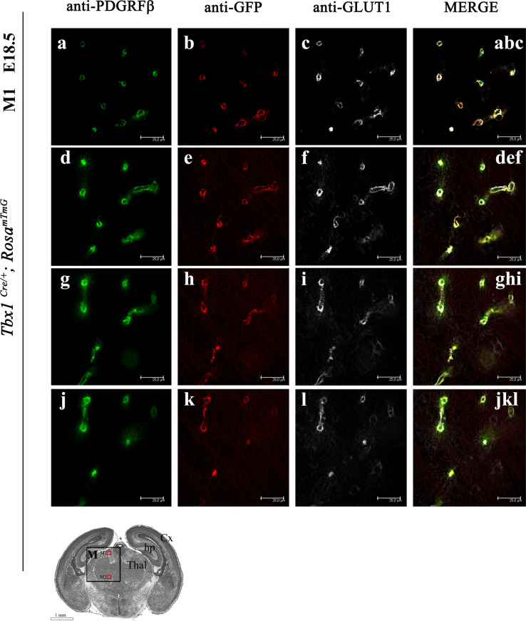
The loss of a single copy of <i>TBX1</i> accounts for most of the clinical signs and symptoms of 22q11.2 deletion syndrome, a common genetic disorder that is characterized by multiple congenital anomalies and brain-related clinical problems, some of which likely have vascular origins. <i>Tbx1</i> mutant mice have brain vascular anomalies, thus making them a useful model to gain insights into the human disease. Here, we found that the main morphogenetic function of TBX1 in the mouse brain is to suppress vessel branching morphogenesis through regulation of <i>Vegfr3</i> We demonstrate that inactivating <i>Vegfr3</i> in the <i>Tbx1</i> expression domain on a <i>Tbx1</i> mutant background enhances brain vessel branching and filopodia formation, whereas increasing <i>Vegfr3</i> expression in this domain fully rescued these phenotypes. Similar results were obtained using an in vitro model of endothelial tubulogenesis. Overall, the results of this study provide genetic evidence that <i>VEGFR3</i> is a regulator of early vessel branching and filopodia formation in the mouse brain and is a likely mediator of the brain vascular phenotype caused by <i>Tbx1</i> loss of function.


Conflict of interest statement

The authors declare that they have no conflict of interest.

Figures

Figure 1.
Figure 1.. Distribution of Tbx1-fated cells in the brain of Tbx1Cre/+; RosamTmG embryos.
(A, B, C, D, E) Representative transverse brain sections of embryos at E9.5 (A) and coronal sections at E10.5 (B) and E11.5 (C, caudal section), (D, medial section), and (E, rostral section) immunostained for GFP (green), which labels Tbx1-fated cells and for endothelial KDR (red). Nuclei (blue) were counterstained with DAPI. White arrowheads in (A) and (B) indicate the perineural vascular plexus and white arrows cells co-expressing GFP and KDR. White boxes in (C, D, E) (merge) are shown as enlarged, single-color channels to the right. (C, D, E, F) Cartoon shows the position of the coronal brain sections shown in panels (C) (caudal, C), (D) (medial, M), and (E) (rostral, R). Green-shaded arrows indicate the relative density of GFP+ cells (Tbx1-fated) along the rostral--> caudal brain axis at E11.5. Scale bars, (A) 250 μm, (B, C, D, E) 500 μm. Abbreviations: hm, head mesenchyme; hb, hindbrain; Cx, cortex; LV, lateral ventricle; BG, basal ganglia; mb, midbrain; Thal, thalamus; Hyp, hypothalamus.
Figure 2.
Figure 2.. Distribution of Tbx1-fated cells in the brain of Tbx1Cre/+; RosamTmG embryos.
(A, B, C, D, E, F, G, H, I, J, K, L) Representative coronal brain sections of embryos at E13.5 and (G, H, I, J, K, L) at E15.5 stained for GFP (green) and endothelial GLUT1 (red). Boxed areas in (A, C, E, G, I, K) are enlarged in the adjacent panels. (M) Cartoon showing the relative density of GFP+ cells (Tbx1-fated) along the rostral--> caudal and dorsal--> ventral axes. Abbreviations: R, rostral; M, medial; C, caudal; Cx, cortex; GE, ganglionic eminences; Thal, thalamus; Hyp, hypothalamus; Pn, pons; sc, superior colliculus.
Figure S1.
Figure S1.. Distribution of Tbx1-fated cells in the brain of Tbx1Cre/+; RosamTmG embryos.
(A, B, C, D, E, F) Distribution of Tbx1-fated cells in rostral (R), medial (M), and caudal (C) coronal brain sections of Tbx1Cre/+; RosamTmG embryos at E18.5 stained for GFP (green) and endothelial GLUT1 (red). Boxed areas in (A, C, E) are enlarged in the adjacent panels. (G, H, I, J, K, L) Endothelial proteins GLUT1 and VEGFR3 co-localize in brain vessels in rostral (R), medial (M), and caudal (C) coronal brain sections of Tbx1Cre/+; RosamTmG embryos at E13.5. Boxed areas in (G, I, K) are enlarged in the adjacent panels. (M, N, O, P, Q, R) Expression of GFP and VEGFR3 in brain vessels in rostral (R), medial (M), and caudal (C) coronal brain sections of Tbx1Cre/+; RosamTmG embryos at E18.5. Boxed areas in (M, O, Q) are enlarged in the adjacent panels. Abbreviations: Cx, cortex; GE, ganglionic eminences; Thal, thalamus; Hyp, hypothalamus; Pn, pons; sc, superior colliculus.
Figure S2.
Figure S2.. High-resolution images do not distinguish between endothelial-derived and pericyte-derived signals in brain vessels.
(A, B, C, D, E, F, G, H, I, J, K, L) Coronal brain sections (medial) of Tbx1Cre/+; RosamTmG embryos at E18.5 stained for PGDFRβ (labels pericytes [green] A, D, G, J), GFP (labels Tbx1-fated cells [red] B, E, H, K), and GLUT1 (labels endothelial cells [white] C, F, I, L). The figure shows optical sections at four different levels (A, D, G, J) within a single representative z-slice.
Figure 3.
Figure 3.. TBX1-GFP and VEGFR3 co-localize in brain endothelial cells.
(A, B, C, D, E, F, G, H, I, J, K, L, M, N, O, P) Representative coronal brain sections of Tbx1Cre/+; RosamTmG embryos at E13.5 and (G, H, I, J, K, L, M, N, O, P) at E15.5 immunostained for GFP (green) and endothelial VEGFR3 (red). Boxed areas in (A, C, E, G, I, K) are enlarged in the adjacent panels. (M, N, O, P) High-magnification images show that endothelial filopodia are labeled by anti-GFP (M, O) but not anti-VEGFR3 (N, P, MN, OP) antibodies. Abbreviations: R, rostral; M, medial; C, caudal; Cx, cortex; GE, ganglionic eminences; Thal, thalamus; Hyp, hypothalamus; Pn, pons; sc, superior colliculus.
Figure 4.
Figure 4.. Tbx1 and Vegfr3 interact genetically to regulate brain vessel and filopodial density.
(A, B, C, D, E) Representative coronal brain sections (medial) of E18.5 embryos immunostained for GLUT1 (green). (F, F’) Cartoon indicates the position of the counting boxes (M1, M2), shown at high magnification in panels M1 (a–e) and M2 (a’–e’), used to quantify vessel branch point (F, F’) and filopodial (f, f’) density in embryos with the indicated genotypes. ***P-value < 0.001, **P-value < 0.01, *P-value < 0.05. Error bars ± SD. Abbreviations: Cx, cortex; hp, hippocampus; Thal, thalamus.
Figure S3.
Figure S3.. Tbx1 and Vegfr3 interact genetically to regulate brain vessel and filopodial density.
(A, B, C, D, E, F, G, H, I, J) Representative coronal brain sections in rostral (A, B, C, D, E) and caudal (F, G, H, I, J) positions of E18.5 embryos stained for endothelial GLUT1. The cartoon of a rostral brain section (R) indicates the position of the counting boxes R1 and R2, which are shown at high magnification in panels R1, a–e and R2, a’–e’. The cartoon showing a caudal brain section (C) indicates the position of the counting boxes C1 and C2, shown at high magnification in panels C1, a–e and C2, a’–e’. Abbreviations: Cx, cortex; GE, ganglionic eminences; sc, superior colliculus; Pn, pons.
Figure 5.
Figure 5.. Vegfr3 overexpression in HUVECs represses formation of endothelial microtubules.
(A) Microtubule hypobranching after transfection with pcDNA3-Vegfr3. (B) Hyperbranching after RNA interference, siRNA VEGFR3. (C) pcDNA3-Vegfr3 rescued hyperbranching caused by TBX1 knockdown. ***P-value < 0.001, **P-value < 0.01, *P-value < 0.05. Error bars ± SD. Scale bar, 200 μm. Abbreviations: EV, empty vector.
Figure S4.
Figure S4.. Vegfr2 expression in cultured brain endothelial cells is Tbx1 dosage– and Vegfr3 dosage–dependent.
(A) Vegfr2 expression in bEND5 cells over-expressing Tbx1. (B) Vegfr2 expression in HUVECs and bEND5 cells over-expressing Vegfr3. (C) Enhanced VEGFR2 expression in HUVECs after TBX1 knockdown returned to control levels of expression in cells co-transfected with TBX1 siRNA and pcDNA3-Vegfr3.
Figure S5.
Figure S5.. Increased Vegfr3 expression induced by Tbx1Cre activation of TgVegfr3 is revealed by qRT–PCR but not by immunofluorescence.
(A, B, C, D, E, F, G, H, I, J, K, L) Representative coronal brain sections of Tbx1Cre/+ (A, B, C, D, E, F) and TgVegfr3;Tbx1Cre/+ (G, H, I, J, K, L) embryos (E18.5) stained for VEGFR3 (red), VEGFR2 (green), and GFP (Tbx1 cell fate, white). The cartoon shows the position of the coronal brain sections (medial, M) shown in panels M1 and M2. (M) Brain expression of Vegfr3 in Tbx1Cre/+ and TgVegfr3;Tbx1Cre/+ embryos at E18.5.
Figure 6.
Figure 6.. Vegfr3 overexpression rescues brain vessel abnormalities in Tbx1 mutants.
(A, B, C, D, E) Representative coronal brain sections (medial) of embryos at E18.5 immunostained for GLUT1 (green). (F) The cartoon indicates the position of the counting boxes (M1, M2), shown at high magnification in panels M1 (a–e) and M2 (a’–e’), that were used to quantify vessel branch point (F) and filopodial (f) density in embryos with the indicated genotypes. ***P-value < 0.001, **P-value < 0.01, *P-value < 0.05. Error bars ± SD. Abbreviations: Cx, cortex; hp, hippocampus; Thal, thalamus.
Figure S6.
Figure S6.. Vegfr3 overexpression rescues brain vessel abnormalities in Tbx1 mutants.
(A, B, C, D, E, F, G, H, I, J) Representative coronal brain sections, in rostral (A, B, C, D, E) and caudal (F, G, H, I, J) positions, of embryos at E18.5 immunostained for endothelial GLUT1. The cartoon showing a rostral brain section (R) indicates the position of the counting boxes R1 and R2, which are shown at high magnification in panels R1, a–e and R2, a’–e’. The cartoon showing a caudal brain section (C) indicates the position of the counting boxes C1 and C2, shown at high magnification in panels C1, a–e and C2, a’–e’. Abbreviations: Cx, cortex; GE, ganglionic eminences; sc, superior colliculus; Pn, pons.

References

    1. Benedito R, Rocha SF, Woeste M, Zamykal M, Radtke F, Casanovas O, Duarte A, Pytowski B, Adams RH (2012) Notch-dependent VEGFR3 upregulation allows angiogenesis without VEGF-VEGFR2 signalling. Nature 484: 110–114. 10.1038/nature10908 - DOI - PubMed
    1. Chen L, Mupo A, Huynh T, Cioffi S, Woods M, Jin C, McKeehan W, Thompson-Snipes L, Baldini A, Illingworth E (2010) Tbx1 regulates Vegfr3 and is required for lymphatic vessel development. J Cell Biol 189: 417–424. 10.1083/jcb.200912037 - DOI - PMC - PubMed
    1. Cioffi S, Martucciello S, Fulcoli FG, Bilio M, Ferrentino R, Nusco E, Illingworth E (2014) Tbx1 regulates brain vascularization. Hum Mol Genet 23: 78–89. 10.1093/hmg/ddt400 - DOI - PubMed
    1. Dumont DJ, Jussila L, Taipale J, Lymboussaki A, Mustonen T, Pajusola K, Breitman M, Alitalo K (1998) Cardiovascular failure in mouse embryos deficient in VEGF receptor-3. Science 282: 946–949. 10.1126/science.282.5390.946 - DOI - PubMed
    1. Hogan KA, Ambler CA, Chapman DL, Bautch VL (2004) The neural tube patterns vessels developmentally using the VEGF signaling pathway. Development 131: 1503–1513. 10.1242/dev.01039 - DOI - PubMed

Publication types

Substances

LinkOut - more resources