Review
. 2022 Nov;43(11):886-900.
doi: 10.1016/j.it.2022.09.002. Epub 2022 Oct 7.

Linking nutrient sensing, mitochondrial function, and PRR immune cell signaling in liver disease

Affiliations
Review

Linking nutrient sensing, mitochondrial function, and PRR immune cell signaling in liver disease

Claudia Kemper et al. Trends Immunol. 2022 Nov.

Abstract

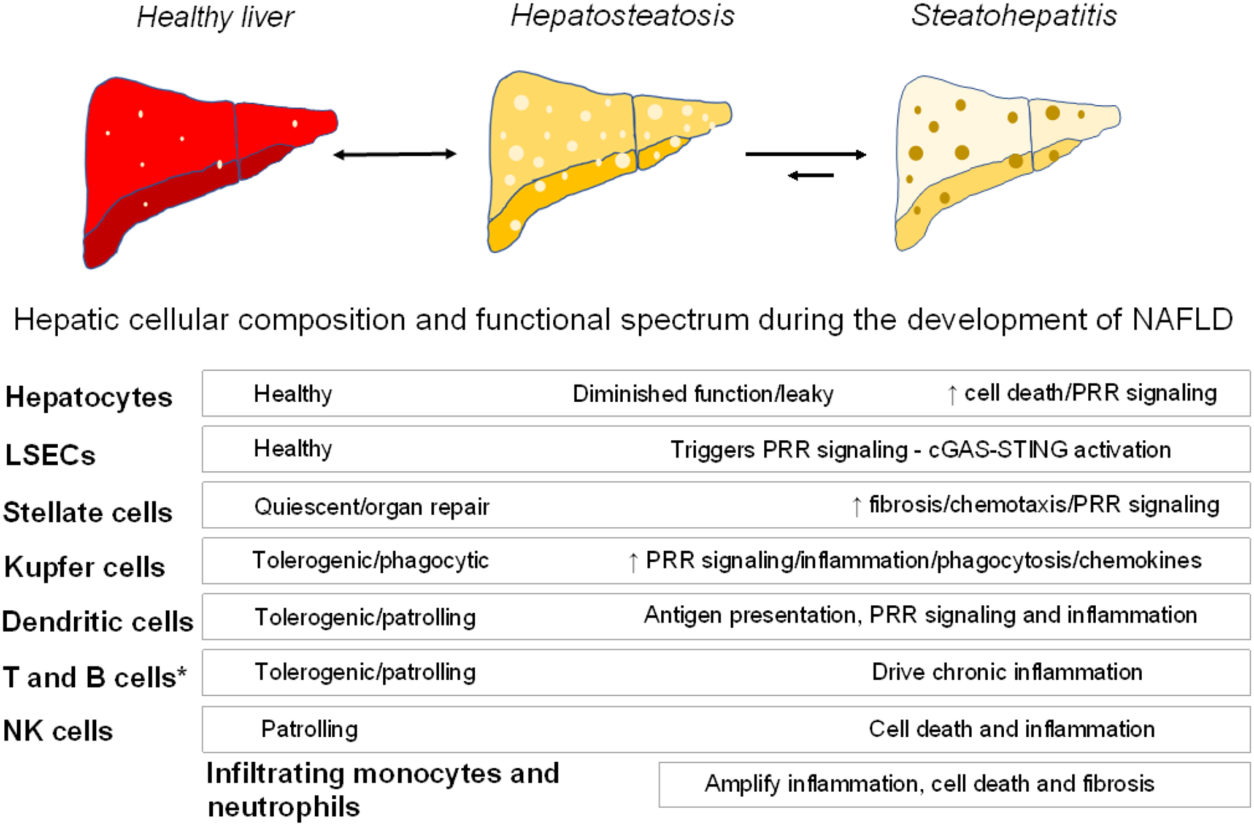
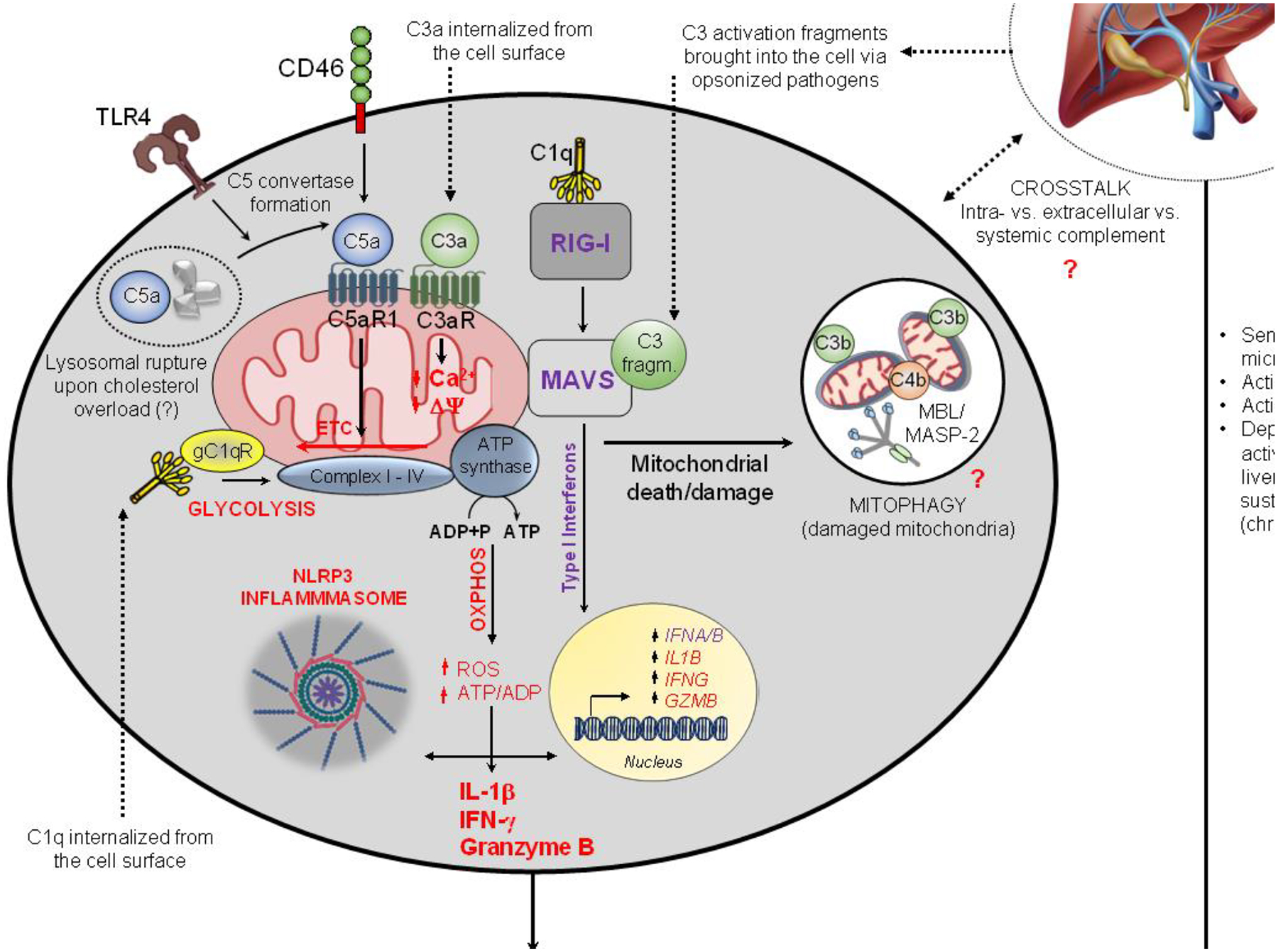
Caloric overconsumption in vertebrates promotes adipose and liver fat accumulation while perturbing the gut microbiome. This triad triggers pattern recognition receptor (PRR)-mediated immune cell signaling and sterile inflammation. Moreover, immune system activation perpetuates metabolic consequences, including the progression of nonalcoholic fatty liver disease (NAFLD) to nonalcoholic hepatic steatohepatitis (NASH). Recent findings show that sensing of nutrient overabundance disrupts the activity and homeostasis of the central cellular energy-generating organelle, the mitochondrion. In parallel, whether caloric excess-initiated PRR signaling and mitochondrial perturbations are coordinated to amplify this inflammatory process in NASH progression remains in question. We hypothesize that altered mitochondrial function, classic PRR signaling, and complement activation in response to nutrient overload together play an integrated role across the immune cell landscape, leading to liver inflammation and NASH progression.

Keywords: cardiometabolic disease; complement; liver; mitochondria; pattern recognition receptors.


Conflict of interest statement

Declaration of interests No interests are declared.

Figures

Figure 1.
Figure 1.. Liver and immune cell contributions to NAFLD
Shown is a schematic of various cell populations in the mammalian liver with their functional contributions to maintaining a healthy organ and their presumed role in NAFLD transitions from hepatosteatosis to steatohepatitis [7]. Hepatosteatosis has a high potential to revert to the healthy state with reversal of risk factors (indicated by a double-sided arrow). The capacity to reverse NASH is much more limited (indicated by a shorter arrow) [96]. The bulk of the pathophysiologic effects of the various cell types signal the transition from hepatosteatosis to steatohepatitis. LSECs, liver sinusoidal epithelial cells. *, includes αβ CD4+ and CD8+ T cells (resident memory T cell subset), γδ T cells, invariant natural killer T cells (iNKT cells), mucosal-associated invariant T cells (MAIT), innate lymphoid cells (ILCs), and B cell plasma cells [84].
Figure 2.
Figure 2.. Model of mammalian cell mitochondrial-PRR crosstalk perturbations in NAFLD/NASH
In mammals, high fat diet-induced mitochondrial perturbations include changes in morphology (fission/fusion), function (metabolism), fidelity and integrity (mtDNA release). These in turn, engage cell-intrinsic PRRs, which cumulatively induce pro-inflammatory cytokine production, release of DAMPs, and cell death (left side – see text for details) [7]. Nutrient overload–mediated mitochondrial dysfunction is most prominent in hepatocytes but is operational to various degrees across all liver cells [7]. The exogenous effects of cell-intrinsic PRR-driven cell activation/death (for example, in hepatocytes) on other cells is depicted on the right side in red underneath each cell type in a simplified fashion (e.g., IL-1β production and NLRP3 release by hepatocytes undergoing pyroptosis induce TGF-β in stellate cells, which supports fibrosis). Mitochondrial dysfunction and specific PRR engagement in specialized liver cells are listed within each cell type and known cross-communications between cells are indicated by red-arrows. The consequences of these inflammatory pathways emanating from different cell types comprise various components of inflammatory signaling during the progression to NASH [84]. *Of the broad range of resident and incoming immune cells with mitochondrial and PRR activation hallmarks, monocytes and macrophages are the best understood. cGAS-STING, cyclic GMP-AMP synthase and stimulator of interferon genes pathway; ECV, extracellular vesicle; FFAs, free fatty acids; Inflammas., inflammasomes; LDs, lipid droplets; LSEC; liver sinusoidal endothelial cell; Mito-Dysf., mitochondrial dysfunction; mtDNA, mitochondrial DNA; Oxphos; oxidative phosphorylation; ROS, reactive oxygen species; TLR9, toll-like receptor 9. Some icons/cell types were imported from BioRender (https://biorender.com/).
Figure 3.
Figure 3.. Proposed model of human complosome-PRR signaling axes in mitochondrial function
In this model, circulating and intracellular complement contribute to the amplification of inflammation seen in NAFLD [95]. Liver-derived complement, activated by microbial components released from compromised intestinal barriers, can aggravate human disease by sustaining vasculature and hepatocyte activation [97]. Intracellularly active complement can engage the mitochondrial C5aR1 which increases ROS production, reverses direction of the ETC and induces NLRP3 inflammasome assembly and IL-1β production [65,71]. TLR4 fosters cell-autonomous C5a generation via increased C3/C5 convertase formation and human monocytes can also activate intracellular C4 [65]. The RIG-I/MAVS axis is activated by cell-intrinsic C1q and C3 activation fragments introduced via opsonized pathogens and triggers type I IFN production [68]. Internalized C1q and C3a impact on ROS production and mitochondrial membrane potential, respectively, which compromise mitochondrial integrity and overall sustain cell-intrinsic DAMP presence [70,72]. Complosome-mediated mitophagy may mitigate this process by removal of injured mitochondria. The shown complosome perturbations can contribute to pathological cell changes in infection and sterile inflammation [65,98]; whether they also operate in liver cells and/or contribute to NASH remains to be assessed. ADP, adenosine diphosphate; ATP, adenosine triphosphate; CTL, cytotoxic T lymphocyte; gC1qR, globular-head C1q receptor; MAVS, mitochondrial anti-viral signaling protein; NAFLD, non-alcoholic fatty liver disease; NASH, non-alcoholic steatohepatitis; NLRP3, NOD-, LRR- and pyrin domain-containing protein 3 inflammasome; Oxphos, oxidative phosphorylation; RIG-I, retinoic-inducible gene I; ROS, reactive oxygen species; Th1, T helper type 1 cell; TLR4, toll-like receptor 4. Some icons were imported from BioRender (https://biorender.com/).
Figure 4.
Figure 4.. Potential novel therapeutic intervention points for mitochondrial-PRR axis perturbations in NAFLD/NASH
Current NAFLD/NASH interventions aim to reduce cellular lipid accumulation by means of diet, bariatric surgery, and pharmacological inhibition of adipogenesis [99]. Increasing cellular NAD+ concentrations via oral provision of the NAD precursor nicotinamide riboside is currently being explored in the clinic (Clinicaltrials.gov NCT02835664). Targeting PRRs associated with mitochondrial dysfunction, induction of cytokines/chemokines, and/or cell death may be an additional means to reduce or revert the general pro-inflammatory state of liver cells in NAFLD/NASH (see text). However, such approaches need to be carefully explored because the nutrient-mitochondria-PRRs pathways are likely operative across cell subpopulations and may serve distinct functions within specialized cells, including the control of pathways contributing to cell survival, homeostasis, and regeneration. cGAS-STING, cyclic GMP-AMP synthase, and stimulator of interferon genes pathway; LDs, lipid droplets; NAD+, nicotinamide adenine dinucleotide; NLRP3, NOD-, LRR- and pyrin domain-containing protein 3 inflammasome; PPAR γ, peroxisome proliferator activated receptor γ; RIG-I, retinoic-inducible gene I; ROS, reactive oxygen species; TLR, toll-like receptor. Some icons/cell types were imported from BioRender (https://biorender.com/).

References

    1. O’Neill S and O’Driscoll L (2015) Metabolic syndrome: a closer look at the growing epidemic and its associated pathologies. Obes Rev 16, 1–12. 10.1111/obr.12229 - DOI - PubMed
    1. Anstee QM et al. (2019) From NASH to HCC: current concepts and future challenges. Nat Rev Gastroenterol Hepatol 16, 411–428. 10.1038/s41575-019-0145-7 - DOI - PubMed
    1. Estes C et al. (2018) Modeling the epidemic of nonalcoholic fatty liver disease demonstrates an exponential increase in burden of disease. Hepatology 67, 123–133. 10.1002/hep.29466 - DOI - PMC - PubMed
    1. Baker PR 2nd and Friedman JE (2018) Mitochondrial role in the neonatal predisposition to developing nonalcoholic fatty liver disease. The Journal of clinical investigation 128, 3692–3703. 10.1172/JCI120846 - DOI - PMC - PubMed
    1. Kramer DA et al. (2018) Fasting and refeeding induces changes in the mouse hepatic lipid droplet proteome. Journal of proteomics 181, 213–224. 10.1016/j.jprot.2018.04.024 - DOI - PubMed

Publication types

LinkOut - more resources