. 2022 Oct 10;13(1):5859.
doi: 10.1038/s41467-022-33605-1.

Scleral PERK and ATF6 as targets of myopic axial elongation of mouse eyes

Affiliations

Scleral PERK and ATF6 as targets of myopic axial elongation of mouse eyes

Shin-Ichi Ikeda et al. Nat Commun. .

Abstract

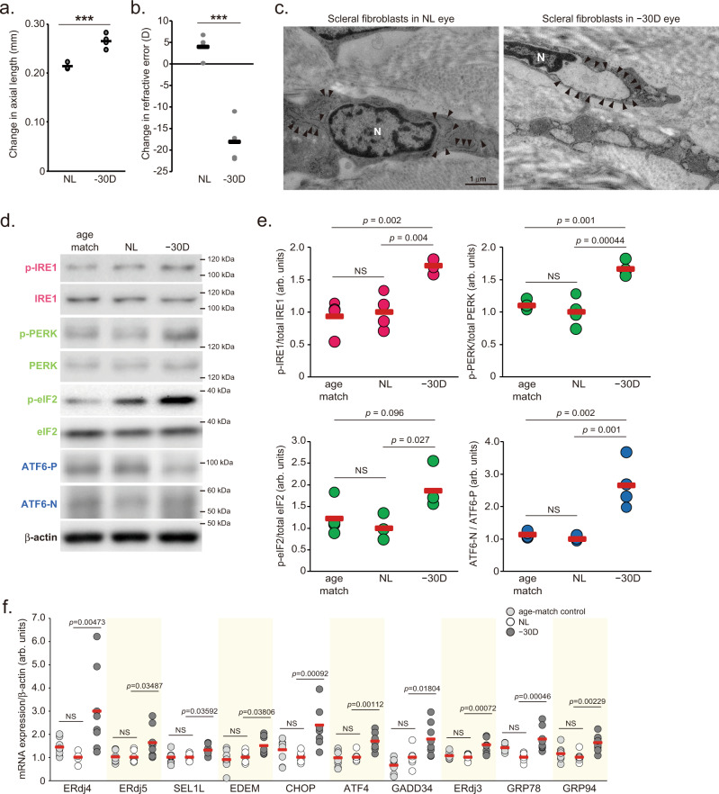
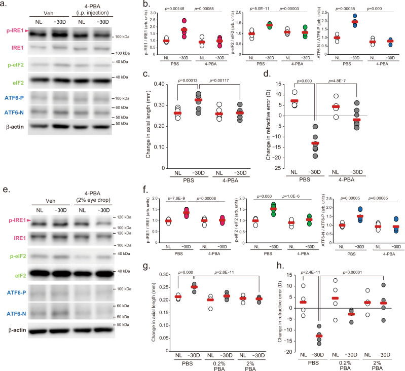
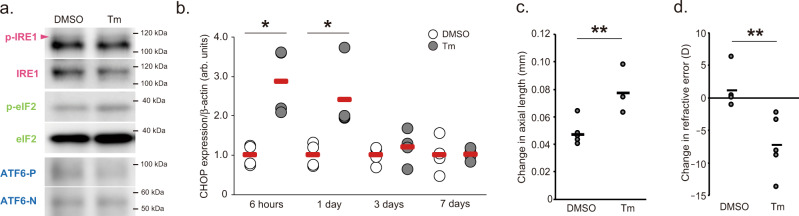
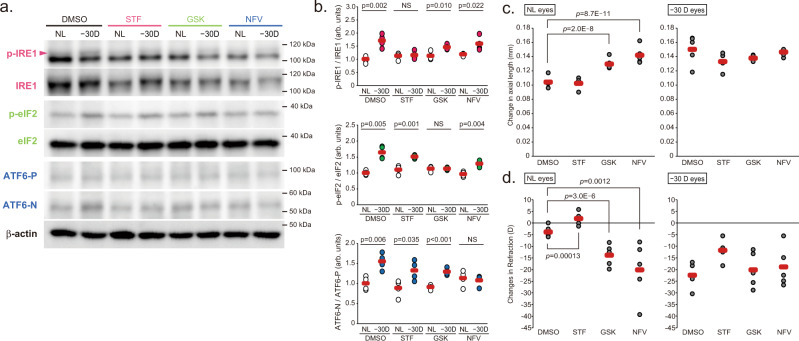
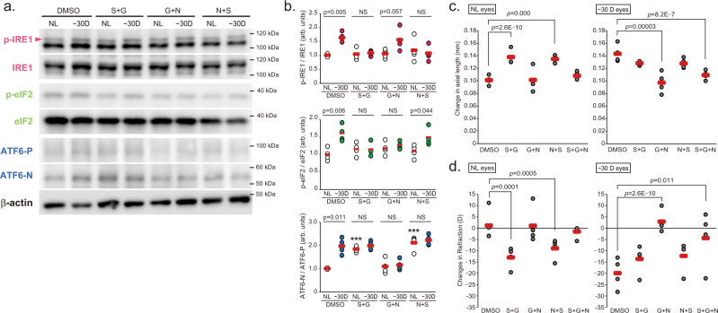
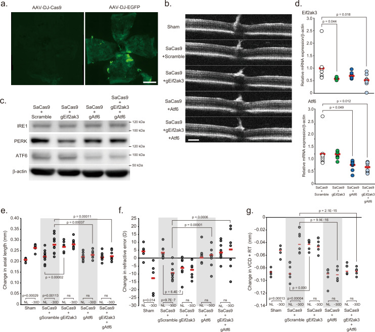
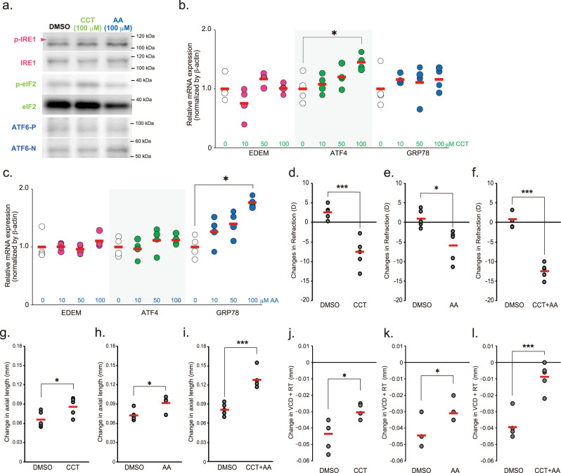
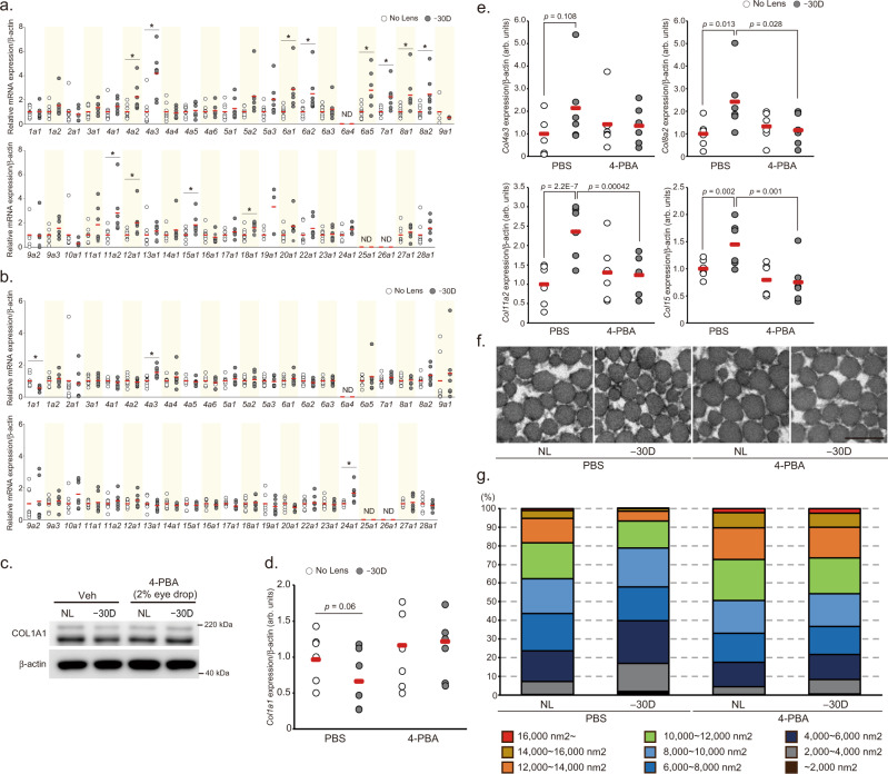
Axial length is the primary determinant of eye size, and it is elongated in myopia. However, the underlying mechanism of the onset and progression of axial elongation remain unclear. Here, we show that endoplasmic reticulum (ER) stress in sclera is an essential regulator of axial elongation in myopia development through activation of both PERK and ATF6 axis followed by scleral collagen remodeling. Mice with lens-induced myopia (LIM) showed ER stress in sclera. Pharmacological interventions for ER stress could induce or inhibit myopia progression. LIM activated all IRE1, PERK and ATF6 axis, and pharmacological inhibition of both PERK and ATF6 suppressed myopia progression, which was confirmed by knocking down above two genes via CRISPR/Cas9 system. LIM dramatically changed the expression of scleral collagen genes responsible for ER stress. Furthermore, collagen fiber thinning and expression of dysregulated collagens in LIM were ameliorated by 4-PBA administration. We demonstrate that scleral ER stress and PERK/ATF6 pathway controls axial elongation during the myopia development in vivo model and 4-PBA eye drop is promising drug for myopia suppression/treatment.


Conflict of interest statement

We report grants from Tsubota Laboratory and Novartis during the course of the study. In addition, K.T., T.K., S.I., and X.J. have been internationally applying for a patent WO2018/164113, which has already been registered in Japan. The sponsors had no control over the experiments, interpretation, writing, or publication of this work. K.T. reports serving as chief executive officer for Tsubota Laboratory, a company producing myopia-related devices. H.T., T.K., and K.T. own unlisted stocks of Tsubota Laboratory. The other authors declare no competing interests.

Figures

Fig. 1
Fig. 1. Lens-induced myopia induces axial elongation and endoplasmic reticulum stress in the sclera.
a Changes in axial length during 3-week lens-induced myopia (LIM) in C57BL6J mice (n = 4). NL: No Lens control, −30D: minus 30 D lens-wearing eye. ***p = 0.00062; Student’s two-tailed t-test. b Changes in refraction during 3-week LIM in C57BL6J mice (n = 4); ***p = 0.00029; Student’s two-tailed t-test. c Transmission electron microscopy images of no lens (NL)-wearing (left panels) and minus 30 D lens-wearing (right panels) sclerae. Representative images of three biologically independent samples; N indicates nucleus and allow heads indicate rough endoplasmic reticulum. Scale bars: 1.0 µm. d Immunoblots showing ER sensor protein activation evaluated by phosphorylation levels of IRE1, PERK, eIF2α (downstream of PERK), and the ATF6 precursor (ATF6-P) and cleaved form of ATF6 (ATF6-N). Representative blots from four independent experiments are shown. Age-match: age-matched non-treated control, NL: No Lens-wearing eye, −30D: minus 30 D lens-wearing eye. e Densitometric quantification of blots in Fig. 1d using ImageJ. NL group was assigned a value of 1.0. Each group were n = 4 (each sample pooled 3 sclerae). The p values were determined by one-way ANOVA with Tukey HSD (two-tailed). NS: Not Significant. f UPR target gene expression level determined by quantitative PCR in age-matched control (right gray), No-Lens control (white) and LIM (gray) sclerae for 3-week LIM (n = 7 or 8). NL group was assigned a value of 1.0. The p values were determined by one-way ANOVA with Tukey HSD (two-tailed). Source data are provided as a Source Data file.
Fig. 2
Fig. 2. Attenuation of scleral ER stress is sufficient to suppress myopia development.
a Intraperitoneal (i.p.) injection of 4-phenylbutyric acid (4-PBA; 200 mg/kg/day) suppresses Lens-induced myopia (LIM)-induced activation in UPR branch determined by Western blotting in sclerae. NL: No Lens control, −30D: minus 30 D lens-wearing eye. b Densitometric quantification of blots in Fig. 2a using ImageJ. PBS-NL group was assigned a value of 1.0. Each group were n = 4 (each sample pooled 3 sclerae). The p values were determined by two-tailed Generalized Estimating Equations. c LIM-induced axial elongation is inhibited by 4-PBA administration for 3 weeks (n = 6 per group); **p < 0.01 (p = 0.001), ***p < 0.001; two-tailed Generalized Estimating Equations. d LIM-induced myopic shift in refraction is inhibited by 4-PBA administration for 3 weeks (n = 6 per group); ***p < 0.001; two-tailed Generalized Estimating Equations. e Eye drop administration of 4-PBA (2% solution in PBS) during LIM period (to both eyes) suppresses LIM-induced activation in UPR branch determined by Western blotting in sclerae. f Densitometric quantification of blots in Fig. 2e using ImageJ.PBS-NL group was assigned a value of 1.0. Each group were n = 4 (each sample pooled 3 sclerae). ***p < 0.001; two-tailed Generalized Estimating Equations. g 4-PBA instillation suppresses axial elongation in a dose-dependent manner (n = 4 per group); ***p < 0.001; two-tailed Generalized Estimating Equations. h 4-PBA instillation suppresses myopic shift in refraction in a dose-dependent manner (n = 4 per group); ***p < 0.001; two-tailed Generalized estimating equations. Source data are provided as a Source Data file.
Fig. 3
Fig. 3. Induction of scleral ER stress results in the development of myopia.
a A single tunicamycin (Tm; 50 µg/mL) instillation induces acute IRE1, eIF2, and ATF6 activation (1 hour after instillation) determined by Western blotting (n = 4, each sample pooled 3 sclerae). b A single Tm (50 µg/mL) instillation induces acute ER stress, evaluated as CHOP mRNA expression by qPCR (n = 4 per group). DMSO group in each time point were assigned a value of 1.0. *p < 0.05 compared to each control; Student’s two-tailed t-test (Exact p values are as follows: 6 h, p = 0.017; 1 day, p = 0.024). c A single Tm (50 µg/mL) instillation induces axial elongation (n = 5 per group); **p < 0.01; Student’s two-tailed t-test (p = 0.003). d A single Tm instillation induces myopic shift in refraction (n = 5 per group); **p < 0.01; Student’s two-tailed t-test (p = 0.009). Source data are provided as a Source Data file.
Fig. 4
Fig. 4. Inhibition of a single UPR pathway with specific inhibitors disrupts the refractive condition of the mouse eyes.
a Effect of instillation of IRE1 inhibitor STF080310 (STF; 100 μM), PERK inhibitor GSK2656157 (GSK; 100 μM), and ATF6 inhibitor nelfinavir (NFV; 100 μM) on activation of the UPR pathways by LIM in sclerae. NL: No Lens control, −30D: minus 30 D lens-wearing eye. b Densitometric quantification of blots in Fig. 3a using ImageJ. DMSO-NL group was assigned a value of 1.0. Each group were n = 4 (each sample pooled 3 sclerae). The p values were determined by two-tailed Generalized Estimating Equations for four groups comparison (DMSO vs STF vs GSK vs NFV in each eyes) and Student’s two-tailed t-test for no lens (NL) and −30D lens (−30D) eyes comparison in each treatments. NS: Not Significant. c Effects of STF, GSK, and NFV instillation on axial elongation in NL and −30 D eyes (n = 5 per group); ***p < 0.001 compared to the DMSO group; two-tailed Generalized Estimating Equations. d Effects of STF, GSK, and NFV instillation on refraction in NL and −30 D eyes (n = 5 per group); **p < 0.01 (p = 0.001), ***p < 0.001compared to the DMSO group; two-tailed Generalized Estimating Equations. Source data are provided as a Source Data file.
Fig. 5
Fig. 5. Inhibition of both PERK and ATF6 pathways suppresses myopia progression caused by negative lens wearing.
a Effects of combined STF, GSK, and NFV instillation (S + G: STF and GSK, G + N: GSK and NFV, N + S: NFV and STF; 100 μM each) on LIM-induced activation of IRE1, eIF2, and ATF6 in sclerae. NL: No Lens control, −30D: minus 30 D lens-wearing eye. b Densitometric quantification of blots in Fig. 5a using ImageJ. DMSO-NL group was assigned a value of 1.0. Each group were n = 4 (each sample pooled 3 sclerae). The p values were determined by two-tailed Generalized Estimating Equations for four groups comparison (DMSO vs S + G vs G + N vs N + S in each eye) and Student’s two-tailed t-test for NL and −30D eyes comparison in each treatment. NS: Not Significant. c Effects of combined STF, GSK, and NFV instillation on axial elongation in NL and −30 D eyes (n = 4 per group); ***p < 0.001 compared to the DMSO group; two-tailed Generalized Estimating Equations. d Effects of combined STF, GSK, and NFV instillation on refraction in NL and −30 D eyes (n = 4 per group); *p < 0.05 (p = 0.011), **p < 0.01 (p = 0.005), ***p < 0.001 compared to the DMSO group; two-tailed Generalized Estimating Equations. Source data are provided as a Source Data file.
Fig. 6
Fig. 6. Effect of AAV-based gene knockdown of PERK and ATF6 in sclera on myopia development.
a Distribution of EGFP expression in the scleral whole-mount 28 days after sub-tenon’s capsule injection of adeno-associated virus serotype DJ (AAV-DJ)-EGFP or AAV-DJ-SaCas9 (as non-fluorescence control); scale bar, 1 mm. b Representative Optical Coherence Tomography images 28 days after of each AAV injection; scale bar, 100 µm. c Expression level of IRE1, PERK, and ATF6 protein 28 days after of AAV-SaCas9 with AAV-gRNA against Eif2ak3 (Gene symbol of PERK, gEig2ak3) and/or Atf6 (gAtf6) injections. d Scleral Eif2ak3 and Atf6 knockdown by AAV-DJ-CRISPR/Cas9 injection into sub-tenon’ capsule. After 28 days injection, gene expression in the sclera determined by qPCR (n = 9, respectively). SaCas9 + gScramble group was assigned a value of 1.0. The p values were determined by one-way ANOVA with Tukey HSD. e Effects of scleral Eif2ak3 and/or Atf6 knockdown on axial elongation (sham: n = 5, gScramble: n = 11, gEif2ak3: n = 9, gAtf6: n = 8, gEif2ak3 + gAtf6: n = 8). The p values were determined by two-tailed Generalized Estimating Equations for five groups comparison (sham vs gScramble vs gEif2ak3 vs gAtf6 vs gEif2ak3 + gAtf6) and Student’s two-tailed t-test for two groups comparison (NL vs −30D). NL: No Lens control, −30D: minus 30 D lens-wearing eye. f Effects of scleral Eif2ak3 and/or Atf6 knockdown on refraction (sham: n = 5, gScramble: n = 9, gEif2ak3: n = 9, gAtf6: n = 8, gEif2ak3 + gAtf6: n = 9). The p values were determined by two-tailed Generalized Estimating Equations for five groups comparison and Student’s two-tailed t-test for two groups comparison. g Effects of scleral Eif2ak3 and/or Atf6 knockdown on VCD + RT (sham: n = 5, gScramble: n = 7, gEif2ak3: n = 7, gAtf6: n = 7, gEif2ak3 + gAtf6: n = 7). The p values were determined by two-tailed Generalized Estimating Equations for five groups comparison and Student’s two-tailed t-test for two groups comparison. Source data are provided as a Source Data file.
Fig. 7
Fig. 7. Effect of PERK and ATF6 agonizts instillation on myopia development.
a Effect of PERK agonist CCT020312 (CCT) and ATF6 agonist AA147 (AA) instillation on IRE1, eIF2 and ATF6 activity in sclera. One hour after eye drop, the sclerae were harvested and subjected to Western blotting. Each compound specifically activated each pathway. b GRP78, ATF4, and EDEM gene expression induced by CCT. Each zero group was assigned a value of 1.0. *p = 0.029, compared to 0 (as a control) group; one-way ANOVA with Tukey HSD (n = 4, respectively). c GRP78, ATF4, and EDEM gene expression induced by AA. Each zero group was assigned a value of 1.0. *p = 0.013, compared to 0 (as a control) group; one-way ANOVA with Tukey HSD (n = 4, respectively). d Effects of 1-week instillation of CCT (100 μM) instillation on refraction; ***p = 0.00055; Student’s two-tailed t-test. e Effects of 1-week instillation of AA (100 μM) instillation on refraction; *p = 0.016; Student’s two-tailed t-test. f Effects of 1-week instillation of CCT plus AA (100 μM, respectively) instillation on refraction; ***p = 0.00001; Student’s two-tailed t-test. g Effects of 1-week instillation of CCT instillation on axial length; *p = 0.041; Student’s two-tailed t-test. h Effects of 1-week instillation of AA instillation on axial length; *p = 0.015; Student’s two-tailed t-test. i Effects of 1-week instillation of CCT plus AA instillation on axial length; ***p = 0.00039; Student’s two-tailed t-test. j Effects of 1-week instillation of CCT instillation on VCD + RT. *p = 0.036; Student’s two-tailed t-test. k Effects of 1-week instillation of AA instillation on VCD + RT. *p = 0.024; Student’s two-tailed t-test. l Effects of 1-week instillation of CCT plus AA instillation on axial length; ***p = 0.0004; Student’s two-tailed t-test. Source data are provided as a Source Data file.
Fig. 8
Fig. 8. Scleral ER stress is associated with collagen remodeling.
a Effect of 1 week LIM on the expression of 43 collagen genes in C57BL6J mouse sclerae (NL: n = 8, −30D: n = 7). No Lens group was assigned a value of 1.0. *p < 0.05 compared to each No Lens group; Student’s two-tailed t-test (Exact p values are as follows. 4a2: p = 0.023, 4a3: p = 0.001, 6a1: p = 0.008, 6a2: p = 0.027, 6a5: p = 0.024, 7a1: p = 0.009, 8a1: p = 0.041, 8a2: p = 0.024, 11a2: p = 0.022, 12a1: p = 0.048, 15a1: p = 0.036, 18a1: p = 0.038). b Effect of 3 weeks LIM on the expression of 43 collagen genes in C57BL6J mouse sclerae (NL: n = 8, −30D: n = 7). No Lens group was assigned a value of 1.0. *p < 0.05 compared to each No Lens group; Student’s two-tailed t-test (Exact p values are as follows. 1a1: p = 0.012, 4a3: p = 0.009, 24a1: p = 0.017). c 4-PBA instillation cancels LIM-induced decrease in COL1A1 protein expression in sclerae (n = 4, each sample pooled 3 sclerae). NL: No Lens control, −30D: minus 30 D lens-wearing eye. d The effect of LIM and 4-PBA instillation on Col1a1 mRNA expression in sclerae (n = 6, respectively). The p values were determined by Generalized Estimating Equations. e 4-PBA instillation cancels LIM-induced increase in Col4a3, Col8a2, Col11a2, and Col15a1 mRNA expression evaluated by qPCR in sclerae (n = 6 per group); The p values were determined by two-tailed Generalized Estimating Equations. f Transmission electron microscopy images of sclerae collagen fiber in No lens control (NL) and −30 D lens worn (−30D) sclera of PBS or 4-PBA instilled C57BL6J mouse. Representative images of three biologically independent samples; scale bar, 200 nm. g 100% stacked bar charts of scleral collagen fiber area (n = 3 biologically independent samples). Five images from one sclera. Fiber area was measured using ImageJ software. Source data are provided as a Source Data file.

References

    1. Dolgin E. The myopia boom. Nature. 2015;519:276–278. doi: 10.1038/519276a. - DOI - PubMed
    1. Flitcroft DI. The complex interactions of retinal, optical and environmental factors in myopia aetiology. Prog. Retin Eye Res. 2012;31:622–660. doi: 10.1016/j.preteyeres.2012.06.004. - DOI - PubMed
    1. Tideman JW, et al. Association of axial length with risk of uncorrectable visual impairment for europeans with myopia. JAMA Ophthalmol. 2016;134:1355–1363. doi: 10.1001/jamaophthalmol.2016.4009. - DOI - PubMed
    1. Leo SW. Current approaches to myopia control. Curr. Opin. Ophthalmol. 2017;28:267–275. doi: 10.1097/ICU.0000000000000367. - DOI - PubMed
    1. Gentle A, Liu Y, Martin JE, Conti GL, McBrien NA. Collagen gene expression and the altered accumulation of scleral collagen during the development of high myopia. J. Biol. Chem. 2003;278:16587–16594. doi: 10.1074/jbc.M300970200. - DOI - PubMed

Publication types