Review
. 2023 Feb;22(2):127-144.
doi: 10.1038/s41573-022-00571-8. Epub 2022 Oct 10.

Drugging p53 in cancer: one protein, many targets

Affiliations
Review

Drugging p53 in cancer: one protein, many targets

Ori Hassin et al. Nat Rev Drug Discov. 2023 Feb.

Abstract

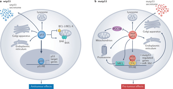
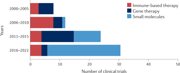
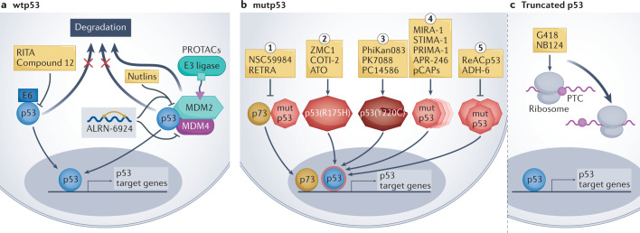
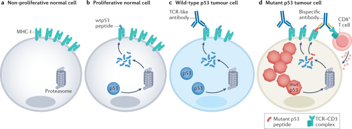
Mutations in the TP53 tumour suppressor gene are very frequent in cancer, and attempts to restore the functionality of p53 in tumours as a therapeutic strategy began decades ago. However, very few of these drug development programmes have reached late-stage clinical trials, and no p53-based therapeutics have been approved in the USA or Europe so far. This is probably because, as a nuclear transcription factor, p53 does not possess typical drug target features and has therefore long been considered undruggable. Nevertheless, several promising approaches towards p53-based therapy have emerged in recent years, including improved versions of earlier strategies and novel approaches to make undruggable targets druggable. Small molecules that can either protect p53 from its negative regulators or restore the functionality of mutant p53 proteins are gaining interest, and drugs tailored to specific types of p53 mutants are emerging. In parallel, there is renewed interest in gene therapy strategies and p53-based immunotherapy approaches. However, major concerns still remain to be addressed. This Review re-evaluates the efforts made towards targeting p53-dysfunctional cancers, and discusses the challenges encountered during clinical development.


Conflict of interest statement

M.O. consults for Quintrigen.

Figures

Fig. 1
Fig. 1. Tumour-suppressive effects of wild-type p53 and oncogenic effects of mutant p53.
a | Wild-type p53 (wtp53) acts predominantly as a transcription factor to restrict cancer cell proliferation and survival. Many non-transcriptional effects are also involved. Wtp53 can promote mitochondrially induced apoptosis by interacting with multi-domain members of the apoptosis regulator BCL-2 family (such as BCL-2 and BCL-X), unleashing the activity of pro-apoptotic BH3-only proteins such as BAK and BAX. Wtp53 can also increase Ca2+ load upon stress, resulting in induction of apoptosis. Additionally, wtp53 is an important regulator of autophagy. b | Mutant p53 (mutp53) can modulate transcription by piggybacking on other transcription factors (TFX) and can also promote cancer by non-transcriptional mechanisms. Mutp53 can inhibit peroxisome proliferator-activated receptor-γ coactivator 1α (PGC1α), a master regulator of mitochondrial biogenesis and oxidative phosphorylation. Moreover, in contrast to wtp53, mutp53 inhibits endoplasmic reticulum (ER) stress-induced apoptosis by modulating the unfolded protein response (UPR), which increases cell survival upon ER stress. Mutp53 also induces the transcription of many genes that encode proteasome subunits. This transcriptional activation, mediated by the binding of mutp53 to the transcription factor nuclear factor erythroid 2-related factor 2 (NRF2), results in elevated proteasome activity and enhanced degradation of tumour suppressor proteins. Both wtp53 and mutp53 impinge on multiple cellular organelles and compartments, and can also elicit non cell-autonomous effects through secretion of various molecules, both soluble and those carried by exosome or extracellular vesicle. The presence of wtp53 inhibits the expression of Golgi scaffolding proteins, thus inhibiting secretory vesicle biogenesis in the Golgi apparatus, while mutp53 can modulate Golgi apparatus function by inducing microRNA miR-30d expression through interacting with the hypoxia responsive factor HIF1α. Consequently, miR-30d modulates tubulo-vesiculation of the Golgi apparatus, promoting vesicular trafficking and secretion. RE, response element.
Fig. 2
Fig. 2. Numbers of p53-targeted clinical trials by year and treatment category.
Clinical trials with p53-targeted therapies initiated after 1 January 2000 were stratified by year blocks and category. Gene therapy clinical trials were popular before the year 2000 (12 clinical trials initiated), but their number declined sharply soon thereafter, owing to mounting concerns about the safety of this strategy. These numbers increased again in the course of 2011–2015, mostly reflecting the clinical trials driven by Shenzhen SiBiono GeneTech and several trials of SGT-53 (SynerGene Therapeutics). Immune-based clinical trials targeting p53 were rather uncommon before 2000 (two clinical trials). With the introduction of new anticancer vaccination approaches, the number of relevant p53-based clinical trials has increased. Presently, most p53-based immunotherapy clinical trials use a combination of immune checkpoint inhibition and a p53-activating agent (either gene therapy or small molecules). It is expected that, owing to the growing interest in bispecific antibodies and T cell receptor (TCR)-like antibodies (see Fig. 4), p53-centric clinical trials that use these strategies will become more popular in the coming years. Visibly, the biggest increase in p53-based clinical trials in the past decade involved small-molecule drugs. This may be attributed, at least in part, to the emergence of new screening methods and improved compound libraries, along with better understanding of the deregulation of p53 in cancer.
Fig. 3
Fig. 3. p53-based small molecules for cancer therapy.
a | In p53-wild-type (wtp53) tumours, small-molecule drug development is mainly focused on inhibiting or degrading negative regulators of p53, including MDM2, MDM4 and human papillomavirus (HPV) E6. Such inhibition increases p53 protein abundance and wtp53 activity in the cancer cells, promoting the expression of p53 target genes. ALRN-6924 is a stapled peptide that blocks both MDM2–p53 and MDM4–p53 interactions. p53-activating proteolysis targeting chimeras (PROTACs) work by targeting MDM2 for ubiquitylation by particular E3 ligases, resulting in MDM2 degradation. b | In tumours expressing missense mutant p53 (mutp53) proteins, drug development aims to restore wtp53 conformation and/or inhibit gain-of-function activities of mutp53 such as inhibition of p73. From left to right: (1) small molecules such as RETRA (reactivation of transcriptional reporter activity) or NSC59984, which inhibit the interaction of mutp53 with p73, unleash p73 and enable it to enter the nucleus and transactivate target genes that partly overlap with p53 target genes. (2) Some small molecules (arsenic trioxide (ATO), ZMC1) act predominantly on structural p53 mutants (such as p53(R175H)) to restore wtp53 conformation and induce p53 target gene expression. (3) The p53(Y220C) mutant has an accessible crevice near the site of mutation, which can be targeted by small molecules to thermodynamically stabilize the mutant protein and shift it towards a wild-type-like state. (4) Many compounds (such as APR-246 and pCAPs) target a broad spectrum of p53 mutants to restore a wtp53-like structure, thus enabling p53 target gene activation (for a more detailed mechanistic description see ref.). (5) Some small molecules — such as ReACp53 or ADH-6 — act by inhibiting mutp53 aggregation, restoring wtp53-like structure and activating p53 target genes. c | Other small molecules inhibit the recognition of premature termination codons (PTCs), enabling translational readthrough and synthesis of full-length p53 protein in cells that harbour truncating TP53 mutations. The overarching goal of all these drugs is to restore the expression of wtp53 target genes as a means to induce cancer cell death or replicative senescence, thereby curtailing tumour growth. RITA, reactivation of p53 and induction of tumour cell apoptosis.
Fig. 4
Fig. 4. New antibody-based strategies to target p53 in cancer cells.
a | In the non-proliferative compartment of normal tissues, TP53 is usually silent, and therefore cells do not produce p53 protein and do not present p53-derived peptides on their surface major histocompatibility complex (MHC) class I. b | Proliferating normal cells produce low amounts of p53 protein and present small amounts of p53-derived peptides on their MHC class I (MHC-I). c | In cancer cells expressing wild-type p53 (wtp53), oncogenic stress upregulates p53 mRNA synthesis and translation, causing a more pronounced presentation of p53-derived peptides. These differences in quantity and quality of presented peptides provide the rationale for developing antibody-based strategies to target selectively the cancer cells. T cell receptor (TCR)-like antibodies recognize p53-derived epitopes displayed on MHC-I on the cell surface, triggering an immune response. d | In cancer cells that harbour TP53 missense mutations, p53 protein levels are even more elevated; consequently, such cells may present increased amounts of peptides derived from non-mutated regions of the protein (wtp53 peptides in blue), along with neopeptides comprising the mutated sequence (mutp53 peptide, in red). Bispecific antibodies can be engineered to recognize a neoantigen derived from mutp53 and the TCR–CD3 complex on CD8+ T cells, which results in selective cytotoxicity against mutp53-expressing cancer cells.
Fig. 5
Fig. 5. p53 can influence immunotherapy by modulating the tumour immune microenvironment.
In p53-wild-type (wtp53) tumours, the tumour immune microenvironment has a predominantly anti-tumoural effect. This can be attributed to several mechanisms. For example, wtp53 can upregulate the expression of UL16-binding protein 1 (ULBP1) and ULBP2 and microRNA miR-34a. ULBP1 and ULBP2 are activating ligands for natural killer (NK) cells, which enhances their cytotoxic activity. miR-34 blocks the translation of PDL1 mRNA and drives its degradation; consequently, PDL1 protein levels are downregulated in cancer cells that retain wtp53, sensitizing these tumour cells to CD8+ T cell-mediated killing. Moreover, wtp53 can promote the secretion of anti-tumoural cytokines such as tumour necrosis factor (TNF), which further enhances the protective effects of the tumour immune microenvironment. In TP53-mutated cells, ULBP1 and ULBP2 are downregulated and PDL1 is upregulated, causing resistance to killing by NK and CD8+ T cells, respectively. Furthermore, loss of p53 function can drive secretion of WNT ligands, which bind to their cognate receptors on macrophages, causing them to secrete IL-1β, which attracts pro-tumoural cells such as neutrophils. Mutp53 proteins promote the secretion of exosomes that can deliver microRNAs such as miR-1246, which convert anti-tumoural M1 macrophages to pro-tumoural M2 macrophages. Such pro-tumoural macrophages secrete IL-1β, which attracts pro-tumoural regulatory T (Treg) cells that dampen the anti-tumoural response and protect the tumour from immune elimination. MHC-I, major histocompatibility complex class I; RE, response element; TCR, T cell receptor.
Fig. 6
Fig. 6. p53-based genetic therapies.
Wild-type p53 (wtp53)-encoding DNA and RNA can be introduced into cancer cells by several approaches, including recombinant viruses and nanoparticles. This drives p53 expression and transcription of wtp53 target genes, resulting in anticancer effects. In TP53-mutated tumours, delivery of CRISPR–Cas9 together with suitable guide RNA (gRNA) might potentially enable base editing, restoring wild-type TP53 sequence.

References

    1. Barbosa A, et al. Gene panel tumor testing in ovarian cancer patients significantly increases the yield of clinically actionable germline variants beyond BRCA1/BRCA2. Cancers. 2020;12:2834. doi: 10.3390/cancers12102834. - DOI - PMC - PubMed
    1. Hoe KK, Verma CS, Lane DP. Drugging the p53 pathway: understanding the route to clinical efficacy. Nat. Rev. Drug Discov. 2014;13:217–236. doi: 10.1038/nrd4236. - DOI - PubMed
    1. Sabapathy K, Lane DP. Therapeutic targeting of p53: all mutants are equal, but some mutants are more equal than others. Nat. Rev. Clin. Oncol. 2018;15:13–30. doi: 10.1038/nrclinonc.2017.151. - DOI - PubMed
    1. Bykov VJN, Eriksson SE, Bianchi J, Wiman KG. Targeting mutant p53 for efficient cancer therapy. Nat. Rev. Cancer. 2017;182:89–102. - PubMed
    1. Cheok CF, Lane DP. Exploiting the p53 pathway for therapy. Cold Spring Harb. Perspect. Med. 2017;7:a026310. doi: 10.1101/cshperspect.a026310. - DOI - PMC - PubMed

Publication types

Substances