. 2023 Apr;44(4):822-831.
doi: 10.1038/s41401-022-01001-6. Epub 2022 Oct 10.

Hyperthermia promotes degradation of the acute promyelocytic leukemia driver oncoprotein ZBTB16/RARα

Affiliations

Hyperthermia promotes degradation of the acute promyelocytic leukemia driver oncoprotein ZBTB16/RARα

Qian-Qian Wang et al. Acta Pharmacol Sin. 2023 Apr.

Abstract

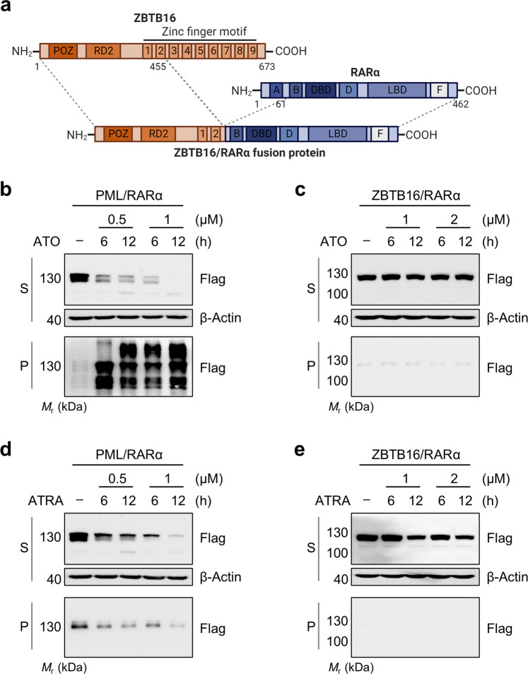
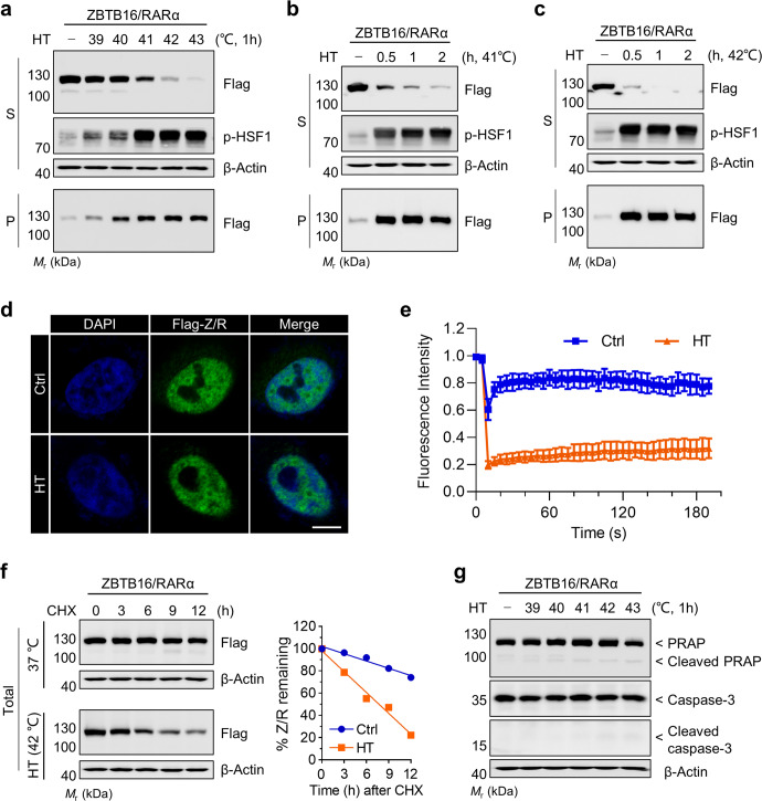
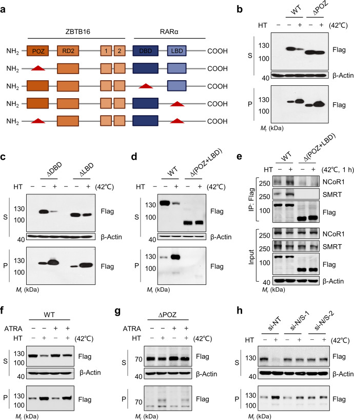
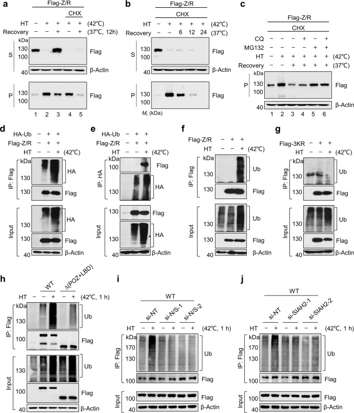
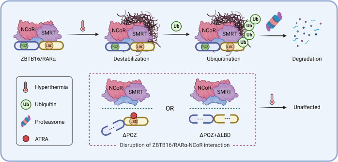
The acute promyelocytic leukemia (APL) driver ZBTB16/RARα is generated by the t(11;17) (q23;q21) chromosomal translocation, which is resistant to combined treatment of all-trans retinoic acid (ATRA) and arsenic trioxide (ATO) or conventional chemotherapy, resulting in extremely low survival rates. In the current study, we investigated the effects of hyperthermia on the oncogenic fusion ZBTB16/RARα protein to explore a potential therapeutic approach for this variant APL. We showed that Z/R fusion protein expressed in HeLa cells was resistant to ATO, ATRA, and conventional chemotherapeutic agents. However, mild hyperthermia (42 °C) rapidly destabilized the ZBTB16/RARα fusion protein expressed in HeLa, 293T, and OCI-AML3 cells, followed by robust ubiquitination and proteasomal degradation. In contrast, hyperthermia did not affect the normal (i.e., unfused) ZBTB16 and RARα proteins, suggesting a specific thermal sensitivity of the ZBTB16/RARα fusion protein. Importantly, we found that the destabilization of ZBTB16/RARα was the initial step for oncogenic fusion protein degradation by hyperthermia, which could be blocked by deletion of nuclear receptor corepressor (NCoR) binding sites or knockdown of NCoRs. Furthermore, SIAH2 was identified as the E3 ligase participating in hyperthermia-induced ubiquitination of ZBTB16/RARα. In short, these results demonstrate that hyperthermia could effectively destabilize and subsequently degrade the ZBTB16/RARα fusion protein in an NCoR-dependent manner, suggesting a thermal-based therapeutic strategy that may improve the outcome in refractory ZBTB16/RARα-driven APL patients in the clinic.

Keywords: ZBTB16/RARα fusion protein; acute promyelocytic leukemia; arsenicals; hyperthermia; nuclear receptor corepressors; proteolysis.


Conflict of interest statement

The authors declare no competing interests.

Figures

Fig. 1
Fig. 1. Effect of ATO and ATRA on the stability and degradation of the PML/RARα and ZBTB16/RARα fusion proteins.
Schematic representation of the ZBTB16, RARα, and oncogenic ZBTB16/RARα (Z/R) fusion proteins with functional domains and breakpoints (a). PML/RARα- and Z/R-transfected HeLa cells were exposed to ATO (0.5, 1 µM) for 6 and 12 h (b and c) or ATRA (1, 2 µM) for 6 and 12 h (d and e). Protein expression in soluble (S) and pellet (P) fractions of RIPA lysate was analyzed by Western blotting with anti-Flag and anti-β-actin monoclonal antibodies.
Fig. 2
Fig. 2. Effect of hyperthermia on destabilization of the ZBTB16/RARα fusion protein.
Exogenous Z/R fusion protein-expressing HeLa cells were exposed to hyperthermia in a temperature-dependent (a) and time-dependent manner (b and c). The changes in the Z/R fusion protein in the supernatant (S) and pellet fractions (P) of RIPA lysate were determined by Western blotting. Phosphorylated HSF1 (pHSF1) protein was used as a positive control for the heat shock response. Morphological changes in the Z/R oncoprotein with or without hyperthermia were determined by confocal microscopy (d). The scale bar is 5 µm. Dynamic changes in the Z/R protein by hyperthermia were determined by FRAP (e). The normal and hyperthermia-treated cells were exposed to 10 µg/mL CHX for the indicated time intervals. Then, the Z/R protein levels were analyzed by Western blotting (left panel) and quantified by ImageJ (right panel) (f). The apoptosis-associated proteins PRAP and Caspase-3 were measured by Western blotting (g). HT indicates hyperthermia.
Fig. 3
Fig. 3. The NCoR1/SMRT interaction is responsible for hyperthermia-mediated ZBTB16/RARα fusion protein destabilization.
HeLa cells were transiently transfected with four different truncated Z/R plasmids, including single deletion of POZ, LBD, DBD, or double deletion of POZ and LBD (a), and then subjected to hyperthermia. Stability changes of Z/R and its mutants were determined by Western blots (b, c, d). The interaction of NCoR1/SMRT with Z/R and its mutants was analyzed by an immunoprecipitation (IP) assay using an anti-Flag antibody (e). Wild-type (f) and POZ domain-truncated (g) Z/R-overexpressing HeLa cells were pretreated with ATRA (2 µM) for 4 h and then subjected to hyperthermia at 42 °C for 1 h. Z/R proteins in soluble (S) and pellet (P) fractions of RIPA lysate were determined by Western blots. Effect of the corepressor NCoR1 and SMRT knockdown by siRNA on hyperthermia-induced stability changes of Z/R (h). si-NT, nontargeting siRNA; si-N/S, si-NCoR1, and si-SMRT. HT indicates hyperthermia.
Fig. 4
Fig. 4. Hyperthermia promotes ubiquitination and proteasomal degradation of the ZBTB16/RARα fusion protein.
Z/R-overexpressing HeLa cells were pretreated with or without 10 µg/ml cycloheximide (CHX) for 1 h, followed by exposure to hyperthermia at 42 °C for 1 h. Afterward, the cells were allowed to recover at 37 °C for 12 h in the presence or absence of CHX (a). Likewise, cells were also recovered at 37 °C in the presence of CHX in a time-dependent manner (b). The abovementioned cells were recovered at 37 °C in the presence of MG132 (10 µM) or CQ (20 µM) to determine the pathway of hyperthermia-induced insoluble Z/R protein (i.e., in pellet) degradation (c). Z/R protein changes in the supernatants (S) and pellet fractions (P) were assessed by Western blot with an anti-Flag antibody. Additionally, ubiquitination of the Z/R protein by hyperthermia was assessed. HeLa cells cotransfected with HA-Ub and Flag-Z/R were exposed to hyperthermia. Ubiquitination of Z/R was analyzed by immunoprecipitation (IP) with anti-Flag (d) and anti-HA (e) antibodies. Wild-type (f), three lysine residue mutants (e.g., ubiquitin-binding sites; K242R, K387R, K396R) (g), POZ and LBD domain-deleted Flag-Z/R (h) were transfected into HeLa cells. NCoR1/SMRT (i) and SIAH2 (j) were silenced in the Z/R-overexpressing HeLa cells by siRNAs. These transfected HeLa cells were subjected to hyperthermia at 42 °C for 1 h, followed by immunoprecipitation with an anti-Flag antibody. The ubiquitination changes were analyzed by Western blots. si-NT, nontargeting siRNA; HT indicates hyperthermia.
Fig. 5
Fig. 5. Schematic diagram of the mechanisms by which hyperthermia degrades the ZBTB16/RARα fusion protein.
Upon hyperthermia treatment, the Z/R fusion protein was destabilized in an NCoR-dependent manner and then ubiquitinated, eventually being degraded via the proteasome pathway.

Similar articles

Cited by

References

    1. Bradner JE, Hnisz D, Young RA. Transcriptional Addiction in cancer. Cell. 2017;168:629–43. doi: 10.1016/j.cell.2016.12.013. - DOI - PMC - PubMed
    1. Zhuang JJ, Liu Q, Wu DL, Tie L. Current strategies and progress for targeting the “undruggable” transcription factors. Acta Pharmacol Sin. 2022. 10.1038/s41401-021-00852-9. - PMC - PubMed
    1. Hasserjian RP, Nardi V. Bedside to bench and back: identifying a new clinically relevant driver in pediatric acute myeloid leukemia. Blood Cancer Discov. 2022;3:173–5. doi: 10.1158/2643-3230.BCD-22-0004. - DOI - PMC - PubMed
    1. Wang ZY, Chen Z. Acute promyelocytic leukemia: from highly fatal to highly curable. Blood. 2008;111:2505–15. doi: 10.1182/blood-2007-07-102798. - DOI - PubMed
    1. Jiao B, Ren ZH, Liu P, Chen LJ, Shi JY, Dong Y, et al. 8-CPT-cAMP/all-trans retinoic acid targets t(11;17) acute promyelocytic leukemia through enhanced cell differentiation and PLZF/RARα degradation. Proc Natl Acad Sci USA. 2013;110:3495–500. doi: 10.1073/pnas.1222863110. - DOI - PMC - PubMed

MeSH terms