. 2022 Oct 10;5(1):1077.
doi: 10.1038/s42003-022-03983-9.

Specific disruption of the ventral anterior temporo-frontal network reveals key implications for language comprehension and cognition

Affiliations

Specific disruption of the ventral anterior temporo-frontal network reveals key implications for language comprehension and cognition

Evie Kourtidou et al. Commun Biol. .

Abstract

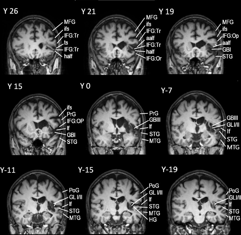
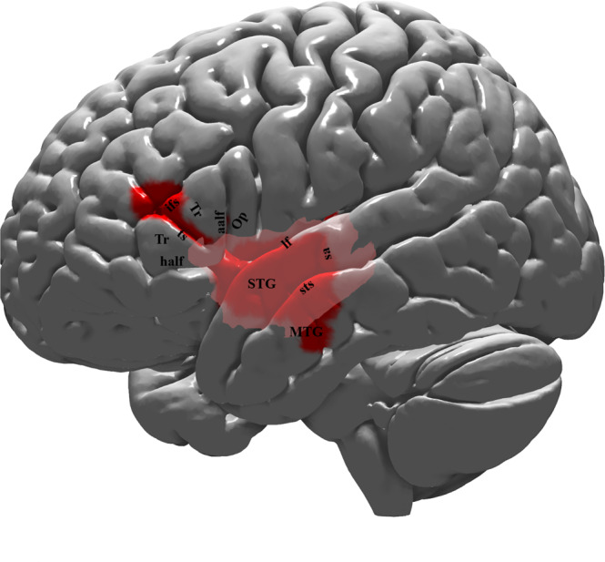
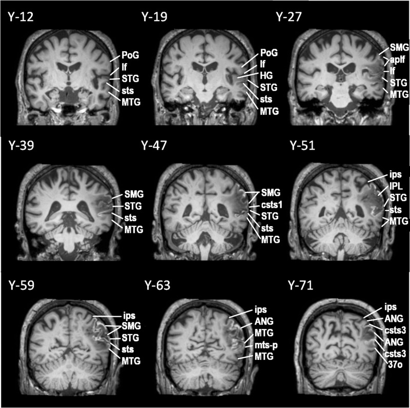
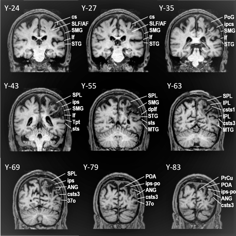
Recent investigations have raised the question of the role of the anterior lateral temporal cortex in language processing (ventral language network). Here we present the language and overall cognitive performance of a rare male patient with chronic middle cerebral artery cerebrovascular accident with a well-documented lesion restricted to the anterior temporal cortex and its connections via the extreme capsule with the pars triangularis of the inferior frontal gyrus (i.e. Broca's region). The performance of this unique patient is compared with that of two chronic middle cerebral artery cerebrovascular accident male patients with damage to the classic dorsal posterior temporo-parietal language system. Diffusion tensor imaging is used to reconstruct the relevant white matter tracts of the three patients, which are also compared with those of 10 healthy individuals. The patient with the anterior temporo-frontal lesion presents with flawless and fluent speech, but selective impairment in accessing lexico-semantic information, in sharp contrast to the impairments in speech, sentence comprehension and repetition observed after lesions to the classic dorsal language system. The present results underline the contribution of the ventral language stream in lexico-semantic processing and higher cognitive functions, such as active selective controlled retrieval.


Conflict of interest statement

The authors declare no competing interests.

Figures

Fig. 1
Fig. 1. Successive coronal MRI images in MNI stereotaxic coordinates (Y) depicting the lesion of patient AA.
The lesion appears at Y35 and continues as far as Y20, including a part of the pars triangularis (area 45) and the white matter below (see Y26, Y21). At Y19, the lesion also includes part of the caudate, the adjacent internal capsule (IC), the external capsule (EC), and the temporo-frontal extreme capsule fasciculus (TFexcF), just below insular gyrus Brevis I (GBI). Subcortically, there is damage to the claustrum, putamen, and the anterior part of the insula (GBI) under which courses the TFexcF. From Y15 to Y7, the lesion includes a small part of the anterior STG. Moving on posteriorly, between Y3 and Y-3, one can observe damage to the TFexcF which courses between the claustrum and the anterior insula (gyrus Brevis III; GBIII), the STG and a small part of MTG, including both banks of the superior temporal sulcus (sts) (see Y0). At Y-7, one observes that the lesion is located anterior to the level of the sulcus acousticus (sa) that lies on the lateral STG (see also Fig. 2). Subcortically, the lesion includes part of the caudate, IC, EC, TFexcF between the claustrum and insular gyrus Brevis III and gyrus Longus I & II, STG, sts and the whole MTG. The temporal lesion is visible as far posterior as Y-15 where it is restricted to the insula, caudate, putamen, claustrum, EC, TFexcF, and at Y-19 to putamen and lower insula, leaving Heschl’s gyrus and the surrounding temporal areas completely spared. Posterior to Y-20, no lesion is detected. Brain areas are topologically defined according to the atlas of the morphology of the human cerebral cortex in the MNI Stereotaxic Space. aalf ascending anterior ramus of the lateral fissure; GBI gyrus brevis I of insula; GBIII gyrus brevis III of insula; GLI gyrus longus I of insula; GLII gyrus longus II of insula; half horizontal ascending ramus of the lateral fissure; HG Heschl’s gyrus; IFG:Tr inferior frontal gyrus, pars triangularis; IFG:Op inferior frontal gyrus, pars opercularis, Or pars orbitalis; Tr pars triangularis; ifs inferior frontal sulcus; lf lateral fissure; MFG middle frontal gyrus; MTG middle temporal gyrus; PoG postcentral gyrus; PrG precentral gyrus; sts superior temporal sulcus; ts triangular sulcus.
Fig. 2
Fig. 2. Lateral view of the left hemisphere lesion of the patient with damage to the ventral language network (patient AA).
The cortical lesion is marked by the red color and occupies the superior and middle temporal gyri (STG and MTG) anterior to the sulcus acousticus (sa), as well as the pars triangularis (area 45) of the inferior frontal gyrus. Note that the posterior parts of the superior and middle temporal gyri (i.e. the classical Wernicke area) and also the cortex of the pars opercularis (area 44) are spared. Thus, the lesion is restricted to the ventral language stream. Subcortically, the lesion is represented by the pale pink color. The lesion of the patient was reconstructed in MNI stereotaxic space and projected on the standard average MNI brain,. Abbreviations: aalf ascending anterior ramus of the lateral fissure; half horizontal ascending ramus of the lateral fissure; ifs inferior frontal sulcus; lf lateral fissure; MTG middle temporal gyrus; Op pars opercularis (area 44); sa sulcus acousticus; STG superior temporal gyrus; sts superior temporal sulcus; Tr pars triangularis (area 45); ts triangular sulcus.
Fig. 3
Fig. 3. Successive coronal MRI images in MNI stereotaxic coordinates (Y) depicting the lesion of patient MM with damage to the dorsal posterior language region.
The lesion first appears below the central sulcus, just posterior to the sulcus acousticus, at Y-12, where a small part of the STG is damaged. At approximately Y-19, there is damage to both the planum temporale, where HG lies and the lateral part of the STG, including both banks of the sts (involving the classic Wernicke region). At Y-27, there is complete damage to HG, planum temporale, the entire STG and both banks of sts. At this level, one observes damage to the parietal operculum and the anterior part of SMG (area PF). At Y-39, there is still damage to the entire STG, including both banks of the sts. The parietal operculum, and the ventral part of SMG are completely destroyed, as well as the underlying white matter, which affected fibers from both the SLF III and AF. Further posteriorly, at Y-47, the entire SMG, STG and both banks of sts, as well as the underlying white matter is destroyed. At Y-51, the lesion includes the entire STG and both banks of the sts. The lesion extends from the ips, down to the posterior temporal lobe. Thus, all of the SMG was damaged including the adjacent STG and the upper part of adjacent MTG. At Y-59, again there is damage in the SMG gyrus and the underlying white matter. The damage extends from the ips as far as the posteriormost part of the adjacent MTG. At Y-63, the lesion extends below the ips including the ANG and MTG, until the mts-p. Between Y-71 and Y-79, the lesion includes the ANG from the intraparietal sulcus as far ventral as the csts-3. Brain areas are topologically defined according to the atlas of the morphology of the human cerebral cortex in the MNI Stereotaxic Space. ANG angular gyrus; aplf ascending posterior ramus of the lateral fissure; csts1 caudal superior temporal sulcus, branch 1; csts3 caudal superior temporal sulcus, branch 3; HG Heschl’s gyrus; IPL inferior parietal lobule; ips intraparietal sulcus; lf lateral fissure; MTG middle temporal gyrus; mts-p posterior middle temporal sulcus; PoG postcentral gyrus; SMG supramarginal gyrus; STG superior temporal gyrus; sts superior temporal sulcus.
Fig. 4
Fig. 4. Lateral view of the left hemisphere lesion of patient MM with damage to the dorsal posterior language region.
The area affected by the lesion is marked with the blue color. The lesion includes the posterior temporal region, i.e. clearly posterior to the level of the sulcus acousticus (sa). It also includes the Heschl’s gyrus (HG) region, which lies within the lateral fissure, and a part of the posterior middle temporal gyrus (pMTG). Thus, the posterior temporal region (Wernicke’s area) is damaged. The lesion extends to the supramarginal (SMG) and angular (ANG) gyri of the inferior parietal lobule. Thus, the lesion is restricted to the dorsal posterior language network, sparing all cortical areas associated with the ventral language network, i.e. the anterior to intermediate temporal areas and their connections via the TFexcF to area 45 on the IFG. The lesion was reconstructed in MNI stereotaxic space and projected on the standard average MNI brain,. Abbreviations: ANG angular gyrus; csts1 caudal superior temporal sulcus, first segment; csts2 caudal superior temporal sulcus, second segment (angular sulcus); csts3 caudal superior temporal sulcus, third segment (anterior occipital sulcus); ipcs inferior postcentral sulcus; ips intraparietal sulcus; lf lateral fissure; pMTG posterior middle temporal gyrus; pSTG posterior superior temporal gyrus; sa sulcus acousticus; SMG supramarginal gyrus; sts superior temporal sulcus.
Fig. 5
Fig. 5. Successive coronal MRI images in MNI stereotaxic coordinates (Y) depicting the lesion of patient TA with damage to the dorsal posterior language region.
The lesion starts posterior to the central sulcus (cs) and after Y-24. At Y-27, the white matter below SMG and postcentral gyrus areas 2, 1, 3b is damaged affecting SLF III fibers (originating from the SMG), SLF II fibers (originating from ANG), and AF fibers (originating from Tpt. At Y 31, a small area of anterior SMG is added to the white matter lesion. At Y-35 and Y-39, the lesion includes SMG and the white matter below. At Y-43, the whole SMG, the white matter below and a small part of area Tpt, that is the caudal STG, is damaged, leaving primary acoustic areas on Heschl’s gyrus intact. Further posteriorly, at Y-47, -51, -55 as well as -59, damage continues to include the whole SMG and SPL (Superior Parietal Lobe), occupying a larger part of STG (see Y-55). At Y-63, superior and inferior parietal lobule areas, including areas PFm and PG, as well as the white matter below, until csts3 are affected. At Y-67 till Y-71, the superior and inferior parietal lobule areas, including ANG, as well as the underlying white matter until the area just below csts2, are lesioned (see Y-69). At Y-75, Y-79, and Y-83, the precuneus, superior and inferior parietal lobule areas until csts3 are affected, while at Y-83, the lesion includes the areas of POA and the whole ANG until csts3. The lesion continues further posteriorly to parieto-occipital areas, affecting even occipital areas such as area 19, in Y-98, where the lesion ends. Brain areas are topologically defined according to the atlas of the morphology of the human cerebral cortex in the MNI Stereotaxic Space. 3a: area 3a; 37o: lateral occipitoparietal area 37; AF Arcuate Fasciculus; ANG angular gyrus; cs central sulcus; csts1, csts3 caudal superior temporal sulcus, 1st and 3rd ramus; IPL inferior parietal lobule; ipcs inferior postcentral sulcus; ips intraparietal sulcus; ips-po intraparietal sulcus, paroccipital part; lf lateral fissure; MTG middle temporal gyrus; POA parieto-occipital arcus; PoG postcentral gyrus; PrCu precuneus; SLFII/III superior longitudinal fasciculus, subdivision II/III; SMG supramarginal gyrus; SPL superior parietal lobule; STG superior temporal gyrus; sts superior temporal sulcus; Tpt caudal superior temporal cortical region.
Fig. 6
Fig. 6. Lateral view of the left hemisphere lesion of patient TA.
The area invaded by the lesion is marked with the yellow color and involves mostly the parietal lobe, including some adjacent posterior temporal and occipital areas. Thus, the lesion is restricted to the dorsal posterior language network, sparing all cortical areas associated with the ventral language network, such as the pars triangularis (area 45) of the inferior frontal gyrus and the anterior temporal areas. The lesion was reconstructed in MNI stereotaxic space and projected on the standard average MNI brain,. Abbreviations: ANG angular gyrus; csts1 caudal superior temporal sulcus, first segment; csts2 caudal superior temporal sulcus, second segment (angular sulcus); csts3 caudal superior temporal sulcus, third segment (anterior occipital sulcus); ips intraparietal sulcus; ips-po intraparietal sulcus, paroccipital segment; lf lateral fissure; pSTG posterior superior temporal gyrus; PoG postcentral gyrus; SMG supramarginal gyrus; spcs superior postcentral sulcus; sps superior parietal sulcus.
Fig. 7
Fig. 7. Individual language and cognitive profiles for the three patients.
The vertical axis shows percentiles up to the 50th in all graphs. For each neuropsychological test, raw scores were transformed into percentiles on the basis of the corresponding normative studies (cited in Methods). For speech rate in the stroke story and cookie theft picture description, percentiles were calculated based on a group of healthy participants sampled from the project “Investigation of cortical surface patterns and their relation with speech metrics and performance in neuropsychological assessment in healthy participants” carried out at Aeginition Hospital in Athens, School of Medicine, Greece (research protocol approval ID: ΩΟΞΛ46Ψ98N2-7PN, July 2017). In a, performance of patient TA is represented with yellow. In b, performance of patient MM is represented with blue and in (c), performance of patient AA is represented with red. In (d), all profiles are shown for comparison. As one can observe in (c), the differential performance of AA in specific domains is manifested, i.e. impaired performance in tests involving selective retrieval vs. non-impaired performance in tests measuring other cognitive processes (points within the inner polygon correspond to impaired performance, below the 5th percentile). In (d), the combined profiles illustrate the differences in the performance of AA (red polygon) compared to MM (blue polygon) and TA (yellow polygon). While AA shows generally preserved language and cognitive abilities, his scores in measures requiring selective retrieval (i.e. PPVT-R, COWF-s, COWF-Ph) is significantly decreased, manifesting lower performance compared to MM and TA. However, the two posterior CVA patients despite their generally decreased performance, scored higher than AA in procedures requiring selective retrieval. Repetition cannot be visualized in the above graphs, because Tsapkini and colleagues did not report percentiles for the BDAE repetition subscale alone. Nevertheless, we clarify in the text that repetition skills of AA were intact in contrast to the other two patients. ACi auditory comprehension index; BNT Boston Naming Test; PPVT-R Peabody vocabulary test-revised; COWF-s semantic subscale of Controlled Oral Word Fluency; COWF-ph phonemic subscale of Controlled Oral Word Fluency; DS-f Digit Span forward condition; DS-b Digit Span backward condition; StrSt Stroke Story speech rate (words/minute); CTP Cookie Theft Picture speech rate (words/minute).

References

    1. Geschwind N. The organization of language and the brain. Science. 1970;170:940–944. doi: 10.1126/science.170.3961.940. - DOI - PubMed
    1. Patterson K, Nestor PJ, Rogers TT. Where do you know what you know? The representation of semantic knowledge in the human brain. Nat. Rev. Neurosci. 2007;8:976–987. doi: 10.1038/nrn2277. - DOI - PubMed
    1. Schwartz MF, et al. Anterior temporal involvement in semantic word retrieval: Voxel-based lesion-symptom mapping evidence from aphasia. Brain. 2009;132:3411–3427. doi: 10.1093/brain/awp284. - DOI - PMC - PubMed
    1. Mesulam M, Thompson C, Weintraub S, Rogalski E. The Wernicke conundrum and the anatomy of language comprehension in primary progressive aphasia. Brain. 2015;138:2423–2437. doi: 10.1093/brain/awv154. - DOI - PMC - PubMed
    1. Gorno-Tempini ML, et al. Classification of primary progressive aphasia and its variants. Neurology. 2011;76:1006–1014. doi: 10.1212/WNL.0b013e31821103e6. - DOI - PMC - PubMed

Publication types