. 2022 Oct 10;26(1):55.
doi: 10.1186/s40824-022-00299-x.

Contact osteogenesis by biodegradable 3D-printed poly(lactide-co-trimethylene carbonate)

Affiliations

Contact osteogenesis by biodegradable 3D-printed poly(lactide-co-trimethylene carbonate)

Mohamad Nageeb Hassan et al. Biomater Res. .

Abstract

Background: To support bone regeneration, 3D-printed templates function as temporary guides. The preferred materials are synthetic polymers, due to their ease of processing and biological inertness. Poly(lactide-co-trimethylene carbonate) (PLATMC) has good biological compatibility and currently used in soft tissue regeneration. The aim of this study was to evaluate the osteoconductivity of 3D-printed PLATMC templates for bone tissue engineering, in comparison with the widely used 3D-printed polycaprolactone (PCL) templates.

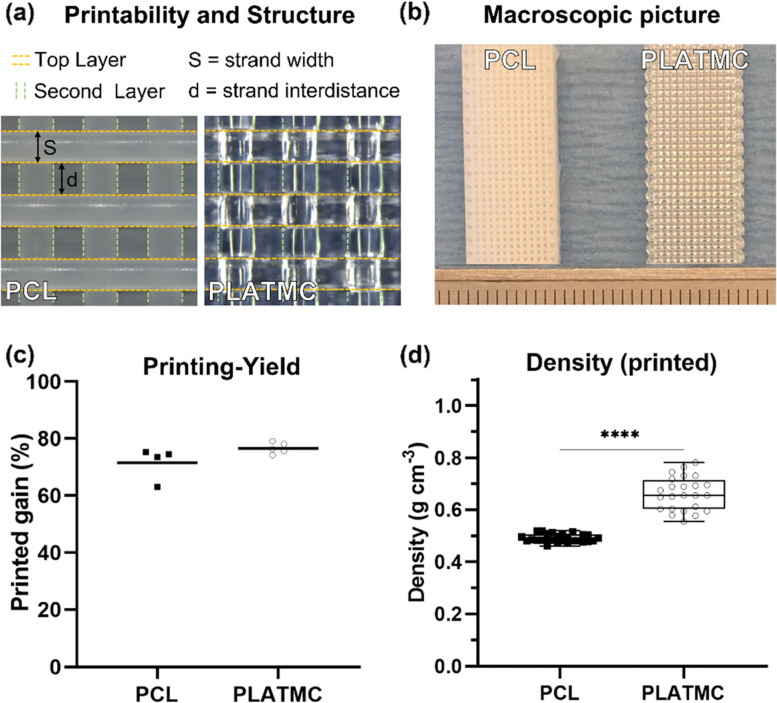
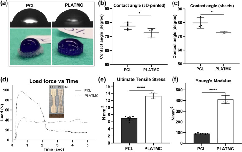
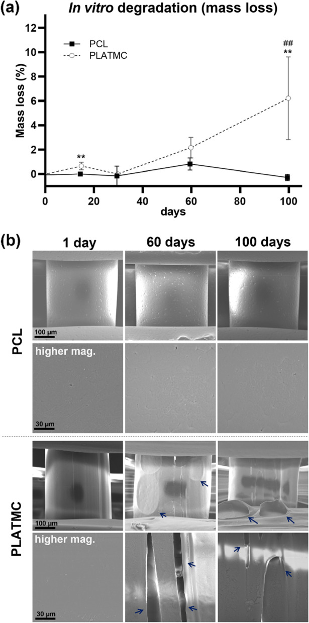
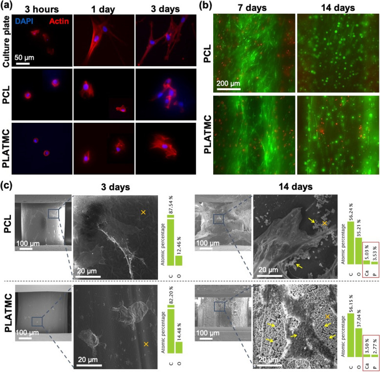
Methods: The printability and physical properties of 3D-printed templates were assessed, including wettability, tensile properties and the degradation profile. Human bone marrow-derived mesenchymal stem cells (hBMSCs) were used to evaluate osteoconductivity and extracellular matrix secretion in vitro. In addition, 3D-printed templates were implanted in subcutaneous and calvarial bone defect models in rabbits.

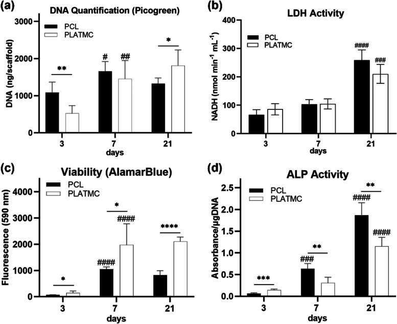
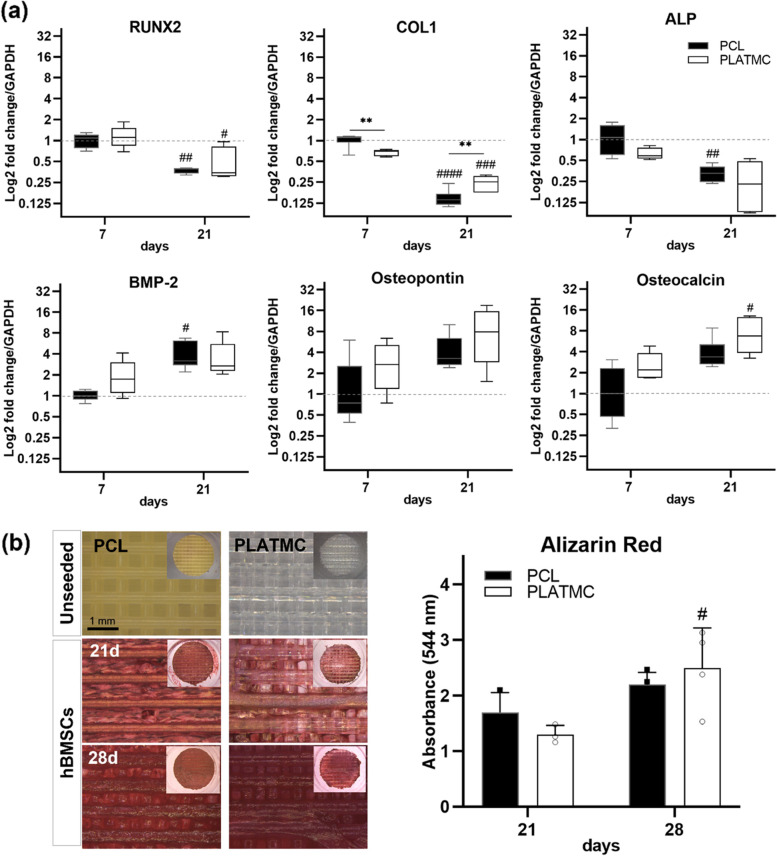
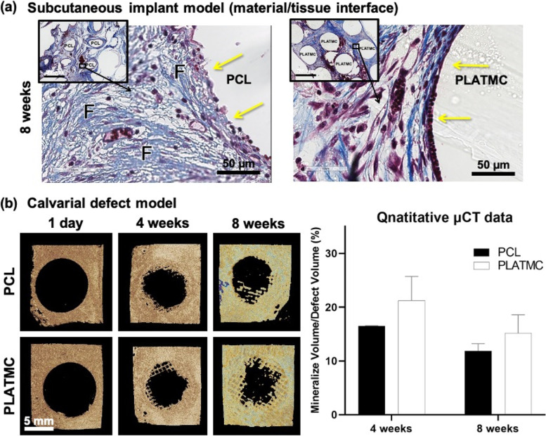
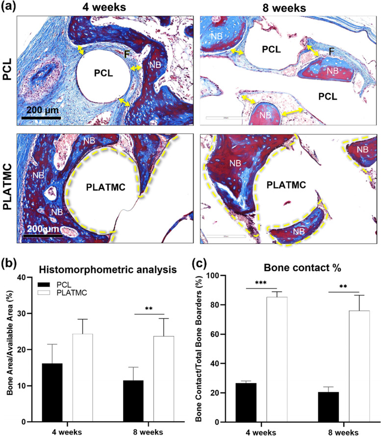
Results: Compared to PCL, PLATMC exhibited greater wettability, strength, degradation, and promoted osteogenic differentiation of hBMSCs, with superior osteoconductivity. However, the higher ALP activity disclosed by PCL group at 7 and 21 days did not dictate better osteoconductivity. This was confirmed in vivo in the calvarial defect model, where PCL disclosed distant osteogenesis, while PLATMC disclosed greater areas of new bone and obvious contact osteogenesis on surface.

Conclusions: This study shows for the first time the contact osteogenesis formed on a degradable synthetic co-polymer. 3D-printed PLATMC templates disclosed unique contact osteogenesis and significant higher amount of new bone regeneration, thus could be used to advantage in bone tissue engineering.

Keywords: 3D-printing; ALP activity; Degradation; Osteoconduction; Poly(lactide-co-trimethylene carbonate); Polycaprolactone; Printability.


Conflict of interest statement

The authors declare no financial or commercial conflicts of interest.

Figures

Fig. 1
Fig. 1
Printability of the 3D-printed PCL and PLATMC, and their calculated printing-yield and density of the printed templates. a microscopic pictures to the printed structures, marked with dashed lines to track the strands in the top two layers, on which the strand width (diameter) and strand interdistance were measured to determine the printability of each polymer. b macroscopic pictures for the printed structures, scale bar in mm. c graph for the mean printing-yield (n = 4), and (d) box plots for the density of the printed templates (n = 25). The statistical significance between the groups is marked with Asterisks (*), **** p < 0.0001
Fig. 2
Fig. 2
Physical characterization of the 3D-printed PCL and PLATMC, in terms of wettability and mechanical properties. a micrographs for contact angle measurement (top), and macroscopic images for the hydrophilic behavior using a drop of dye/water (bottom raw). b and c charts for the contact angle measurements of PLATMC versus PCL in 3D-printed (b) and casted sheet forms (c), respectively. d load force vs time curves, with inset photographs for the printed samples prepared according to ASTM-D638. e and f column charts of the mean ultimate tensile stress, and Young’s modulus, respectively. Note the significant higher wettability and tensile strength of PLATMC. * p > 0.0332, **** p < 0.0001
Fig. 3
Fig. 3
In vitro degradation of the 3D-printed PCL and PLATMC in PBS at 37 °C monitored up to 100 days. a line-graph for the mass loss quantification. Note the significant higher degradation rate of PLATMC compared to the undetectable degradation of PCL, while significance between each time point and the previous time point in the same group is marked with hash symbol (#), **p > 0.0021. b SEM micrographs for the printed templates at 1, 60 and 100 days, with signs of degradation marked with blue arrows
Fig. 4
Fig. 4
hBMSCs attachment, viability and ECM secretion on 3D-printed PCL and PLATMC: a microscopic images showing cytoskeleton immunofluorescence staining after 3 h, 1 day, and 3 days compared to culture plate surface (control); F-actin filaments stained by Phalloidin (red) and nuclei stained by DAPI (blue). b Live/dead stain for seeded cells after 7 and 14 days (z-stacked images). c SEM showing cell adhesion (3 days), and ECM deposition (14 days) and the corresponding EDX characterization to the substrate surface marked with (x). Note the abundant secretion of micron-sized globular accretions marked by YELLOW arrows on PLATMC compared to PCL (14 days), with their Ca and P contents characterized by EDX
Fig. 5
Fig. 5
SEM micrographs analyzing the remarkable globular accretions of the cement line matrix, totally covering, and anchored to the surface of PLATMC templates. a General view of the globular layer secreted by seeded hBMSCs on the surface of 3D-printed PLATMC templates at 14 days. At higher magnifications, the surface is totally covered with globular (vesicular) layer in addition to layers of homogenous structural matrix on the top of the globular layer. b SEM micrographs of PCL and PLATMC samples, at 21 and 28 days after Alizarin red dye and mineralized ECM extraction, showing the persistent anchorage of globular accretions (1–2 µm diameter/each) to PLATMC surface, while no remaining matrix or cells were noticed on PCL. Not the cells/matrix anchored to the top of the globular accretions (Green arrowheads) and the connecting fibrillar collagen (ORANGE arrowheads)
Fig. 6
Fig. 6
Quantitative analysis of cellular proliferation and activity of hBMSCs seeded on 3D-printed PCL and PLATMC at 3, 7 and 21 days, represented as column charts showing: a cell proliferation characterized by DNA quantification using Picogreen assay; b apoptotic tendency characterized by LDH activity assay; c cell metabolic activity characterized by alamarBlue assay; and d ALP activity. Note the higher proliferation rate and viability on PLATMC, while less ALP activity compared to PCL. Statistical significance between each time point and the previous time point in the same group is marked with hash symbol (#), while significance between the groups is marked with Asterisks (*) at p < 0.05; * p > 0.0332, ** p > 0.0021, *** p > 0.0002, **** p < 0.0001
Fig. 7
Fig. 7
Osteogenic differentiation of hBMSCs seeded on 3D-printed PCL and PLATMC characterized by gene expression of osteogenic markers and Alizarin red staining. a box plots representing the gene expression of selected osteogenic markers at 7 and 21 days. b left-hand side shows micrographs of the mineralization stained by Alizarin red at 21 and 28 days, compared with unseeded templates (blank), while the inset pictures show the gross view. A column chart is plotted on the right-hand side representing the quantified optical density of the dissolved stain of each group subtracted from blanks (unseeded templates). Statistical significance between each time point and the previous time point in the same group is marked with hash symbol (#), while significance between the groups is marked with Asterisks (*) at p < 0.05; * p > 0.0332, ** p > 0.0021, *** p > 0.0002, **** p < 0.0001
Fig. 8
Fig. 8
Summary of the outcomes from in vivo implantation of 3D-printed PCL and PLATMC templates (in rabbits); in subcutaneous model and in calvarial defect model. a representative histological micrographs of the subcutaneously implanted templates focusing on the material/tissue interface at 8 weeks as indicated by YELLOW arrows (scale bar = 50 µm), stained with Massons’ trichrome, while the inset figures represent the overall view at lower magnification (scale bar = 500 µm); (F) represents fibrous connective tissues. b µCT reconstructed pictures of the calvarial defect model at 4 and 8 weeks, while a bar chart is plotted on the right-hand side representing their quantified mineralized volume/total defect volume (n = 4 /group/timepoint)
Fig. 9
Fig. 9
Summary of the histological outcomes of 3D-printed PCL and PLATMC templates implanted in the calvarial defect model (in rabbits). a histological micrographs (stained with Masson’s trichrome) at 4 and 8 weeks (scale bar = 200 µm) showing the interface of new bone with template strands. Note the direct contact (contact osteogenesis) of the new formed bone on PLATMC. b and c represents the quantitative histomorphometric analysis and bone contact (%) calculation, respectively. (F) represents fibrous connective tissues; (YELLOW dashed Line) represents areas of contact osteogenesis (present only at PLATMC); (NB) represents areas of new bone; (YELLOW double arrow) represents the characterized gap (fibrous connective tissue) at material/tissue interface (present only at PCL)

References

    1. Wang W, Yeung KWK. Bone grafts and biomaterials substitutes for bone defect repair: a review. Bioact Mater. 2017;2(4):224–247. doi: 10.1016/j.bioactmat.2017.05.007. - DOI - PMC - PubMed
    1. Carrel J-P, Wiskott A, Moussa M, Rieder P, Scherrer S, Durual S. A 3D printed TCP/HA structure as a new osteoconductive scaffold for vertical bone augmentation. Clin Oral Implants Res. 2016;27(1):55–62. doi: 10.1111/clr.12503. - DOI - PubMed
    1. Davies JE. Understanding peri-implant endosseous healing. J Dent Educ. 2003;67(8):932–49. doi: 10.1002/j.0022-0337.2003.67.8.tb03681.x. - DOI - PubMed
    1. Tamimi F, Torres J, Al-Abedalla K, Lopez-Cabarcos E, Alkhraisat MH, Bassett DC, et al. Osseointegration of dental implants in 3D-printed synthetic onlay grafts customized according to bone metabolic activity in recipient site. Biomaterials. 2014;35(21):5436–5445. doi: 10.1016/j.biomaterials.2014.03.050. - DOI - PubMed
    1. Davies JE. Mechanisms of endosseous integration. Int J Prosthodont. 1998;11(5):391–401. - PubMed