. 2022 Oct 10;15(1):144.
doi: 10.1186/s13045-022-01366-5.

CXCL13 chemokine is a novel player in multiple myeloma osteolytic microenvironment, M2 macrophage polarization, and tumor progression

Affiliations

CXCL13 chemokine is a novel player in multiple myeloma osteolytic microenvironment, M2 macrophage polarization, and tumor progression

Katia Beider et al. J Hematol Oncol. .

Abstract

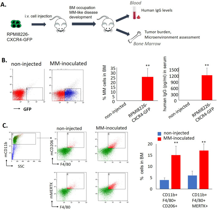
Background: We assessed the mechanism by which multiple myeloma (MM) shapes the bone marrow (BM) microenvironment and affects MΦ polarization.

Methods: In vivo xenograft model of BM-disseminated human myeloma, as well as analysis of MM cell lines, stromal components, and primary samples from patients with MM, was utilized.

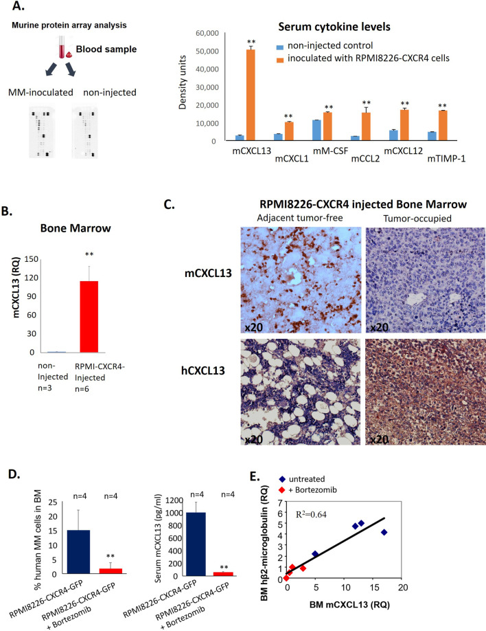
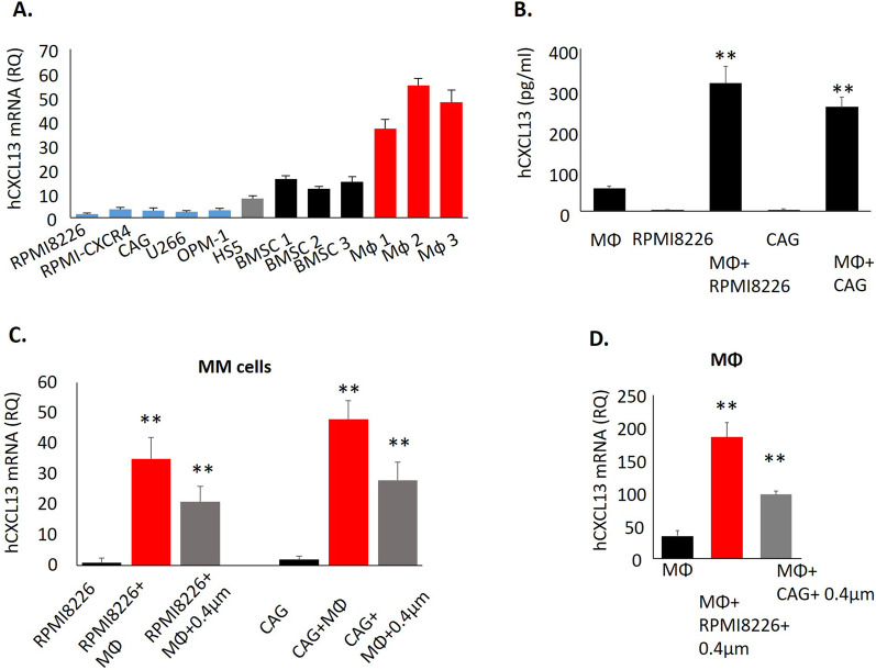
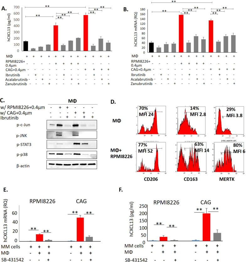
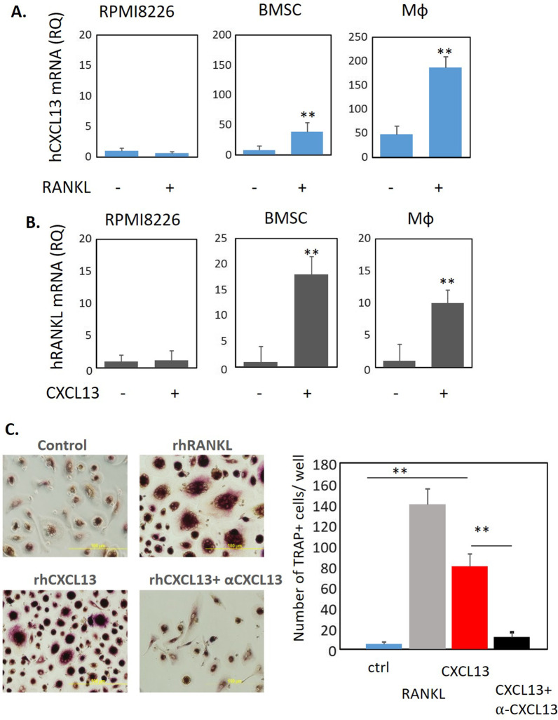
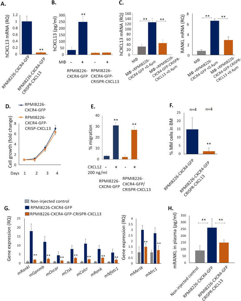
Results: Analysis of the BM from MM-bearing mice inoculated with human CXCR4-expressing RPMI8226 cells revealed a significant increase in M2 MΦ cell numbers (p < 0.01). CXCL13 was one of the most profoundly increased factors upon MM growth with increased levels in the blood of MM-bearing animals. Myeloid cells were the main source of the increased murine CXCL13 detected in MM-infiltrated BM. MM cell lines induced CXCL13 and concurrent expression of M2 markers (MERTK, CD206, CD163) in co-cultured human MΦ in vitro. Interaction with MΦ reciprocally induced CXCL13 expression in MM cell lines. Mechanistically, TGFβ signaling was involved in CXCL13 induction in MM cells, while BTK signaling was implicated in MM-stimulated increase of CXCL13 in MΦ. Recombinant CXCL13 increased RANKL expression and induced TRAP+ osteoclast (OC) formation in vitro, while CXCL13 neutralization blocked these activities. Moreover, mice inoculated with CXCL13-silenced MM cells developed significantly lower BM disease. Reduced tumor load correlated with decreased numbers of M2 MΦ in BM, decreased bone disease, and lower expression of OC-associated genes. Finally, higher levels of CXCL13 were detected in the blood and BM samples of MM patients in comparison with healthy individuals.

Conclusions: Altogether, our findings suggest that bidirectional interactions of MΦ with MM tumor cells result in M2 MΦ polarization, CXCL13 induction, and subsequent OC activation, enhancing their ability to support bone resorption and MM progression. CXCL13 may thus serve as a potential novel target in MM.


Conflict of interest statement

The authors declare no competing financial interests.

Figures

Fig. 1
Fig. 1
M2 macrophages are increased in BM of MM-inoculated mice. A Schematic representation of xenograft systemic model of CXCR4-driven MM with BM involvement. NSG mice were i.v. inoculated with RPMI8226-CXCR4-GFP cells. Non-injected NSG mice were used as controls. B MM tumor load was measured by enumeration of GFP+ human MM cells in the BM and by evaluation of human IgG levels in the blood of non-injected (n = 5) or inoculated animals (n = 5). Data are presented as mean ± SE, **p < 0.01. C Presence of M2 murine MΦ in the BM of non-injected controls (n = 5) or MM-inoculated animals (n = 5), evaluated by flow cytometry. Representative plots for gating strategy evaluating the expression of murine CD11b, F4/80, CD206, and MERTK markers. Bars showing mean of triplicates ± STDEV, **p < 0.01
Fig. 2
Fig. 2
Murine CXCL13 is increased in the blood and BM of MM-inoculated mice. A Evaluation of cytokine levels in the serum of MM-inoculated (pooled from 3 mice) and non-injected control (pooled from 3 mice) animals using cytokine array. B Levels of mCXCL13 in the BM of MM-inoculated (n = 6) and control mice (n = 3), evaluated by qRT-PCR. Data are presented as the mean of triplicates ± STDEV (**p < 0.01). C Immunohistochemistry for mCXCL13 and human CXCL13. D, E RPMI8226-CXCR4-GFP cells were injected i.v. into NSG mice. Mice were either untreated (control group, n = 4) or treated with subcutaneous injections of bortezomib (1 mg/kg, twice a week) (n = 4). D Tumor burden (%GFP + cells) in BM and serum levels of murine CXCL13 were evaluated on day 24. E Co-expression of human β2-microglobulin and murine Cxcl13 in the BM of MM-inoculated mice, evaluated by qRT-PCR
Fig. 3
Fig. 3
Interaction between MM cells and macrophages up-regulates CXCL13 in both cell populations. A CXCL13 mRNA expression in human MM cell lines, primary BMSC, and peripheral-blood generated MΦ, evaluated by qRT-PCR. Data are presented as the mean of triplicates ± STDEV (**p < 0.01). B Peripheral-blood-derived MΦ were cultured in the absence or presence of MM cell lines RPMI8226 or CAG (direct co-culture) for 48 h. Levels of secreted CXCL13 in the culture medium were evaluated by ELISA. Data are presented as the mean of triplicates ± STDEV (**p < 0.01). C CXCL13 mRNA levels in MM cells RPMI8226 and CAG, cultured in the absence or presence of peripheral-blood derived MΦ, either in direct contact co-culture or separated by 0.4 µm membrane, evaluated by qRT-PCR. D CXCL13 mRNA levels in peripheral-blood generated MΦ, cultured in the absence or presence of RPMI8226 and CAG cells, separated by a 0.4 µm membrane, evaluated by qRT-PCR. Data are presented as the mean of triplicates ± STDEV (**p < 0.01)
Fig. 4
Fig. 4
An increase in CXCL13 upon the interaction between MM cells and macrophages is mediated by BTK and TGFβ signaling. A Peripheral-blood-derived MΦ were cultured in the absence or presence of MM cells RPMI8226 (direct co-culture) with or without ibrutinib (20 µM), acalabrutinib (20 µM) and zanubrutinib (20 µM) for 48 h. The levels of secreted CXCL13 in the culture medium were evaluated by ELISA. Data are presented as the mean of triplicates ± STDEV (**p < 0.01). B Peripheral-blood derived MΦ were cultured in the absence or presence of MM cell lines RPMI8226 and CAG, separated by 0.4 µm membrane with or without ibrutinib (20 µM), acalabrutinib (20 µM) and zanubrutinib (20 µM) for 48 h and subjected to subsequent analysis. Expression levels of CXCL13 mRNA in MΦ cells evaluated by qRT-PCR. C MM-induced signaling in MΦ co-cultured in 0.4 µm transwells for 48 h, evaluated by Western blot analysis. Blots were probed for p-c-Jun, p-JNK, p-STAT3 and p-p38. β-actin was used as an internal control. Representative data from at least two independent experiments are shown. D Peripheral-blood-derived MΦ were cultured in the absence or presence of MM cells RPMI8226 (direct co-culture) for 48 h and subjected to flow cytometry analysis. Expression levels of cell surface CD206, CD163, and MERTK on gated CD11b+ cells are depicted in representative histograms. Quantification of geometric mean fluorescence intensity (MFI) of surface markers and percentage of positive cells was performed. E CXCL13 mRNA levels in MM cells RPMI8226 and CAG, cultured in the absence or presence of peripheral-blood derived MΦ, treated with SB-431542 (20 µM) for 48 h, evaluated by qRT-PCR. F Peripheral-blood-derived MΦ were cultured in the absence or presence of MM cells RPMI8226 and CAG (direct co-culture) with or without SB-431542 (20 µM) for 48 h. Levels of secreted CXCL13 in the culture medium were evaluated by ELISA. Data are presented as the mean of triplicates ± STDEV (**p < 0.01)
Fig. 5
Fig. 5
Reciprocal regulation of CXCL13 in RANKL in BMSC and Mф. Induction of osteoclast generation by CXCL13. RPMI8226 cells, primary BMSC or peripheral blood-generated macrophages were A treated with RANKL (100 ng/ml) for 48 h and mRNA level of CXCL13 was tested by qPCR, and B treated with CXCL13 (500 ng/ml) for 48 h and mRNA levels of RANKL was tested by qPCR. Data are presented as the mean of triplicates ± STDEV (**p < 0.01). C Human peripheral blood mononuclear cells from healthy donors were grown in the presence of recombinant human M-CSF (100 ng/ml) and RANKL (100 ng/ml) or CXCL13 (500 ng/ml) −/+ anti-CXCL13 (10 µg/ml) for 2 weeks. TRAP staining was performed and multinucleated TRAP+ cells were enumerated
Fig. 6
Fig. 6
CXCL13 silencing in MM cells inhibits MM growth in vivo and suppresses MM-associated osteoclastogenesis in murine BM. A CXCL13 mRNA levels in RPMI8226-CXCR4-GFP and RPMI8226-CXCR4-GFP-CRISPR-CXCL13 cells evaluated by qRT-PCR. B CXCL13 secreted levels in conditioned medium of RPMI8226-CXCR4-GFP and RPMI8226-CXCR4-GFP-CRISPR-CXCL13 cells, cultured alone or co-cultured with peripheral-blood derived MΦ. C Peripheral-blood derived MΦ were cultured in the absence or presence of MM cell lines RPMI8226-CXCR4-GFP and RPMI8226-CXCR4-GFP-CRISPR-CXCL13, separated by 0.4 µm membrane for 48 h and subjected to subsequent analysis. Expression levels of CXCL13 and RANKL mRNA in MΦ cells evaluated by qRT-PCR. D In vitro cell growth kinetics of RPMI8226-CXCR4-GFP and RPMI8226-CXCR4-GFP-CRSIPR-CXCL13 cells, enumerated by FACS. E Transwell migration of RPMI8226-CXCR4-GFP and RPMI8226-CXCR4-GFP-CRSIPR-CXCL13 cells during 4 h in response to CXCL12 (200 ng/mL), evaluated by FACS. F-H NSG mice were i.v. inoculated with RPMI8226-CXCR4-GFP cells (n = 4) or RPMI8226-CXCR4-GFP-CRISPR-CXCL13 cells (n = 4). F MM tumor load was measured by enumeration of GFP+ human MM cells in the BM of inoculated animals. Data are presented as mean ± SE, **p < 0.01. G MNCs from the BM of control (n = 4) and MM-inoculated (n = 5) mice were purified and RNA was extracted. Gene expression of murine factors was evaluated by qRT-PCR. H Murine RANKL levels in plasma of non-inoculated (n = 3), RPMI8226-CXCR4-GFP (n = 4) and RPMI8226-CXCR4-GFP-CRISPR-CXCL13-inoculated (n = 4) animals at day 24 following cell injection. Data are presented as mean ± STDEV, **p < 0.01
Fig. 7
Fig. 7
CXCL13 expression and flow cytometry analysis in primary MM samples. A CXCL13 levels in peripheral blood of MM patients with active disease (n = 32), stable disease (n = 29), or in healthy donors (n = 9), evaluated by ELISA. B CXCL13 levels in BM plasma samples of MM patients (n = 36) and healthy donors (n = 6), evaluated by ELISA. Data are presented as mean ± STDEV, **p < 0.01. C Flow cytometry analysis of cells in normal BM samples (n = 6) and MM BM samples (n = 20). The cells were pre-gated on CD11b+ CD14+ population. The frequency of RANK+ CD51+ cells, MERTK+ cells and CD163+ CD206+ was established (upper panel). The frequency of CD14+ CD16− and CD14+ CD16+ subpopulations, and percentage of MERTK+ cells in both subsets are presented (lower panel). D (i) Spearman’s rho correlation (two-tailed) between the frequency of CD163+ CD206+ and the frequency of RANK+ CD51+ subsets on CD11b+ CD14+ cells in BM samples (normal BM, n = 6; MM BM, n = 20). (ii) Spearman’s rho correlation (two-tailed) between the expression of MERTK and the co-expression of RANK and CD51 on CD11b+ CD14+ cells in BM samples (normal BM, n = 6; MM BM, n = 20). (iii) Spearman’s rho correlation (two-tailed) between CXCL13 protein levels in BM plasma and the frequency of MERTK expression on CD11b+ CD14+ cells in BM samples (normal BM, n = 6; MM BM, n = 20). E Expression of CXCL13 in normal BM samples, BM samples from patients with MM, and plasmacytoma samples, evaluated by immunohistochemical staining. Original magnification of × 400 is shown

References

    1. van de Donk N, Pawlyn C, Yong KL. Multiple myeloma. Lancet. 2021;397:410–427. - PubMed
    1. Kumar SK, Rajkumar V, Kyle RA, et al. Multiple myeloma. Nat Rev Dis Primers. 2017;3:17046. - PubMed
    1. Gulla A, Anderson KC. Multiple myeloma: the (r)evolution of current therapy and a glance into future. Haematologica. 2020;105:2358–2367. - PMC - PubMed
    1. Gay F, Magarotto V, Crippa C, et al. Bortezomib induction, reduced-intensity transplantation, and lenalidomide consolidation-maintenance for myeloma: updated results. Blood. 2013;122:1376–1383. - PubMed
    1. van de Donk NW, Moreau P, Plesner T, et al. Clinical efficacy and management of monoclonal antibodies targeting cd38 and slamf7 in multiple myeloma. Blood. 2016;127:681–695. - PubMed