

Hyperostosis Frontalis Interna and a Question on Its Pathology: A Case Report
- PMID: 36217295
- PMCID: PMC9575136
- DOI: 10.12659/AJCR.937450
Hyperostosis Frontalis Interna and a Question on Its Pathology: A Case Report
Abstract
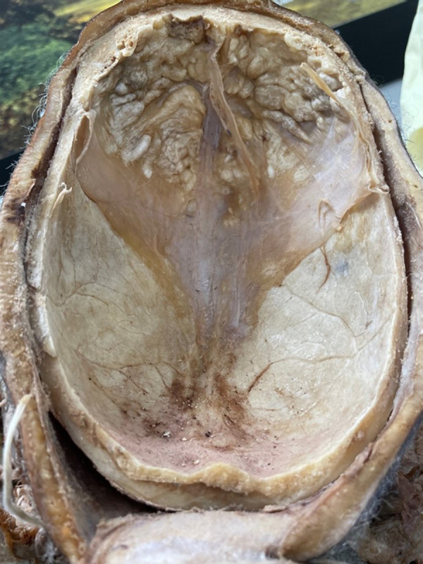
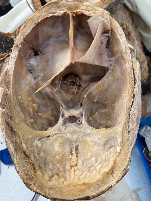
BACKGROUND Hyperostosis frontalis interna is a boney overgrowth of the inner side of the frontal bone of the skull caused by overgrowth of the endocranial surface. It is most often found in women after menopause. It is also associated with hormonal imbalance, being overweight, history of headaches, and neurocognitive degenerative conditions. Female gender, advanced age, extended estrogen stimulation, and elevated leptin levels may also play a role. The thickening is usually confined to the frontal bone, but it can spread as far as the anterior parietal and temporal bones. CASE REPORT During a medical school dissection course, an extensive boney overgrowth in the frontal regions covering the inside of the frontal bone of the skull of a 90-year-old female donor, who died of a cerebrovascular infarction, was identified. This boney overgrowth was mainly confined within the frontal region, but there was some boney overgrowth that extended to the temporal bones. The overgrowth in the endocranium of the temporal bone was not as severe as the overgrowth of the frontal bone. The morphology of the overgrowth was rigid, uneven, and bumpy. Based upon the physical characteristics, we concluded that this presentation was consistent with hyperostosis frontalis interna. CONCLUSIONS Our female donor was found to exhibit a phenomenon which could be clinically underdiagnosed due to its internal nature and asymptomatic presentation. Insight into the potential causes of HFI and its identification during clinical evaluation offers a path for future research to better identify and manage cases of HFI.
Conflict of interest statement
Figures
Similar articles
-
A case of extensive hyperostosis frontalis interna in an 87-year-old female human cadaver.Clin Anat. 2008 Apr;21(3):259-68. doi: 10.1002/ca.20613. Clin Anat. 2008. PMID: 18351650
-
Hyperostosis frontalis interna associated with hypogonadism in an elderly man.Age Ageing. 2006 Mar;35(2):202-3. doi: 10.1093/ageing/afj051. Epub 2006 Jan 23. Age Ageing. 2006. PMID: 16431852
-
Are hyperostosis frontalis interna and leptin linked? a hypothetical approach about hormonal influence on human microevolution.Med Hypotheses. 2002 May;58(5):378-81. doi: 10.1054/mehy.2001.1481. Med Hypotheses. 2002. PMID: 12056872
-
Hyperostosis frontalis interna: case report and review of literature.Ann Clin Lab Sci. 2004 Spring;34(2):206-8. Ann Clin Lab Sci. 2004. PMID: 15228235 Review.
-
[Morgagni-Stewart-Morel syndrome. Case report and review of the literature].Rev Med Inst Mex Seguro Soc. 2016 Sep-Oct;54(5):664-9. Rev Med Inst Mex Seguro Soc. 2016. PMID: 27428347 Review. Spanish.
Cited by
-
Review of imaging modalities and radiological findings of calvarial lesions.World J Radiol. 2025 Jun 28;17(6):107776. doi: 10.4329/wjr.v17.i6.107776. World J Radiol. 2025. PMID: 40606050 Free PMC article. Review.
-
Hyperostosis Fronto-Parieto-Occipitalis: A Cadaveric Case Report.Cureus. 2023 Jul 6;15(7):e41445. doi: 10.7759/cureus.41445. eCollection 2023 Jul. Cureus. 2023. PMID: 37546094 Free PMC article.
References
-
- She R, Szakacs J. Hyperostosis frontalis interna: Case report and review of literature. Ann Clin Lab Sci. 2004;34(2):206–8. - PubMed
-
- Cvetković D, Nikolić S, Brković V, et al. Hyperostosis frontalis interna as an age-related phenomenon – differences between males and females and possible use in identification. Sci Justice. 2019;9(2):172–76. - PubMed
-
- Hershkovitz I, Greenwald C, Rothschild BM, et al. Hyperostosis frontalis inter-na: An anthropological perspective. Am J Phys Anthropol. 1999;109:303–25. - PubMed
-
- Yamakawa K, Mizutani K, Takahashi M, et al. Hyperostosis frontalis interna associated with hypogonadism in an elderly man. Age and ageing. 2006;35(2):202–3. - PubMed
-
- Talarico EF, Jr, Prather AD, Hardt KD. A case of extensive hyperostosis frontalis interna in an 87-year-old female human cadaver. Clin Anat. 2008;21(3):259–68. - PubMed
Publication types
MeSH terms
Substances
LinkOut - more resources
Full Text Sources
