. 2022 Sep 22;25(10):105174.
doi: 10.1016/j.isci.2022.105174. eCollection 2022 Oct 21.

Lessons learned from pre-clinical testing of xenogeneic decellularized esophagi in a rabbit model

Affiliations

Lessons learned from pre-clinical testing of xenogeneic decellularized esophagi in a rabbit model

Edward Hannon et al. iScience. .

Abstract

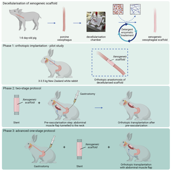
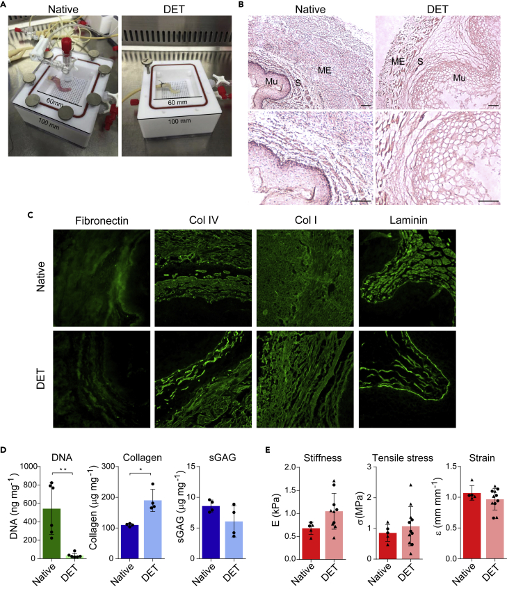
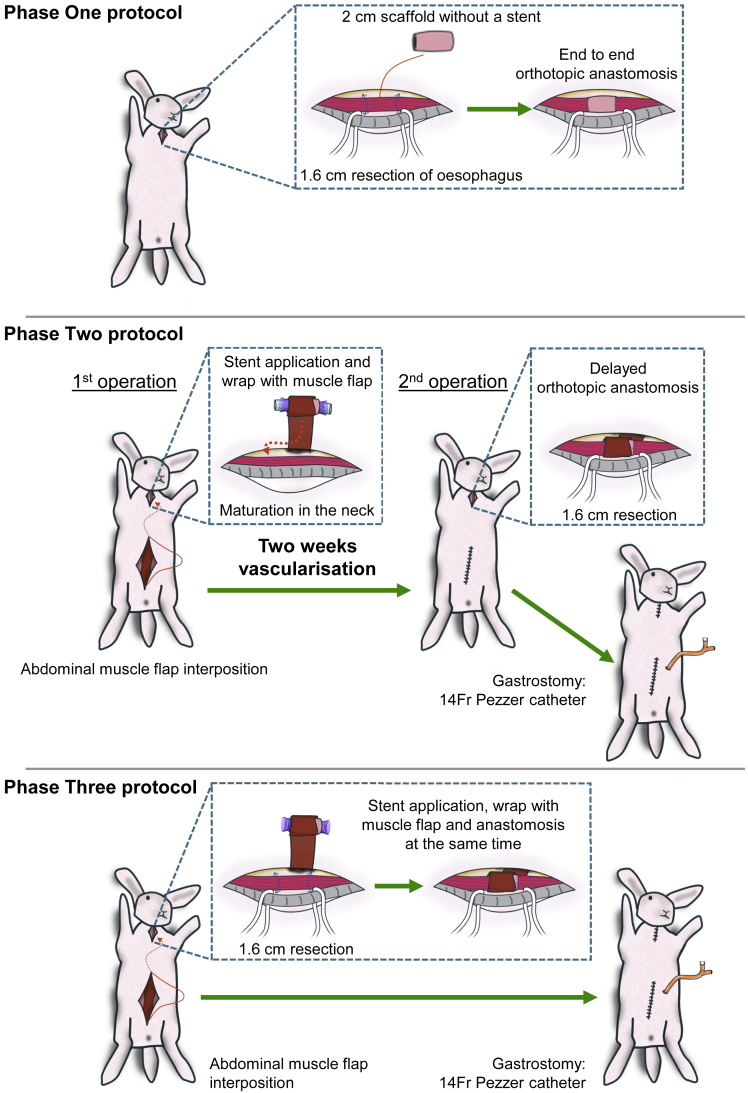
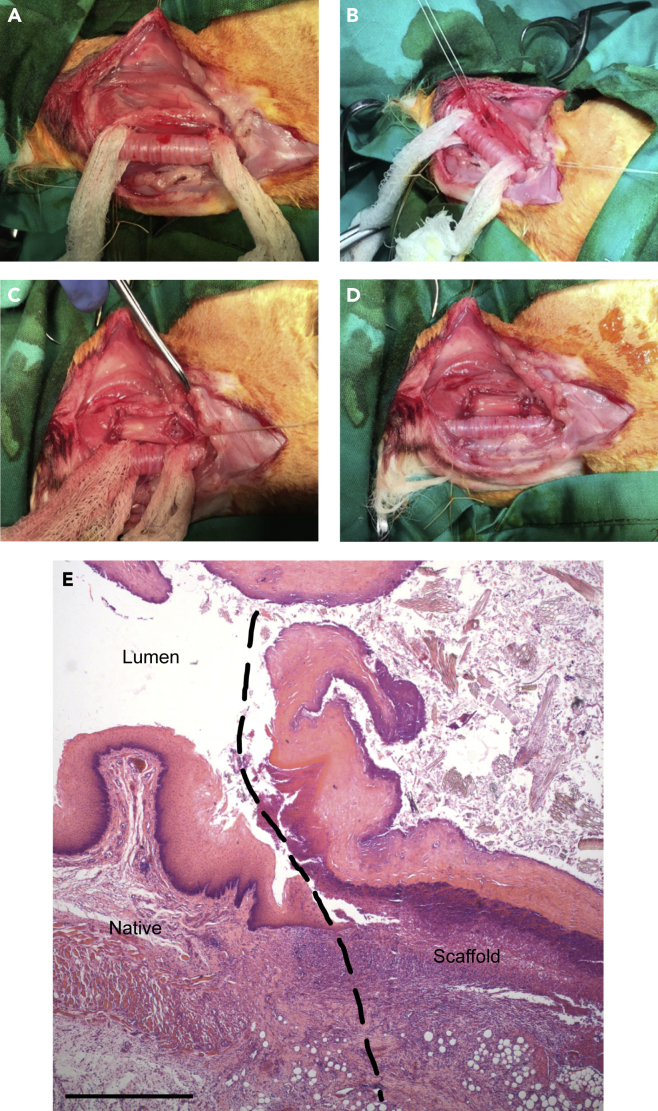
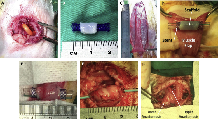
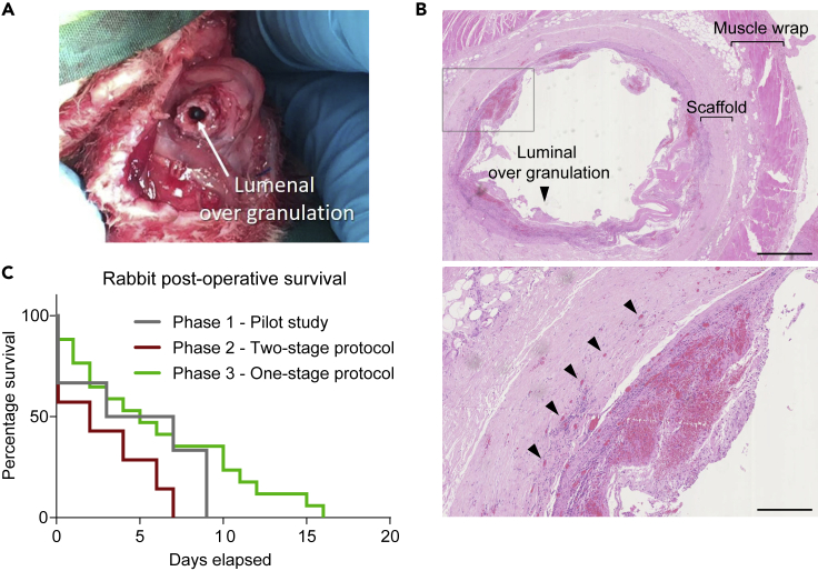
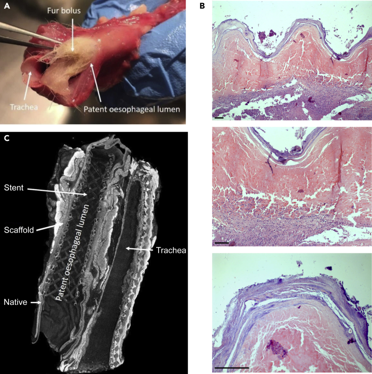
Decellularization of esophagi from several species for tissue engineering is well described, but successful implantation in animal models of esophageal replacement has been challenging. The purpose of this study was to assess feasibility and applicability of esophageal replacement using decellularized porcine esophageal scaffolds in a new pre-clinical model. Following surgical replacement in rabbits with a vascularizing muscle flap, we observed successful anastomoses of decellularized scaffolds, cues of early neovascularization, and prevention of luminal collapse by the use of biodegradable stents. However, despite the success of the surgical procedure, the long-term survival was limited by the fragility of the animal model. Our results indicate that transplantation of a decellularized porcine scaffold is possible and vascular flaps may be useful to provide a vascular supply, but long-term outcomes require further pre-clinical testing in a different large animal model.

Keywords: Biological sciences; Biomedical engineering; Biotechnology; Tissue engineering.


Conflict of interest statement

P.D.C. and L.U. are named inventors of patent application No. PCT/EP2016/071114 and P.D.C. and L.U. are named inventors of UK patent application No. 1708729.7. The remaining authors declare no competing interests.

Figures

None
Graphical abstract
Figure 1
Figure 1
Piglet esophagus decellularization with DET protocol and ECM characterization (A) The esophagi were decellularized in custom chambers. Here a representative esophagus before (native, left) and after (DET, right) the decellularization process is shown. (B) Hematoxylin and eosin (H&E) staining of native (left) and DET tissue (right). ME: muscularis externa. S: submucosa. Mu: mucosa. Scale bar: 100 μm. (C) Immunofluorescence staining for Fibronectin, Collagen IV, Collagen I, and Laminin in native and DET tissue. Scale bar: 50 μm. Both H&E and immunofluorescence show a good preservation of the ECM. (D) DNA quantification shows a significant reduction of DNA following three cycles of DET (p < 0.01, n = 6). Total collagen (p < 0.05, n = 4) and sGAG (n = 4) content were preserved. Bars indicate mean ± SD, circles represent biological replicates. (E) Biomechanical analysis of native and decellularized samples derived from proximal (circle) or distal (triangle) portions of the esophagi. Stiffness (native n = 5; DET n = 10), ultimate tensile stress (native n = 5; DET n = 11), and ultimate strain (native n = 5; DET n = 11) were evaluated. Bars indicate mean ± SD, circles and triangles represent biological replicates of tissue biopsied from the upper and lower thirds of the organ, respectively.
Figure 2
Figure 2
Summary of the three surgery protocols applied in the study Phase One: orthotopic anastomosis of decellularized scaffold. Phase Two: pre-implantation of scaffold in the neck in vascularizing muscle wrap with application of intra-luminal stent followed by orthotopic cervical anastomosis two weeks later. Phase Three: a single-stage anastomosis was performed with a muscle wrap and stent and concomitant gastrostomy formation.
Figure 3
Figure 3
Operative stages of Phase One: esophageal scaffold implantation (A) Trachea exposure. (B) Rabbit esophagus resection. (C) Porcine esophageal scaffold positioning. (D) Orthotopic anastomosis of the scaffold. (E) H&E staining of porcine scaffold following implantation in cervical rabbit esophagus; longitudinal section at the anastomosis level (dashed line) from an animal euthanized at post-operative day nine. Scale bar 250 μm.
Figure 4
Figure 4
Surgical improvements applied from Phase Two (A) Tunneled Stamm gastrostomy formation using a 14Fr Pezzer catheter. (B) Scaffold pre-mounted on stent. (C) An anterior abdominal wall flap was prepared, (D) tunneled to neck and (E) sutured around the scaffold. (F) After two weeks, at the second surgical stage, the vascularized graft was mobilized and (G) anastomosed in orthotopic position.
Figure 5
Figure 5
Second stage of Phase Two procedure and analysis of post-operative survival (A) Macroscopic overgranulation within the lumen of the scaffold. (B) H&E staining of scaffold seven days post second stage orthotopic implantation, sliced transversely across lumen, showing cues of both luminal granulation (arrowhead in top panel) and microvascularization (arrowheads in bottom panel). Top panel scale bar 2.5 mm; bottom panel scale bar 500 μm. (C) Kaplan-Meier survival curve by study groups of Phase One (pilot study, n = 6), Phase Two (two-stage protocol, n = 7) and Phase Three (single-stage protocol, n = 15).
Figure 6
Figure 6
Results of surgery from Phase Three (A) Fur bolus obstruction in autopsy specimens from an animal euthanized at day 15. (B) H&E of porcine scaffolds following single-stage orthotopic anastomosis in cervical rabbit esophagus with vascularizing flap, collected from an animal euthanized at day 16; Top panel 5× magnification, middle panel 10× magnification, bottom panel 20× magnification. Scale bar 50 μm. (C) Micro-CT of en bloc resection of esophagus and trachea after 10 days in vivo demonstrating excellent luminal patency and anastomosis to native esophagus. See also Figure S1 and Video S2.

Similar articles

Cited by

References

    1. Badylak S.F., Vorp D.A., Spievack A.R., Simmons-Byrd A., Hanke J., Freytes D.O., Thapa A., Gilbert T.W., Nieponice A. Esophageal reconstruction with ECM and muscle tissue in a dog model. J. Surg. Res. 2005;128:87–97. doi: 10.1016/j.jss.2005.03.002. - DOI - PubMed
    1. Badylak S., Meurling S., Chen M., Spievack A., Simmons-Byrd A. Resorbable bioscaffold for esophageal repair in a dog model. J. Pediatr. Surg. 2000;35:1097–1103. doi: 10.1053/jpsu.2000.7834. - DOI - PubMed
    1. Boys A.J., Barron S.L., Tilev D., Owens R.M. Building scaffolds for tubular tissue engineering. Front. Bioeng. Biotechnol. 2020;8:1–20. doi: 10.3389/fbioe.2020.589960. - DOI - PMC - PubMed
    1. Catry J., Luong-Nguyen M., Arakelian L., Poghosyan T., Bruneval P., Domet T., Michaud L., Sfeir R., Gottrand F., Cattan P., et al. Circumferential esophageal replacement by a tissue-engineered substitute using mesenchymal stem cells. Cell Transplant. 2017;26:1831–1839. doi: 10.1177/0963689717741498. - DOI - PMC - PubMed
    1. Cesur O., Tanir T.E., Celepli P., Ozarslan F., Hucumenoglu S., Karaibrahimoglu A., Hasirci N. Enhancing esophageal repair with bioactive bilayer mesh containing FGF. Sci. Rep. 2021;11:1–10. doi: 10.1038/s41598-021-98840-w. - DOI - PMC - PubMed

LinkOut - more resources