. 2022 Dec 21;10(6):e0029722.
doi: 10.1128/spectrum.00297-22. Epub 2022 Oct 7.

Comparative Genomic Analysis of Fusobacterium necrophorum Provides Insights into Conserved Virulence Genes

Affiliations

Comparative Genomic Analysis of Fusobacterium necrophorum Provides Insights into Conserved Virulence Genes

Prabha K Bista et al. Microbiol Spectr. .

Abstract

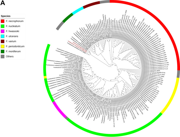
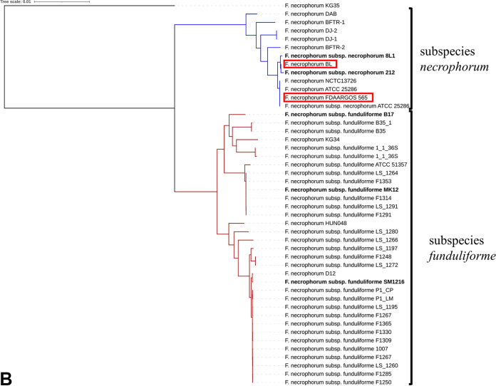
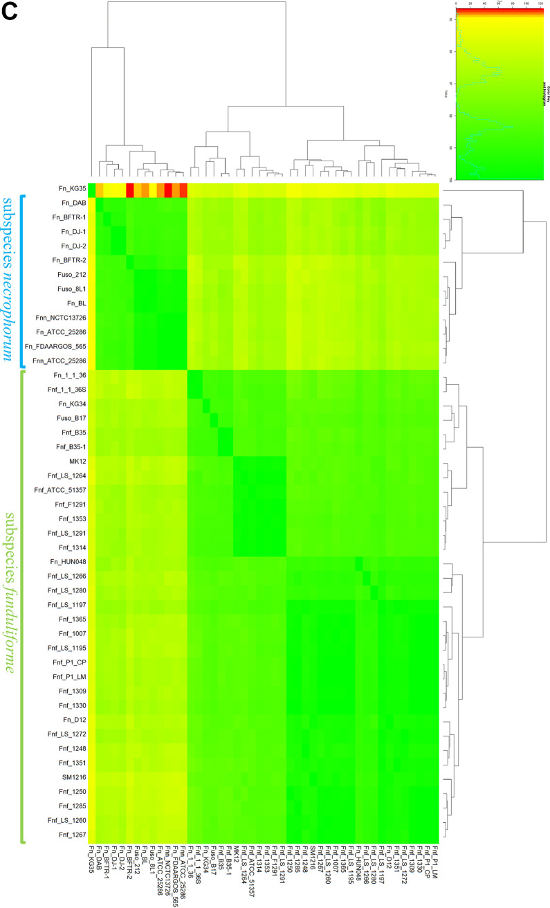
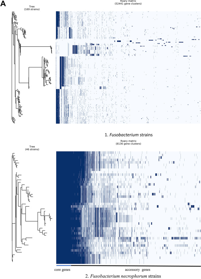
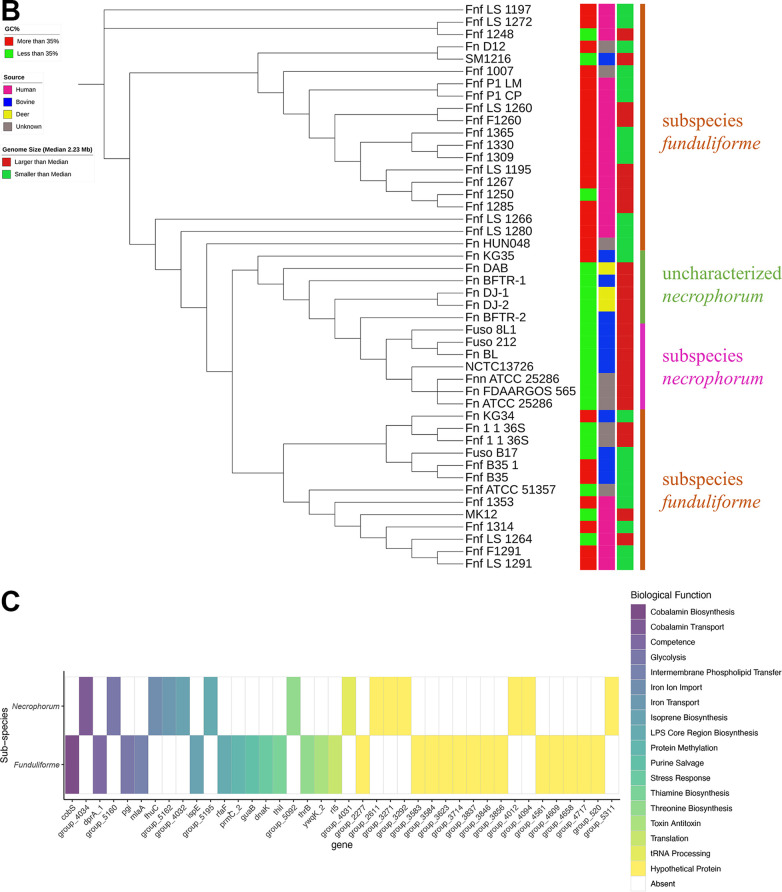
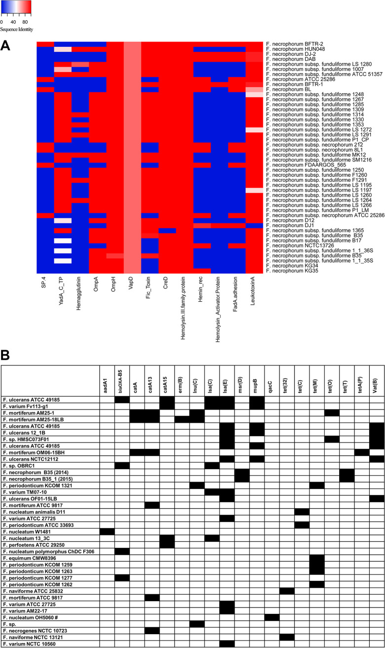
Fusobacterium necrophorum is a Gram-negative, filamentous anaerobe prevalent in the mucosal flora of animals and humans. It causes necrotic infections in cattle, resulting in a substantial economic impact on the cattle industry. Although infection severity and management differ within F. necrophorum species, little is known about F. necrophorum speciation and the genetic virulence determinants between strains. To characterize the clinical isolates, we performed whole-genome sequencing of four bovine isolates (8L1, 212, B17, and SM1216) and one human isolate (MK12). To determine the phylogenetic relationship and evolution pattern and investigate the presence of antimicrobial resistance genes (ARGs) and potential virulence genes of F. necrophorum, we also performed comparative genomics with publicly available Fusobacterium genomes. Using up-to-date bacterial core gene (UBCG) set analysis, we uncovered distinct Fusobacterium species and F. necrophorum subspecies clades. Pangenome analyses revealed a high level of diversity among Fusobacterium strains down to species levels. The output also identified 14 and 26 genes specific to F. necrophorum subsp. necrophorum and F. necrophorum subsp. funduliforme, respectively, which could be essential for bacterial survival under different environmental conditions. ClonalFrameML-based recombination analysis suggested that extensive recombination among accessory genes led to species divergence. Furthermore, the only strain of F. necrophorum with ARGs was F. necrophorum subsp. funduliforme B35, with acquired macrolide and tetracycline resistance genes. Our custom search revealed common virulence genes, including toxins, adhesion proteins, outer membrane proteins, cell envelope, type IV secretion system, ABC (ATP-binding cassette) transporters, and transporter proteins. A focused study on these genes could help identify major virulence genes and inform effective vaccination strategies against fusobacterial infections. IMPORTANCE Fusobacterium necrophorum is an anaerobic bacterium that causes liver abscesses in cattle with an annual incidence rate of 10% to 20%, resulting in a substantial economic impact on the cattle industry. The lack of definite biochemical tests makes it difficult to distinguish F. necrophorum subspecies phenotypically, where genomic characterization plays a significant role. However, due to the lack of a good reference genome for comparison, F. necrophorum subspecies-level identification represents a significant challenge. To overcome this challenge, we used comparative genomics to validate clinical test strains for subspecies-level identification. The findings of our study help predict specific clades of previously uncharacterized strains of F. necrophorum. Our study identifies both general and subspecies-specific virulence genes through a custom search-based analysis. The virulence genes identified in this study can be the focus of future studies aimed at evaluating their potential as vaccine targets to prevent fusobacterial infections in cattle.

Keywords: Fusobacterium necrophorum; pangenome; phylogeny; recombination; subspecies; virulence factors; virulence genes.


Conflict of interest statement

The authors declare no conflict of interest.

Figures

FIG 1
FIG 1
(A) Gene content cladogram of genus Fusobacterium. The unweighted pair group method with arithmetic means (UPGMA) cladogram was constructed based on the dissimilarities among core gene sequence analyses using a UBCG set in 167 Fusobacterium strains. Leptotrichia buccalis DSM 1135 and Cetobacterium somerae ATCC BAA-474 (red font) were used as outgroups. Each colored strip denotes a different Fusobacterium species, as defined in the legend. Gene support index values are given at branching points. (B) Phylogenetic tree of Fusobacterium necrophorum species. Maximum-likelihood phylogenetic tree based on UBCG sequences of 41 RefSeq and five F. necrophorum test strains (bold). The clades are distinctly defined for each subspecies. The blue branch represents the clade for F. necrophorum subsp. necrophorum, and the red branch represents the clade for F. necrophorum subsp. funduliforme. strains BL and FDAARGOS 565 in the red boxes fall into the clade of F. necrophorum subsp. necrophorum, indicating that they are most likely F. necrophorum subsp. necrophorum. Bar, 0.01substitution per nucleotide position. (C) Heat map of ANI of whole-genome comparison of 46 (41 RefSeq sequences and five test strains) F. necrophorum strains. The row and column labels in the heat map correspond to the pairwise alignment of 46 F. necrophorum species. Cells in the heat map correspond to 95% identity and 70% coverage or greater. The identity match ranges from green to yellow to red as identity matches drop to 94.5%. F. necrophorum subsp. necrophorum and F. necrophorum subsp. funduliforme form each group represented in two distinct green blocks in the heat map and clustered together into separate clades in the phylogenetic tree. KG35 had a comparatively small ANI value, with the lowest-identity matches (some values were <95%), as indicated in the red grid. Strains are represented by their acronym and are provided in a supplementary material S1, see Data set S1.
FIG 1
FIG 1
(A) Gene content cladogram of genus Fusobacterium. The unweighted pair group method with arithmetic means (UPGMA) cladogram was constructed based on the dissimilarities among core gene sequence analyses using a UBCG set in 167 Fusobacterium strains. Leptotrichia buccalis DSM 1135 and Cetobacterium somerae ATCC BAA-474 (red font) were used as outgroups. Each colored strip denotes a different Fusobacterium species, as defined in the legend. Gene support index values are given at branching points. (B) Phylogenetic tree of Fusobacterium necrophorum species. Maximum-likelihood phylogenetic tree based on UBCG sequences of 41 RefSeq and five F. necrophorum test strains (bold). The clades are distinctly defined for each subspecies. The blue branch represents the clade for F. necrophorum subsp. necrophorum, and the red branch represents the clade for F. necrophorum subsp. funduliforme. strains BL and FDAARGOS 565 in the red boxes fall into the clade of F. necrophorum subsp. necrophorum, indicating that they are most likely F. necrophorum subsp. necrophorum. Bar, 0.01substitution per nucleotide position. (C) Heat map of ANI of whole-genome comparison of 46 (41 RefSeq sequences and five test strains) F. necrophorum strains. The row and column labels in the heat map correspond to the pairwise alignment of 46 F. necrophorum species. Cells in the heat map correspond to 95% identity and 70% coverage or greater. The identity match ranges from green to yellow to red as identity matches drop to 94.5%. F. necrophorum subsp. necrophorum and F. necrophorum subsp. funduliforme form each group represented in two distinct green blocks in the heat map and clustered together into separate clades in the phylogenetic tree. KG35 had a comparatively small ANI value, with the lowest-identity matches (some values were <95%), as indicated in the red grid. Strains are represented by their acronym and are provided in a supplementary material S1, see Data set S1.
FIG 2
FIG 2
(A) Pangenome alignment of Fusobacterium species. Pangenome analysis was performed using Roary. Pangenome analysis of Fusobacterium (genus level [part 1]) and F. necrophorum (species level [part 2]). The gene matrix shows the presence (blue blocks) and absence (gray areas) of core and accessory genes. In the matrix, genomes are shown as rows and gene clusters as columns. The dendrogram is based on the phylogenetic relatedness of the core genes and accessory gene clusters. (B) F. necrophorum population structure cladogram. The maximum-likelihood tree was constructed based on the pangenome analysis with 826 core genes and calculated based on the GTR+I+G4 model and 100 bootstrap replicates for 46 F. necrophorum strains. The right side of the cladogram includes three color-coded columns representing GC content, source, and genome size of the isolates. The cladogram forms distinct clades of F. necrophorum subsp. funduliforme, F. necrophorum subsp. necrophorum, and F. necrophorum not identified to the subspecies level. (C) F. necrophorum subsp. necrophorum- and F. necrophorum subsp. funduliforme-specific genes. Genes specific to F. necrophorum subsp. necrophorum and F. necrophorum subsp. funduliforme were separated by functional annotation. In the grid, color represents the presence and the absence of color shows the absence of genes. A total of 40 subspecies-specific genes with different biological functions were identified through the UniProt-based search. Nineteen hypothetical genes identified are shown in yellow.
FIG 2
FIG 2
(A) Pangenome alignment of Fusobacterium species. Pangenome analysis was performed using Roary. Pangenome analysis of Fusobacterium (genus level [part 1]) and F. necrophorum (species level [part 2]). The gene matrix shows the presence (blue blocks) and absence (gray areas) of core and accessory genes. In the matrix, genomes are shown as rows and gene clusters as columns. The dendrogram is based on the phylogenetic relatedness of the core genes and accessory gene clusters. (B) F. necrophorum population structure cladogram. The maximum-likelihood tree was constructed based on the pangenome analysis with 826 core genes and calculated based on the GTR+I+G4 model and 100 bootstrap replicates for 46 F. necrophorum strains. The right side of the cladogram includes three color-coded columns representing GC content, source, and genome size of the isolates. The cladogram forms distinct clades of F. necrophorum subsp. funduliforme, F. necrophorum subsp. necrophorum, and F. necrophorum not identified to the subspecies level. (C) F. necrophorum subsp. necrophorum- and F. necrophorum subsp. funduliforme-specific genes. Genes specific to F. necrophorum subsp. necrophorum and F. necrophorum subsp. funduliforme were separated by functional annotation. In the grid, color represents the presence and the absence of color shows the absence of genes. A total of 40 subspecies-specific genes with different biological functions were identified through the UniProt-based search. Nineteen hypothetical genes identified are shown in yellow.
FIG 3
FIG 3
(A) Virulence genes detected in Fusobacterium necrophorum. The heat map shows the percent sequence identity of 13 potential virulence genes compared to the 46 genes in the custom database. The names of F. necrophorum strains are shown on the right, and the virulence genes are below the heat map. Blue indicates ≤40%, white indicates 40 to 50%, and red indicates >50 to 100% sequence identity. Virulence gene acronyms are defined at the bottom. (B) ARGs detected in Fusobacterium. The gene matrix shows the presence and absence of genes detected in each Fusobacterium genome (indicated on the left) using ABRicate. Black indicates the presence and white indicates the absence of antimicrobial resistance genes shown at the tops of the columns. aadA1, aminoglycoside; blaOXA-85, beta-lactam; catA, catA13, catA15, chloramphenicol; erm(B), macrolide; lnu(C), lincosamide; lsa(C), ABC efflux/lincomycin, clindamycin, tiamulin; lsa(E), pleuromutilin, lincosamide, streptogramin A; msr(D), macrolide; mupB, mupirocin; qacC, quaternary ammonium compound; tet(32), tet(C), tet(M), tet(O), tet(T), tetA(P), tetracycline; vat(B), streptogramin.
FIG 4
FIG 4
(A) ClonalFrameML analysis of recombination in F. necrophorum based on 46 genomes mapped to F. necrophorum assembly genomes. In the recombination plot, white vertical bars indicate reconstructed substitutions and dark blue horizontal bars indicate recombination events for each branch of the maximum-likelihood tree. The strain names are on the left, and the sizes and gene positions of the recombination events are shown across the alignment. (B) Phylogenetic tree constructed based on ClonalFrameML analysis of recombination in 46 F. necrophorum genomes, five tests and 41 reference strains. Three distinct clades are formed, with the red and yellow branches representing clade I and III, specific to F. necrophorum subsp. funduliforme, and the blue branch representing clade II, specific to F. necrophorum subsp. necrophorum (clade IIa), with F. necrophorum strains not identified to the subspecies level. Strain KG35 had the longest branch, which indicates significant recombination events. Test strains are in bold. The scale bar represents the length of the branch corresponding to the number of changes per site.

Similar articles

Cited by

References

    1. Finegold SM. 1996. Anaerobic Gram-negative bacilli, 4.11.1–4.11.17. In Baron S (ed), Medical Microbiology, 4th ed. University of Texas Medical Branch at Galveston, Galveston, TX. - PubMed
    1. Riordan T, Wilson M. 2004. Lemierre’s syndrome: more than a historical curiosa. Postgrad Med J 80:328–334. doi:10.1136/pgmj.2003.014274. - DOI - PMC - PubMed
    1. Riordan T. 2007. Human infection with Fusobacterium necrophorum (necrobacillosis), with a focus on Lemierre’s syndrome. Clin Microbiol Rev 20:622–659. doi:10.1128/CMR.00011-07. - DOI - PMC - PubMed
    1. Ramos-Vara JA, Rook J, Scanlan CM, Mugli F, Yamini B. 1997. Fusobacterium necrophorum septicemia in a lamb: pathologic and microbiologic characterization. J Vet Diagn Invest 9:79–82. doi:10.1177/104063879700900115. - DOI - PubMed
    1. Bennett KW, Eley A. 1993. Fusobacteria: new taxonomy and related diseases. J Med Microbiol 39:246–254. doi:10.1099/00222615-39-4-246. - DOI - PubMed

Substances

LinkOut - more resources