. 2022 Dec 21;10(6):e0271622.
doi: 10.1128/spectrum.02716-22. Epub 2022 Oct 11.

SARS-CoV-2-Neutralizing Humoral IgA Response Occurs Earlier but Is Modest and Diminishes Faster than IgG Response

Affiliations

SARS-CoV-2-Neutralizing Humoral IgA Response Occurs Earlier but Is Modest and Diminishes Faster than IgG Response

Yuki Takamatsu et al. Microbiol Spectr. .

Abstract

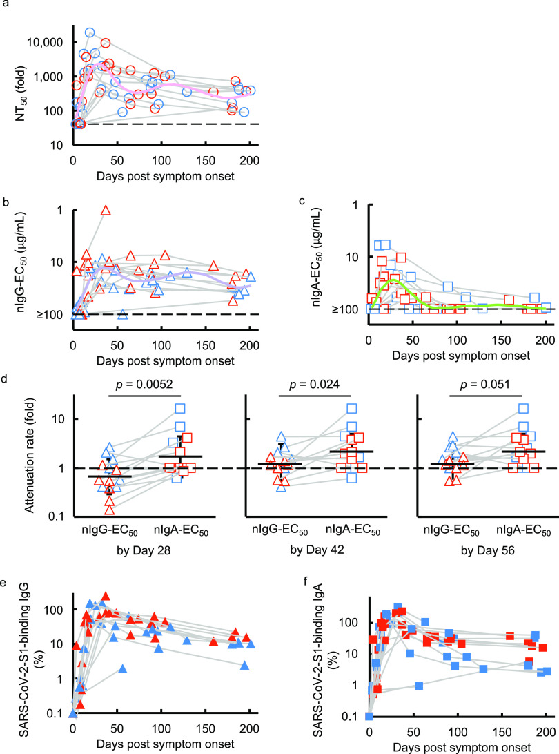
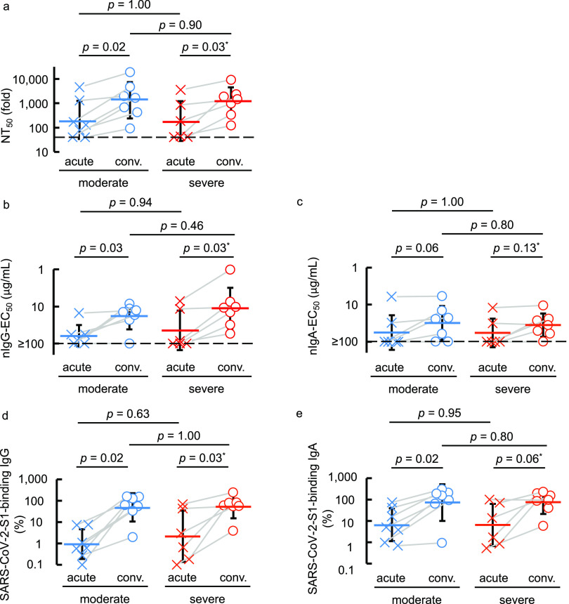
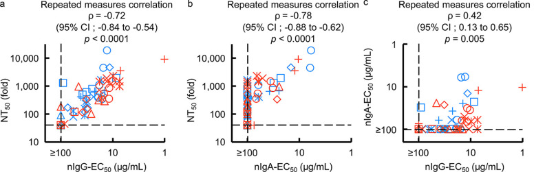
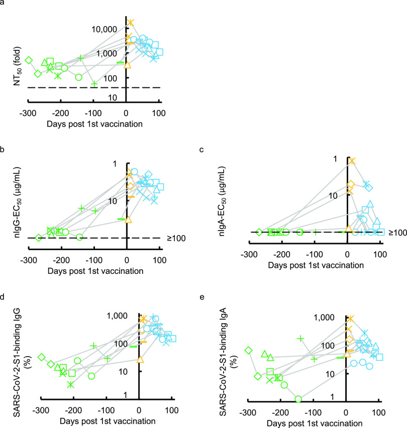
Secretory immunoglobulin A (IgA) plays a crucial role in mucosal immunity for preventing the invasion of exogenous antigens; however, little is understood about the neutralizing activity of serum IgA. Here, to examine the role of IgA antibodies against COVID-19 illnesses, we determined the neutralizing activity of serum/plasma IgG and IgA purified from previously SARS-CoV-2-infected and COVID-19 mRNA vaccine-receiving individuals. We found that serum/plasma IgA possesses substantial but rather modest neutralizing activity against SARS-CoV-2 compared to IgG with no significant correlation with the disease severity. Neutralizing IgA and IgG antibodies achieved the greatest activity at approximately 25 and 35 days after symptom onset, respectively. However, neutralizing IgA activity quickly diminished to below the detection limit approximately 70 days after onset, while substantial IgG activity was observed until 200 days after onset. The total neutralizing activity in sera/plasmas of those with COVID-19 largely correlated with those in purified IgG and purified IgA and levels of anti-SARS-CoV-2-S1-binding IgG and anti-SARS-CoV-2-S1-binding IgA. In individuals who were previously infected with SARS-CoV-2 but had no detectable neutralizing IgA activity, a single dose of BNT162b2 or mRNA-1273 elicited potent serum/plasma-neutralizing IgA activity, but the second dose did not further strengthen the neutralization antibody response. The present data show that the systemic immune stimulation with natural infection and COVID-19 mRNA-vaccines elicits both SARS-CoV-2-specific neutralizing IgG and IgA responses in serum, but the IgA response is modest and diminishes faster than the IgG response. IMPORTANCE Secretory dimeric immunoglobulin A (IgA) plays an important role in preventing the invasion of foreign objects by its neutralizing activity on mucosal surfaces, while monomeric serum IgA is thought to relate to the phagocytic immune system activation. Here, we report that individuals with the novel coronavirus disease (COVID-19) developed both systemic neutralizing IgG (nIgG) and IgA (nIgA) active against severe acute respiratory syndrome coronavirus 2 (SARS-CoV-2). Although the nIgA response was quick and reached the highest activity earlier than the nIgG response, nIgA activity was modest and diminished faster than nIgG activity. In individuals who recovered from COVID-19 but had no detectable nIgA activity, a single dose of COVID-19 mRNA vaccine elicited potent nIgA activity, but the second dose did not further strengthen the antibody response. Our study provides novel insights into the role and the kinetics of serum nIgA against the pathogen in both naturally infected and COVID-19 mRNA vaccine-receiving COVID-19-convalescent individuals.

Keywords: COVID-19; COVID-19 mRNA vaccine; SARS-CoV-2; anti-SARS-CoV-2 immunoglobulin; humoral immunity; immunoglobulin A; immunoglobulin G; neutralizing antibodies.


Conflict of interest statement

The authors declare no conflict of interest.

Figures

FIG 1
FIG 1
Kinetics of SARS-CoV-2-neutralizing activity and S1-binding antibodies. (a to c) VeroE6TMPRSS2 cells were exposed to wild-type SARS-CoV-205-2N with or without various concentrations of diluted sera/plasmas (a), purified IgG (b), or purified IgA (c), and the neutralizing activity and the amounts of S1-binding antibodies were determined. The dashed line denotes the assay limit values (≤40-fold for the panel a and ≥100 μg/mL for panels b and c). Note that the highest viral neutralizing activity of purified IgG and IgA was seen around 35 and 25 days, respectively, after onset. Furthermore, the neutralizing activity of serum IgA diminished much quicker than that of IgG. The colored lines (pink, NT50 for pink; purple, nIgG-EC50; light green, nIgA-EC50) denote the fitted curve. (d) Attenuation rate of the nIgG-EC50 and nIgA-EC50 between the highest neutralizing activity of purified IgG and IgA by day 28, 42, and 56 postonset and neutralizing activity determined latest in the study. (e and f) The kinetics of the amount of S1-binding IgG (e) and IgA (f) are also shown. The amount of S1-binding IgG and IgA increased by approximately day 21 post-symptom onset, followed by a gradual decrease. Note that in contrast, substantial amounts of S1-binding IgG and IgA persisted around 200 days after onset, while the decay occurred more rapidly in IgA. Blue symbols denote the samples collected from individuals with moderate symptoms, while red symbols are those from individuals with severe symptoms.
FIG 2
FIG 2
COVID-19-convalescent individuals possess greater neutralizing activity and SARS-CoV-2-S1-binding antibody levels than those at the acute phase. (a to e) The neutralizing activity of sera/plasmas, purified IgG, and purified IgA (a, b, and c, respectively) and the amounts of S1-binding IgG and IgA (d and e, respectively) were compared between the acute phase (less than 14 days post-symptom onset or when the individual required oxygen treatment) and the convalescent phase (14 days post-symptom onset and beyond which no oxygen was required). Blue symbols denote the samples collected from individuals with moderate symptoms, while red symbols are those from individuals with severe symptoms.
FIG 3
FIG 3
Correlations of purified IgG and IgA neutralizing activities with serum/plasma neutralizing titers. (a and b) The neutralizing activity of purified-IgG (a) (nIgG-EC50) and -IgA (b) (nIgA-EC50) against serum/plasma neutralizing activity (NT50) values are plotted. (c) The nIgA-EC50 values are plotted against the nIgG-EC50 values. Note that a high correlation is observed between NT50 values and nIgG-EC50 values (repeated measures correlation ρ = −0.72 [95% CI, –0.84 to –0.54]) (a) and between the NT50 values and NIgA-EC50 values (ρ = –0.78 [95% CI, –0.88 to –0.62]) (b), while moderate correlation was observed between nIgA-EC50 and nIgG-EC50 (repeated measures correlation ρ = 0.42 [95% CI, 0.13 to 0.65]) (c). Each symbol denotes the sample from one individual. Blue symbols denote the samples collected from individuals with moderate symptoms, while red symbols denote those from individuals with severe symptoms.
FIG 4
FIG 4
Kinetics of neutralizing activity and S1-binding antibody levels before and after COVID-19 mRNA vaccination. (a to e) The kinetics of neutralizing activity (a, b, and c) and S1-binding antibody levels (d and e) in eight previously COVID-experienced individuals who received the COVID-19 mRNA vaccine are shown. Note that all the values significantly rose after the first dose of the vaccine. Also note that none of the participants had detectable nIgA-EC50 values (≥100 μg/mL) before vaccination (c). On the other hand, all of them had low to high levels of nIgA-EC50 after a single dose of the vaccine, and such levels quickly decreased (c), and their S1-binding IgA levels persisted after the two doses of vaccine in the study period (e). The dashed line denotes the assay detection limit (≤40-fold dilution for NT50 and ≥100 μg/mL for nIgG-EC50 and nIgA-EC50). Green symbols denote the samples collected before COVID-19 mRNA vaccination, while yellow and light-blue symbols denote samples collected after the first and second doses, respectively. Each symbol denotes the sample from one individual.

Update of

Similar articles

Cited by

References

    1. Cerutti A, Rescigno M. 2008. The biology of intestinal immunoglobulin A responses. Immunity 28:740–750. doi:10.1016/j.immuni.2008.05.001. - DOI - PMC - PubMed
    1. Cerutti A, Cols M, Gentile M, Cassis L, Barra CM, He B, Puga I, Chen K. 2011. Regulation of mucosal IgA responses: lessons from primary immunodeficiencies. Ann N Y Acad Sci 1238:132–144. doi:10.1111/j.1749-6632.2011.06266.x. - DOI - PMC - PubMed
    1. Bäckhed F, Ley RE, Sonnenburg JL, Peterson DA, Gordon JI. 2005. Host-bacterial mutualism in the human intestine. Science 307:1915–1920. doi:10.1126/science.1104816. - DOI - PubMed
    1. Mazanec MB, Coudret CL, Fletcher DR. 1995. Intracellular neutralization of influenza virus by immunoglobulin A anti-hemagglutinin monoclonal antibodies. J Virol 69:1339–1343. doi:10.1128/JVI.69.2.1339-1343.1995. - DOI - PMC - PubMed
    1. Stiehm ER. 2008. The four most common pediatric immunodeficiencies. J Immunotoxicol 5:227–234. doi:10.1080/15476910802129646. - DOI - PubMed

Publication types