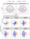
Letter to the Editor: Expected Improvement in Structure-Function Agreement With Macular Displacement Models
- PMID: 36219162
- PMCID: PMC9580223
- DOI: 10.1167/tvst.11.10.14
Letter to the Editor: Expected Improvement in Structure-Function Agreement With Macular Displacement Models
Conflict of interest statement
Disclosure:
Figures


Comment in
-
Author Response: Expected Improvement in Structure-Function Agreement With Macular Displacement Models.Transl Vis Sci Technol. 2022 Oct 3;11(10):15. doi: 10.1167/tvst.11.10.15. Transl Vis Sci Technol. 2022. PMID: 36219161 Free PMC article. No abstract available.
Comment on
-
Clinical Evaluations of Macular Structure-Function Concordance With and Without Drasdo Displacement.Transl Vis Sci Technol. 2022 Apr 1;11(4):18. doi: 10.1167/tvst.11.4.18. Transl Vis Sci Technol. 2022. PMID: 35438719 Free PMC article.
Similar articles
-
Macular edema in branch retinal vein occlusion: types and treatment.Ophthalmic Surg. 1989 Jan;20(1):26-32. Ophthalmic Surg. 1989. PMID: 2927879
-
Retinal edema. Introduction to the First International Cystoid Macular Edema Symposium.Surv Ophthalmol. 1984 May;28 Suppl:433-6. doi: 10.1016/0039-6257(84)90224-8. Surv Ophthalmol. 1984. PMID: 6463845 No abstract available.
-
[Relationship between vitrectomy and the morphology and function of the retina].Nippon Ganka Gakkai Zasshi. 2003 Dec;107(12):836-64; discussion 865. Nippon Ganka Gakkai Zasshi. 2003. PMID: 14733133 Review. Japanese.
-
Retinal astrocytic hamartoma with associated macular edema: report of spontaneous resolution of macular edema as a result of increasing hamartoma calcification.Semin Ophthalmol. 2007 Jul-Sep;22(3):171-3. doi: 10.1080/08820530701501006. Semin Ophthalmol. 2007. PMID: 17763239
-
[Retinal complications of cataract surgery].J Fr Ophtalmol. 2000 Jan;23(1):88-95. J Fr Ophtalmol. 2000. PMID: 10733361 Review. French.
Cited by
-
Rapid Campimetry in glaucoma - correspondence with standard perimetry and OCT.Sci Rep. 2024 Oct 25;14(1):25400. doi: 10.1038/s41598-024-75037-5. Sci Rep. 2024. PMID: 39455627 Free PMC article.
References
-
- Jansonius NM, Schiefer J, Nevalainen J, Paetzold J, Schiefer U. A mathematical model for describing the retinal nerve fiber bundle trajectories in the human eye: average course, variability, and influence of refraction, optic disc size and optic disc position. Exp Eye Res. 2012; 105: 70–78. - PubMed
Publication types
MeSH terms
LinkOut - more resources
Full Text Sources
Medical

