. 2023 Dec;22(6):1137-1151.
doi: 10.1007/s12311-022-01488-z. Epub 2022 Oct 11.

PTRH2 is Necessary for Purkinje Cell Differentiation and Survival and its Loss Recapitulates Progressive Cerebellar Atrophy and Ataxia Seen in IMNEPD Patients

Affiliations

PTRH2 is Necessary for Purkinje Cell Differentiation and Survival and its Loss Recapitulates Progressive Cerebellar Atrophy and Ataxia Seen in IMNEPD Patients

Sylvie Picker-Minh et al. Cerebellum. 2023 Dec.

Abstract

Hom ozygous variants in the peptidyl-tRNA hydrolase 2 gene (PTRH2) cause infantile-onset multisystem neurologic, endocrine, and pancreatic disease. The objective is to delineate the mechanisms underlying the core cerebellar phenotype in this disease. For this, we generated constitutive (Ptrh2LoxPxhCMVCre, Ptrh2-/- mice) and Purkinje cell (PC) specific (Ptrh2LoxPxPcp2Cre, Ptrh2ΔPCmice) Ptrh2 mutant mouse models and investigated the effect of the loss of Ptrh2 on cerebellar development. We show that Ptrh2-/- knockout mice had severe postnatal runting and lethality by postnatal day 14. Ptrh2ΔPC PC specific knockout mice survived until adult age; however, they showed progressive cerebellar atrophy and functional cerebellar deficits with abnormal gait and ataxia. PCs of Ptrh2ΔPC mice had reduced cell size and density, stunted dendrites, and lower levels of ribosomal protein S6, a readout of the mammalian target of rapamycin pathway. By adulthood, there was a marked loss of PCs. Thus, we identify a cell autonomous requirement for PTRH2 in PC maturation and survival. Loss of PTRH2 in PCs leads to downregulation of the mTOR pathway and PC atrophy. This suggests a molecular mechanism underlying the ataxia and cerebellar atrophy seen in patients with PTRH2 mutations leading to infantile-onset multisystem neurologic, endocrine, and pancreatic disease.

Keywords: Ataxia; Cell survival; IMNEPD; PTRH2; Purkinje cells; mTOR.


Conflict of interest statement

The authors declare no competing interests.

Figures

Fig. 1
Fig. 1
Ptrh2−/− mice are normal at birth but at P5 show runting syndrome and impaired cerebellar development (AD = P0) (EH = P5) (A) P0 pup Ptrh2 +/+ (left) and Ptrh2−/− (right). P0 mid-sagittal cerebellum section from (B) Ptrh2 +/+ and (C) Ptrh2−/− mice (HE, scalebar 200 µm). (D) Quantitation of cerebellar volume at P0. (E) P5 pups, Ptrh2 +/+ (left) and Ptrh2−/− (right). P5 mid-sagittal cerebellum section from (F) Ptrh2 +/+ and (G) Ptrh2.−/− mice (HE, scalebar 200 µm). (H) Quantitation of cerebellar volume at P5. Quantitation based on n = 6 animals/group. ns not significant, * p < 0.05
Fig. 2
Fig. 2
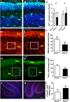
P5 Ptrh2−/− mice have reduced cell proliferation in the EGL and reduced number of neurons in the IGL. Ki67 IHC (green) in (A) Ptrh2+/+ and (B) Ptrh2−/− cerebellum (scalebar 25 µm). (C) Quantitation of Ki67 + cells in the EGL of the anterior (A) and posterior (P) lobes. MBH2 IHC (red) in (D) Ptrh2+/+ and (E) Ptrh2−/− cerebellum (scalebar 50 µm). Box denotes region where cells were counted. (F) Quantitation of MBH2 + cells. (positive cells per 2 × 2 cm viewing field). TLX3 IHC (green) in (G) Ptrh2+/+ and (H) Ptrh2−/− cerebellum (scalebar 50 µm). Box denotes region where cells were counted. (I) Quantitation of TLX3 + cells in the posterior cerebellum (positive cells per 400 × 600Px viewing field) Calbindin (red) Cleaved caspase3 (green) IHC (J) Ptrh2+/+ and (K) Ptrh2.−/− cerebellum (scalebar 100 µm). (L) Quantitation of Caspase 3 + cells in the IGL. Blue = DAPI. Quantitation based on n = 6 animals/group. *p < 0.05 **p < 0.01, ***p < 0.0001
Fig. 3
Fig. 3
P5 Ptrh2−/− mice have abnormal PC morphology. IHC for Calbindin (red) and FoxP4 (green) in (A) Ptrh2+/+ and (C) Ptrh2−/− cerebella. Detail of calbindin stained PCs at higher magnification (B) Ptrh2+/+ (D) Ptrh2.−/− cerebella. (a and c: scalebar 50 µm, b and d: detail of PC morphology). (E) Quantitation of PC surface area (n = 6 animals/group). *p < 0.05
Fig. 4
Fig. 4
PC culture from Ptrh2−/− mice are abnormal. IHC for Calbindin (red) and GFAP (green) in DIV12 mixed cerebellar cultures from (A) Ptrh2+/+ (B) Ptrh2−/− mice. (C) Quantitation of PC soma size (n = 6, number of cells = approx. 60 per genotype). Morphology of PC from DIV12 cultures from (D) Ptrh2+/+ (E) Ptrh2.−/− mice. (F) Quantitation of somatic + dendritic area of PCs, n = 6 number of cells = approx. 60 per genotype, *p < 0.05
Fig. 5
Fig. 5
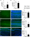
SHH signaling is reduced in the cerebellum of Ptrh2−/− mice. Quantitative PCR for Shh at (A) P0 and (B) P4 (P0 (n = 3/group) and P4 (n = 3/group)). (C) SHH western blot from P5 cerebellum with representative blot showed above graph (n = 3/group). SHH (green) IHC in P5 cerebella (D) Ptrh2+/+ and (E) Ptrh2−/− mice. SHH images merged with DAPI (blue) (F) Ptrh2+/+ and (G) Ptrh2−/− (n = 6). Quantitative PCR for Gli1 at (H) P0 and (I) P4 (P0 (n = 3) and P4 (n = 3)). GLI1 (green) IHC merged with DAPI (blue) in P5 cerebella (J) Ptrh2+/+ and (K) Ptrh2−/− mice (n = 5). Scalebar 100 µm
Fig. 6
Fig. 6
Ptrh2ΔPC mice show adult cerebellar atrophy. H&E staining of midsagittal section at P5 showing cerebellum of (A) Ptrh2+/+ and (B) Ptrh2ΔPC mice. (C) Ptrh2+/+ (top) and Ptrh2ΔPC (bottom) mice at 8 weeks. H&E staining of midsagittal section at 8 weeks (D) Ptrh2+/+ and (E) Ptrh2.ΔPC (H&E, scalebar 400 µm) (F) quantitation of cerebellar volume (n = 5) *p < 0.05
Fig. 7
Fig. 7
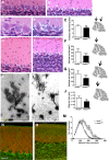
Purkinje cell degeneration in the adult Ptrh2ΔPC mice. H&E staining showing PC layer in (A) Ptrh2 +/+ and (B) Ptrh2ΔPC mice. (C) Quantitation of cell density in two different areas (scalebar 20 µm). H&E staining for PC soma quantitation (D) Ptrh2 +/+ and (E) Ptrh2ΔPC mice (F and G) quantitation of PC soma size in two different lobes indicated by black arrow (scalebar 20 µm). H&E staining of molecular layer in (H) Ptrh2 +/+ and (I) Ptrh2ΔPC mice (scalebar 20 µm). (J) Quantitation of height of the molecular layer. (n = 6/group). Golgi staining of a PC in (K) Ptrh2 +/+ and (L) Ptrh2ΔPC mice and (M) Sholl analysis of dendrites (n = 30 cells/6 animals, mean ± SEM; *p < 0.05, **p < 0.01). Calbindin (red) and GFAP (green) IHC in 8-week-old (N) Ptrh2 +/+ and (O) Ptrh2.ΔPC mice. (Scalebar 50 µm). *p < 0.05, **p < 0.01, ***p < 0.0001
Fig. 8
Fig. 8
Adult Ptrh2ΔPC mice develop motor deficits. Footprint analysis in 6-month-old mice. Footprint pattern in (A) Ptrh2 +/+ and (B) Ptrh2ΔPC mice. Stride length of (C) forepaws and (D) hindpaws. (E) Forepaw (FP)/hindpaw (HP) overlap. (F) Forebase width, (G) hindbase width (n = 22). Composite phenotype score of (H) 6-month-old mice (n = 9) and (I) 18-month-old mice (n = 15). Calbindin IHC of 18-month-old mice (J) Ptrh2 +/+ and (K) Ptrh2.ΔPC (scalebar 100 µm). (ns, not significant *p < 0.05, **p < 0.01, ***p < 0.0001)
Fig. 9
Fig. 9
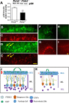
Downregulation of pS6 in Ptrh2−/− and Ptrh2ΔPC mice. (A) Western blot for pS6 from P5 cerebella with representative blot shown above. IHC for Calbindin (green) and pS6 (red) in P5 cerebellum (BC) Ptrh2 +/+ (DE) Ptrh2ΔPC. Yellow arrows in (D) and (E) point to cells that have downregulated pS6. IHC for calbindin (green) and pS6 (red) in adult cerebellum (F and G) Ptrh2 +/+ (HI) Ptrh2ΔPC mice. Scalebar 50 µm. **p < 0.01. Model of cerebellar development in Ptrh2−/− and Ptrh2ΔPC mice. (J) Ptrh2 expressing PCs are morphologically mature, and the secreted Shh reaches the outer EGL causing Gli mediated signal transduction and proliferation in the GNPs. Post-mitotic GNs migrate along glial fibers to form the IGL layer. (K) In the absence of Ptrh2 expression in PCs, they are morphologically stunted, and although there is no difference in the level (depicted by the yellow arrow width), the Shh morphogenetic gradient (depicted by the length of the yellow arrow) is diminished causing lower Gli mediated signal transduction and proliferation. This leads to lower numbers of GNs in the IGL

Similar articles

Cited by

References

    1. Hu H, et al. Mutations in PTRH2 cause novel infantile-onset multisystem disease with intellectual disability, microcephaly, progressive ataxia, and muscle weakness. Ann Clin Transl Neurol. 2014;1(12):1024–1035. doi: 10.1002/acn3.149. - DOI - PMC - PubMed
    1. Le C, et al. Infantile-onset multisystem neurologic, endocrine, and pancreatic disease: case and review. Canadian J Neurol Sci / J Canadien des Sciences Neurologiques. 2019;46(4):459–463. doi: 10.1017/cjn.2019.35. - DOI - PubMed
    1. Picker-Minh S, et al. Phenotype variability of infantile-onset multisystem neurologic, endocrine, and pancreatic disease IMNEPD. Orphanet J Rare Dis. 2016;11(1):52. doi: 10.1186/s13023-016-0433-z. - DOI - PMC - PubMed
    1. Tallquist MD, Soriano P. Epiblast-restricted Cre expression in MORE mice: a tool to distinguish embryonic vs. extra-embryonic gene function. Genesis. 2000;26(2):113–5. doi: 10.1002/(SICI)1526-968X(200002)26:2<113::AID-GENE3>3.0.CO;2-2. - DOI - PubMed
    1. Sabatini DM. Twenty-five years of mTOR: uncovering the link from nutrients to growth. Proc Natl Acad Sci. 2017;114(45):11818. doi: 10.1073/pnas.1716173114. - DOI - PMC - PubMed