. 2023 Apr 25;7(8):1366-1378.
doi: 10.1182/bloodadvances.2022007169.

Effect of antiplatelet agents and tyrosine kinase inhibitors on oxLDL-mediated procoagulant platelet activity

Affiliations

Effect of antiplatelet agents and tyrosine kinase inhibitors on oxLDL-mediated procoagulant platelet activity

Tony J Zheng et al. Blood Adv. .

Abstract

Low-density lipoprotein (LDL) contributes to atherogenesis and cardiovascular disease through interactions with peripheral blood cells, especially platelets. However, mechanisms by which LDL affects platelet activation and atherothrombosis, and how to best therapeutically target and safely prevent such responses remain unclear. Here, we investigate how oxidized low-density lipoprotein (oxLDL) enhances glycoprotein VI (GPVI)-mediated platelet hemostatic and procoagulant responses, and how traditional and emerging antiplatelet therapies affect oxLDL-enhanced platelet procoagulant activity ex vivo. Human platelets were treated with oxLDL and the GPVI-specific agonist, crosslinked collagen-related peptide, and assayed for hemostatic and procoagulant responses in the presence of inhibitors of purinergic receptors (P2YR), cyclooxygenase (COX), and tyrosine kinases. Ex vivo, oxLDL enhanced GPVI-mediated platelet dense granule secretion, α-granule secretion, integrin activation, thromboxane generation and aggregation, as well as procoagulant phosphatidylserine exposure and fibrin generation. Studies of washed human platelets, as well as platelets from mouse and nonhuman primate models of hyperlipidemia, further determined that P2YR antagonists (eg, ticagrelor) and Bruton tyrosine kinase inhibitors (eg, ibrutinib) reduced oxLDL-mediated platelet responses and procoagulant activity, whereas COX inhibitors (eg, aspirin) had no significant effect. Together, our results demonstrate that oxLDL enhances GPVI-mediated platelet procoagulant activity in a manner that may be more effectively reduced by P2YR antagonists and tyrosine kinase inhibitors compared with COX inhibitors.


Conflict of interest statement

Conflict-of-interest disclosure: The authors declare no competing financial interests.

Figures

None
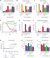
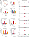
Graphical abstract
Figure 1.
Figure 1.
Elevated oxLDLand enhancedGPVI-mediated procoagulant platelet generation in obese nonhuman primates. Blood was collected from a cohort of lean (n = 6) and diet-induced obese (n = 6) rhesus macaques to prepare serum for analysis of triglycerides (A), total cholesterol (B), high-density lipoprotein cholesterol (C), LDL cholesterol (D), and oxLDL immunoreactivity (E). Citrate-anticoagulated whole blood from lean and obese animals was stimulated with GPVI agonist (0.1 μg/mL CRP-XL) or a combination of GPVI and PAR1 agonist (1 μg/mL CRP-XL, 20 μg/mL TRAP-6) before flow cytometry analysis of platelet α-granule secretion (F) and platelet PS exposure (G). Statistical analysis was performed using a one-way analysis of variance (ANOVA) test and the corresponding P values of significance are as indicated: ∗P < .01, ∗∗P < .001, and ∗∗∗P < .0001. CRP-XL, crosslinked collagen-related peptide.
Figure 2.
Figure 2.
oxLDL potentiates GPVI-mediated platelet aggregation, platelet PS exposure, and platelet-driven fibrin formation. Replicate samples of washed human platelets (2 × 108/mL, in modified HEPES/Tyrode buffer without additional calcium) were incubated with the GPVI-specific agonist CRP-XL) alone (0.1, 0.5, 1 μg/mL), oxLDL alone (20 μg/mL), and oxLDL in combination with CRP-XL. Platelet samples were monitored at 37°C under continuous stirring at 1200 rpm and changes in light transmission were measured (A-D). (E) Washed platelets (2 × 108/mL) were incubated with oxLDL (0, 20 μg/mL) in combination with CRP-XL (0, 0.1, 0.5, 1 μg/mL) and monitored using luciferase enzyme activity (CHRONO-LUME) to detect ATP released as a measure of platelet dense granule secretion. (F) Platelets were stained with APC-CD62P to monitor for platelet surface expression of P-selectin as a marker of α-granule secretion and (G) stained with FITC-PAC-1 to monitor for platelet surface integrin activation. (H) Platelets were incubated with oxLDL (0, 20 μg/mL) in combination with GPVI-specific agonist CRP-XL (0, 0.1, 0.5, 1 μg/mL) before fixation and staining with FITC-lactadherin to monitor for platelet surface PS exposure. (I) Platelets were similarly assessed with FITC-lactadherin staining in response to oxLDL and CRP-XL stimulation after a 10-minute preincubation with CD36 inhibitors Fx-5A (250 μM), sulfo-N-succinimidyl oleate (“sulfo,” 50 μM), or FA6-152 (5 μg/mL). (J) Platelets were incubated with CaCl2 (8.3 mM) and citrated PPP (33% final). Fibrin formation was measured as a change in turbidity at an absorbance of 405 nm. (K) Representative traces are shown and the lag time was calculated. Statistical analysis was performed using a one-way ANOVA test and the corresponding P values of significance are as indicated: ∗P < .05, ∗∗P < .01, and ∗∗∗P < .001. ATP, adenosine triphosphate; CaCl2, calcium dichloride; FITC, fluorescein isothiocyanate; HEPES, N-2-hydroxyethylpiperazine-N′-2-ethanesulfonic acid; PPP, platelet-poor plasma.
Figure 3.
Figure 3.
P2YR antagonists but not COX inhibitors reduce oxLDL potentiation of platelet GPVI responses. (A) Replicate samples (n ≥ 3) of washed human platelets (2 × 108/mL; in modified HEPES/Tyrode buffer without addition of calcium) were treated with P2YR inhibitors ticagrelor (750 nM) and ARC/MRS (10 μM), and COX inhibitors aspirin (1 mM) and indomethacin (10 μM) for 10 minutes and stimulated with oxLDL (20 μg/mL) in combination with GPVI-specific agonist CRP-XL (0.5 μg/mL). Platelet samples were monitored using the luciferase enzyme CHRONO-LUME to detect the luminescence of ATP released as a measure of platelet dense granule secretion. (B) Platelets were stained with APC-CD62P to monitor for platelet surface expression of P-selectin and (C) stained with FITC-PAC-1 to monitor for platelet surface integrin activation. Statistical analysis was performed using a one-way ANOVA test and the corresponding P values of significance are as indicated: ∗∗∗P < .001. (D) Replicate samples (n = 4) of washed human platelets (2 × 108/mL) were treated with P2YR inhibitors ticagrelor (750 nM) and ARC/MRS (10 μM), and COX inhibitors aspirin (1 mM) and indomethacin (10 μM) for 10 minutes and stimulated with oxLDL (20 μg/mL) in combination with GPVI-specific agonist CRP-XL (0.5 μg/mL). Platelet samples were monitored at 37°C under continuous stirring at 1200 rpm and the changes in light transmission were measured. (E) Representative traces of platelet samples treated with each select inhibitor are shown and quantified. (F) Platelets were stained with FITC-lactadherin to monitor for platelet surface PS exposure. (G) Platelet samples were incubated with CaCl2 (8.3 mM) and citrated PPP (33% final). Fibrin formation was measured as a change in turbidity at an absorbance of 405 nm. (H-I) Representative traces are shown and the lag time and time to half the maximum absorbance were calculated. Statistical analysis was performed using a one-way ANOVA test and the corresponding P values of significance are as indicated: ∗P < .05; ns, not significant.
Figure 4.
Figure 4.
Antiplatelet agents and TKIs differentially reduce oxLDL potentiation of platelet GPVI/ITAM signaling. Replicate samples (n = 3) of washed human platelets (1 × 109/mL; in modified HEPES/Tyrode buffer without addition of calcium) were pretreated with P2YR inhibitor ticagrelor, BTK inhibitor ibrutinib (10 μM), or Syk inhibitor R406 (10 μM) for 10 minutes and then stimulated with oxLDL (20 μg/mL) in combination with GPVI-specific agonist CRP-XL (0.5 μg/mL) for 5 minutes at 37°C. After collection into sample buffer, platelet lysates were separated by capillary electrophoresis and stained with indicated antisera for quantitative immunoblot analysis on a ProteinSimple Jess western blotting system. (A) Representative immunoblot analysis and (B) quantitation of signal intensities with indicated antisera; α-tubulin serves as a loading control for total protein levels. Positions of molecular weight (kD) markers are indicated.
Figure 5.
Figure 5.
BTK and Syk inhibitors reduce oxLDL potentiation of platelet hemostatic and procoagulant activity. (A) Replicate samples (n = 4) of washed human platelets (2 × 108/mL; in modified HEPES/Tyrode buffer without addition of calcium) were treated with BTK inhibitor ibrutinib (10 μM) or Syk inhibitor R406 (10 μM) for 10 minutes and stimulated with GPVI-specific agonist CRP-XL (0.5 μg/mL) in combination with oxLDL (20 μg/mL). Platelets were monitored at 37°C under continuous stirring at 1200 rpm and the changes in light transmission were measured. Representative platelet aggregation traces of platelet samples treated with each select inhibitor are shown. (B) Platelets were stained with FITC-lactadherin to monitor for platelet surface PS exposure. Platelet samples were incubated with CaCl2 (8.3 mM) and citrated PPP (33% final). Fibrin formation was measured as a change in turbidity at an absorbance of 405 nm and the lag time (C) was quantified. (D) Platelets were monitored using the luciferase enzyme CHRONO-LUME to detect the luminescence of ATP released as a measure of platelet dense granule secretion, stained with APC-CD62P to monitor for platelet surface expression of P-selectin as a marker of α-granule secretion (E), and stained with FITC-PAC-1 to monitor for platelet surface integrin activation (F). Following the platelet aggregation assays shown in panel A, platelet supernatants were extracted and analyzed for thromboxane generation by ELISA (G). Additional platelet supernatant samples were similarly prepared from platelets treated with oxLDL, CRP-XL, and inhibitors of interest to measure levels of indicated oxylipin metabolite levels with mass spectrometry (H). Statistical analysis was performed using a one-way ANOVA test and the corresponding P values of significance are as indicated: ∗P < .05, ∗∗P < .01, and ∗∗∗P < .001. ELISA, enzyme-linked immunosorbent assay.
Figure 6.
Figure 6.
oxLDL enhances platelet procoagulant activity in vivo. (A) Wild-type C57BL/6J mice (median age, 6 weeks) were intravenously injected with vehicle (phosphate-buffered saline) alone or oxLDL 10 minutes before blood collection into citrate (n = 5 per condition). Whole blood samples were analyzed for platelet-monocyte aggregate formation as previously described., (B) Whole blood samples from vehicle- and oxLDL-injected animals were subsequently stimulated with GPVI agonist (0.1 μg/mL CRP-XL) before flow cytometry analysis of CD62P as a marker of platelet α-granule secretion. (C) Sets of mice (n = 5 each) were administered by vehicle (dimethyl sulfoxide), ticagrelor (100 mg/kg), aspirin (30 mg/kg), or ibrutinib (10 mg/kg) by intraperitoneal injection before oxLDL injection, blood collection, and flow cytometry analysis of FITC-lactadherin to follow platelet PS exposure. (D) Aliquots of citrate-anticoagulated whole blood from obese rhesus macaques (detailed in Figure 1, in previous section) were pretreated with vehicle (dimethyl sulfoxide), ticagrelor, aspirin, or ibrutinib before stimulation with GPVI agonist (0.1 μg/mL CRP-XL), lactadherin-FITC staining, and flow cytometry assessment of platelet PS exposure. Statistical analysis was performed using a one-way ANOVA test and the corresponding P values of significance are as indicated: ∗P < .01.

References

    1. Wendelboe AM, Raskob GE. Global burden of thrombosis: epidemiologic aspects. Circ Res. 2016;118(9):1340–1347. - PubMed
    1. Herrington W, Lacey B, Sherliker P, Armitage J, Lewington S. Epidemiology of atherosclerosis and the potential to reduce the global burden of atherothrombotic disease. Circ Res. 2016;118(4):535–546. - PubMed
    1. Ahmad FB, Anderson RN. The leading causes of death in the US for 2020. JAMA. 2021;325(18):1829–1830. - PMC - PubMed
    1. Tomaiuolo M, Brass LF, Stalker TJ. Regulation of platelet activation and coagulation and its role in vascular injury and arterial thrombosis. Interventional cardiology clinics. 2017;6(1):1. - PMC - PubMed
    1. Podrez EA, Byzova TV, Febbraio M, et al. Platelet CD36 links hyperlipidemia, oxidant stress and a prothrombotic phenotype. Nat Med. 2007;13(9):1086–1095. - PMC - PubMed

Publication types