

Multifocal Sarcoma Mimicking Superficial Vein Thrombosis of the Leg: A Case Report
- PMID: 36219592
- PMCID: PMC9575138
- DOI: 10.12659/AJCR.937317
Multifocal Sarcoma Mimicking Superficial Vein Thrombosis of the Leg: A Case Report
Abstract
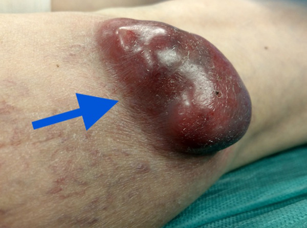
BACKGROUND Leiomyosarcoma is a common tumor found in soft tissue. In relation to the vascular system, leiomyosarcoma appears as the most common malignancy characterized by poor prognosis. Leiomyosarcomas of the leg large vessels often occur late, and their appearance can imitate vein thrombosis with symptoms such as soft tissue swelling or mild pain, and can be misdiagnosed. Peripheral vascular leiomyosarcomas are rare. Especially leiomyosarcomas of the great saphenous vein are uncommon. The tumors develop on the media basis and grow from endovascular to exovascular order. Distant metastasis can be identified and worsen prognosis. CASE REPORT We present a case of a 61-year-old female patient with varicose vein disease complicated by recurrent superficial vein thrombosis. After 2 months of conservative treatment, while waiting for admission to the department of surgery, she developed additional symptoms. Clinical examination on the day of admission revealed several tumors along and near the great saphenous vein on the left limb below the knee. The diagnosis of leiomyosarcoma was confirmed after the surgery, involving excision of the saphenous vein, including tumors formed on its course. Preoperative clinical and ultrasound findings did not suggest malignancy. CONCLUSIONS Leiomyosarcoma of the great saphenous vein is an extraordinarily rare tumor originating from the middle layer of the vessel, mimicking unspecific symptoms and complicating and delaying diagnosis. In every case of vascular or perivascular lesions, a detailed examination and diagnosis it is required, and even unlikely clinical scenarios should be considered.
Conflict of interest statement
Figures


References
-
- Kalodiki E, Stvrtinova V, Allegra C, et al. Superficial vein thrombosis: A consensus statement. Int Angiol. 2012;31(3):203–16. - PubMed
Publication types
MeSH terms
LinkOut - more resources
Full Text Sources
Medical
